- 1Cancer Center, The First Hospital of Jilin University, Changchun, China
- 2International Center of Future Science, Jilin University, Changchun, China
- 3Institute of Translational Medicine, The First Hospital of Jilin University, Changchun, China
- 4Cancer Research Institute of Jilin University, The First Hospital of Jilin University, Changchun, China
Introduction: Cytokine release syndrome (CRS) is one of the leading causes of mortality in patients with COVID-19 caused by the SARS-CoV-2 coronavirus. However, the mechanism of CRS induced by SARS-CoV-2 is vague.
Methods: Using spike protein combined with IL-2, IFN-γ, and TNF-α to stimulate human peripheral blood mononuclear cells (PBMCs) to secrete CRS-related cytokines, the content of cytokines in the supernatant was detected, and the effects of NK, T, and monocytes were analyzed.
Results: This study shows that dendritic cells loaded with spike protein of SARS-CoV-2 stimulate T cells to release much more interleukin-2 (IL-2,) which subsequently cooperates with spike protein to facilitate PBMCs to release IL-1β, IL-6, and IL-8. These effects are achieved via IL-2 stimulation of NK cells to release tumor necrosis factor-α (TNF-α) and interferon-γ (IFN-γ), as well as T cells to release IFN-γ Mechanistically, IFN-γ and TNF-α enhance the transcription of CD40, and the interaction of CD40 and its ligand stabilizes the membrane expression of toll-like receptor 4 (TLR4) that serves as a receptor of spike protein on the surface of monocytes. As a result, there is a constant interaction between spike protein and TLR4, leading to continuous activation of nuclear factor-κ-gene binding (NF-κB). Furthermore, TNF-α also activates NF-κB signaling in monocytes, which further cooperates with IFN-γ and spike protein to modulate NF-κB–dependent transcription of CRS-related inflammatory cytokines.
Discussion: Targeting TNF-α/IFN-γ in combination with TLR4 may represent a promising therapeutic approach for alleviating CRS in individuals with COVID-19.
1 Introduction
COVID-19, caused by SARS-CoV-2, has become a global pandemic since its outbreak in 2019 (1). As of 26 May 2024, the COVID-19 epidemic has caused as many as 13 million deaths worldwide. The clinical manifestations of severe COVID-19 are diverse, with acute respiratory distress syndrome, cytokine release syndrome (CRS), multiple organ failure, and death being the most notable (2, 3). CRS-related cytokines tend to increase progressively with the severity of the disease (4) and may be the leading cause of life-threatening respiratory diseases in patients with severe COVID-19 (5). Interestingly, it has been observed that the concentration of CRS-related cytokines is higher in the plasma of patients with COVID-19 admitted to the intensive care unit (ICU) than in those who are not (6). These cytokines include tumor necrosis factor–α (TNF-α), interleukin-1β (IL-1β), IL-6, IL-10, IL-17, interferon-γ (IFN-γ), and IL-2 (7, 8). However, the interplay between these cytokines remains unclear. Understanding the relationship between various cytokines in CRS is crucial for developing targeted therapies for COVID-19 and cytokine storm syndromes.
Spike protein is one of the four main structural proteins of SARS-CoV-2 (9), which plays a crucial role in the virus’s ability to enter the host cells as it can bind to the ACE2 receptor (10). It also binds directly to pattern recognition receptor TLR4 to activate downstream signaling pathways that upregulate inflammatory factors such as IL-1β and IL-6 (11, 12). Therefore, spike protein may be a critical factor contributing to CRS in patients with COVID-19. The amount of spike protein increases with the increase of viral load. However, clinical studies have shown no significant difference in viral load between patients with severe and mild COVID-19 (13), and the viral load of patients with COVID-19 is relatively high in the initial stage of infection (14, 15). These studies show that the accumulation of viral antigens may not be the leading cause of CRS. These findings indicate that factors other than the viral load may also significantly induce CRS in patients with COVID-19. The intricate interplay between spike protein, immune cells, and cytokines during the CRS remains largely unknown, highlighting the urgent need for further investigation.
During SARS-CoV-2 infection, alveolar macrophages are critical in detecting the virus and producing cytokines and chemokines to recruit innate and adaptive immune cells for virus elimination and disease prevention (16). Of note, macrophages and certain monocyte subsets are pointed out to be the decisive cells of CRS in patients with severe COVID-19 (17). Moreover, the over-activation of the inflammatory response by other immune cells such as neutrophils, dendritic cells (DCs), natural killer (NK) cells, B cells, and T cells can also contribute to CRS in this context (18). Nonetheless, further research is still needed to determine the precise involvement of these cell types in CRS.
In this study, we investigated the mechanism underlying the development of CRS induced by SARS-Cov-2. Our finding suggests a cooperative effect of IL-2 and spike protein in stimulating peripheral blood mononuclear cells (PBMCs) to secrete IL-1β, IL-6, and IL-8. Mechanistically, DCs loaded with spike protein stimulate T cells to secrete IL-2, which subsequently facilitates the production of TNF-α and IFN-γ by NK cells and IFN-γ by T cells. Together, TNF-α and IFN-γ make monocytes more active, and, when stimulated by spike protein of SARS-CoV-2, these cells can release more CRS-related cytokines. Overall, our findings provide insights into the possible mechanism underlying CRS in patients with COVID-19 and illustrate the complex interplay among various cytokines. These findings may pave the way for developing novel therapeutic targets for treating CRS, thereby offering promising avenues for clinical interventions in patients with COVID-19.
2 Materials and methods
2.1 Isolation of PBMCs
The Ethics Committee of the First Hospital of Jilin University approved this study (2020-521). The peripheral blood samples rich in white blood cells were initially obtained from the Changchun Central Blood Station. We recruited 37 donors. Each donor was considered as a separate sample. Each volunteer’s PBMC is considered a biological replicate. All donors who participated in the study were free of SARS-CoV-2 infection, 10 were vaccinated with SARS-CoV-2 vaccine, 5 were not vaccinated when they participated in this study, and the rest were not sure whether to be vaccinated. PBMCs were obtained by Ficoll density gradient centrifugation of lymphocyte separation solution Lymphoprep™ (Axis-Shield, Oslo, Norway). PBMCs were cultured in RPMI-1640 medium (Gibco, Grand Island, NY, USA) in a humidified atmosphere with 5% CO2 at 37°C.
2.2 Spike protein stimulates PBMCs
PBMCs were adjusted to 1 × 10⁶ cells/mL in RPMI-1640 medium, and the PBMCs were inoculated into 24-well plates (NEST Biotechnology Co., Ltd, Wuxi, China) at 400 μL per well. Then, phosphate buffered saline (PBS), spike protein (ACROBiosystems, Beijing, China), IL-2 (100 IU/mL), IL-2 (100 IU/mL) combined with spike protein (10 μM), TNF-α (0.5 ng/mL), TNF-α (0.5 ng/mL) combined with spike protein (10 μM), IFN-γ (1 ng/mL), or IFN-γ (1 ng/mL) combined with spike protein (10 μM) (above cytokines were all from T&L Biological Technology, Beijing, China) was added to PBMCs. After 16 h of incubation, the supernatant was collected for cytokine detection. In some experiments, blocking antibodies against CD40 (10 μg/mL) (Biolegend, San Diego, USA), TNF-α (10 μg/mL) (MedChemExpress, Monmouth Junction, USA), or IFN-γ (10 μg/mL) (eBioscience, San Diego, USA) were added to the cultures.
2.3 Isolation of NK cells, T cells, and monocytes
T cells, NK cells, and monocytes were isolated from PBMCs by EasySep Human T Cell Isolation Kit (StemCell Technologies, Vancouver, Canada), MACSxpress Whole Blood NK Cell Isolation Kit (Miltenyi Biotec, Bergisch Gladbach, Germany), and EasySep Human Monocyte Isolation Kit (StemCell Technologies). Their specific surface antigens can identify monocytes, NK cells, and T cells: CD14+ for monocytes, CD3−CD56+ for NK cells, and CD3+ for T cells. A purity of over 90% can be achieved for each cell type through sorting. Representative purity results are shown in Supplementary Figures S3B–E. In the coculture experiment, the density of monocytes was adjusted to 2 × 10⁵ cells/mL, and NK or T cells were mixed with monocytes of corresponding donors according to the ratio of NK or T cells to monocytes. Cells were inoculated in 24-well plates at 400 μL per well. Then, PBS, spike protein, IL-2, or IL-2 combined with spike protein was added, respectively, and incubated for 16 h. The supernatant was collected, and the cytokines in the supernatant were detected.
2.4 DCs and T-cell coculture
PBMCs were adjusted to 1 × 10⁶ cells/mL in CellGenix® GMP DC serum-free medium (CellGenix, Freiburg, Germany) with GM-CSF (100 ng/mL) and IL-4 (50 ng/mL; both from T&L Biological Technology). Spike protein and TNF-α were added on the fifth day of culture, and DCs were collected on the seventh day. The density of T cells was adjusted to 1 × 10⁶ cells/mL, and DC and T cells were cocultured at a ratio of 1:5 for two days. The supernatant was collected for the detection of IL-2.
2.5 Flow cytometry
The cell density should be adjusted to 1 × 10⁶ cells/mL, and, then, 1 μL of BD GolgiPlug™ (BD Biosciences, San Jose, CA, USA) should be added to each milliliter of the cell suspension. Cells were cultured in an incubator at 37°C for 4 h and washed with PBS once. Following the instructions of BD Cytofix/Cytoperm™ Plus Fixation/Permeabilization Solution Kit (BD Biosciences), cells were fixed, permeabilized, and stained with anti-human TNF-α (Biolegend), anti-human IFN-γ (BD Biosciences), anti-human IL-6 (Biolegend), or isotype control antibodies for 30 min.
To detect CD40 and TLR4 on monocytes, cells were adjusted to 1 × 10⁶ cells/mL and stained with anti-human CD14, anti-human CD40, anti-human TLR4, or isotype control antibodies (the above antibodies were all from Biolegend) at room temperature for 15 min.
PBMCs should be adjusted to a cell density of 2.5 × 10⁶ cells/mL. An equal volume of BD Cytofix™ Fixation Buffer (BD Biosciences) was added to the cell suspension and fixed at 37°C for 10 min. After fixation, the cells were collected and mixed with 200 μL of BD PhosFlow™ Perm Buffer III (BD Biosciences) and then incubated on ice for 30 min. The cells were washed with PBS twice and incubated with anti-human NF-κB p65 (pS529) (BD Biosciences), anti-human CD14, or isotype control antibodies for 30 min at room temperature in the dark.
The antibodies used for flow cytometric staining are 5 µL per million cells in 100 µL staining volume. After washing with PBS once, the cells were analyzed using a FACSAria II flow cytometer (BD Biosciences). The data were analyzed using FlowJo software version 10 (Tree Star, Inc., Ashland, OR, USA).
2.6 Cytokine detection
Cytokines in the supernatants were assayed by Cytometric Bead Array (CBA, BD Biosciences) or respective enzyme-linked immunosorbent assay (ELISA) kit (Biolegend) according to the manufacturer’s instructions.
2.7 Quantitative real-time PCR
PBS, spike protein, IL-2, or IL-2 combined with spike protein was used to stimulate PBMCs for 16 h. Then, monocytes were isolated by a monocyte enrichment kit. RNA of monocytes was extracted by the GeneJET RNA Purification Kit (Thermo Fisher Scientific, Waltham, MA, USA), and cDNA was obtained by Hifair 1st Strand cDNA Synthesis SuperMix for qPCR (Yeasen, Shanghai, China). Quantitative real-time PCR (qPCR) was performed using 2× RealStar Green Fast Mixture (GenStar, Beijing, China) in a CFX384 Real-Time System C1000 Touch Thermal Cycler (Bio-Rad Laboratories, Hercules, CA, USA). The relative transcription levels of the genes of interest were normalized against the expression of glyceraldehyde-3-phosphate dehydrogenase (GAPDH) and calculated using the ΔΔCT method. The sequences of primers used in this study are as follows: CD40, CCTGTTTGCCATCCTCTTGGTG (forward primer) and AGCAGTGTTGGAGCCAGGAAGA (reverse primer); TLR4, AGACCTGTCCCTGAACCCTAT (forward primer) and CGATGGACTTCTAAACCAGCCA (reverse primer); and GAPDH, GTCTCCTCTGACTTCAACAGCG (forward primer) and ACCACCCTGTTGCTGTAGCCAA (reverse primer).
2.8 High-throughput sequencing
PBMCs were stimulated with PBS, spike protein, IL-2, or IL-2 combined with spike protein for 16 h and used for RNA extraction using TRIzol™ reagent (Thermo Fisher Scientific, MA, USA) according to the manufacturer’s instructions. RNA sequencing and bioinformatic analysis were performed at Shanghai Majorbio Bio-Pharm Technology Co., Ltd. (Shanghai, China). RNA sequencing data have been uploaded to the GEO database. GEO accession number is GSE241843.
2.9 Statistical analyses
Statistical analysis was performed by GraphPad Prim 8 (GraphPad Software). First, the Shapiro–Wilk test was used to check if the data followed a normal distribution. If the data followed a normal distribution, then the paired Student’s t-test was used for two data groups, and one-way ANOVA was performed for multiple data groups. However, if the data did not follow a normal distribution, then the Mann–Whitney U-test was used for two data groups, and the Kruskal–Wallis test was used for multiple data groups. Paired Student’s t-test was performed for the comparison of two paired groups. One-way ANOVA or two-way ANOVA was conducted to compare three or more groups. P-value of less than 0.05 was considered statistically significant.
3 Results
3.1 IL-2 cooperates with spike protein to stimulate PBMCs to secrete IL-1β, IL-6, and IL-8
In order to analyze whether spike protein can stimulate PBMCs to secrete IL-6 or IL-1β, PBMCs were treated with different concentrations of spike protein, and a dose-dependent increase in the secretion of IL-6 and IL-1β was observed (Figure 1A). Hereafter, 10 nM spike protein was used for further experiments unless otherwise specified. The stimulatory effect of spike protein was further confirmed by increased secretion of IL-1β, IL-6, and IL-8 upon treatment with spike protein in PBMCs (Supplementary Figure S1). However, no significant changes were observed in the expressions of other cytokines such as IL-2, IL-4, IL-5, IL-10, IL-12, IL-17, TNF-α, IFN-α, or IFN-γ (Supplementary Figure S1). These results suggest that spike protein alone cannot stimulate PBMCs to release various inflammatory factors. This phenomenon differs from the induction of diverse cytokine release in severe patients with COVID-19 (6), indicating that other antigens of COVID-19 or other immune cells may be involved in the process of CRS in the human body.
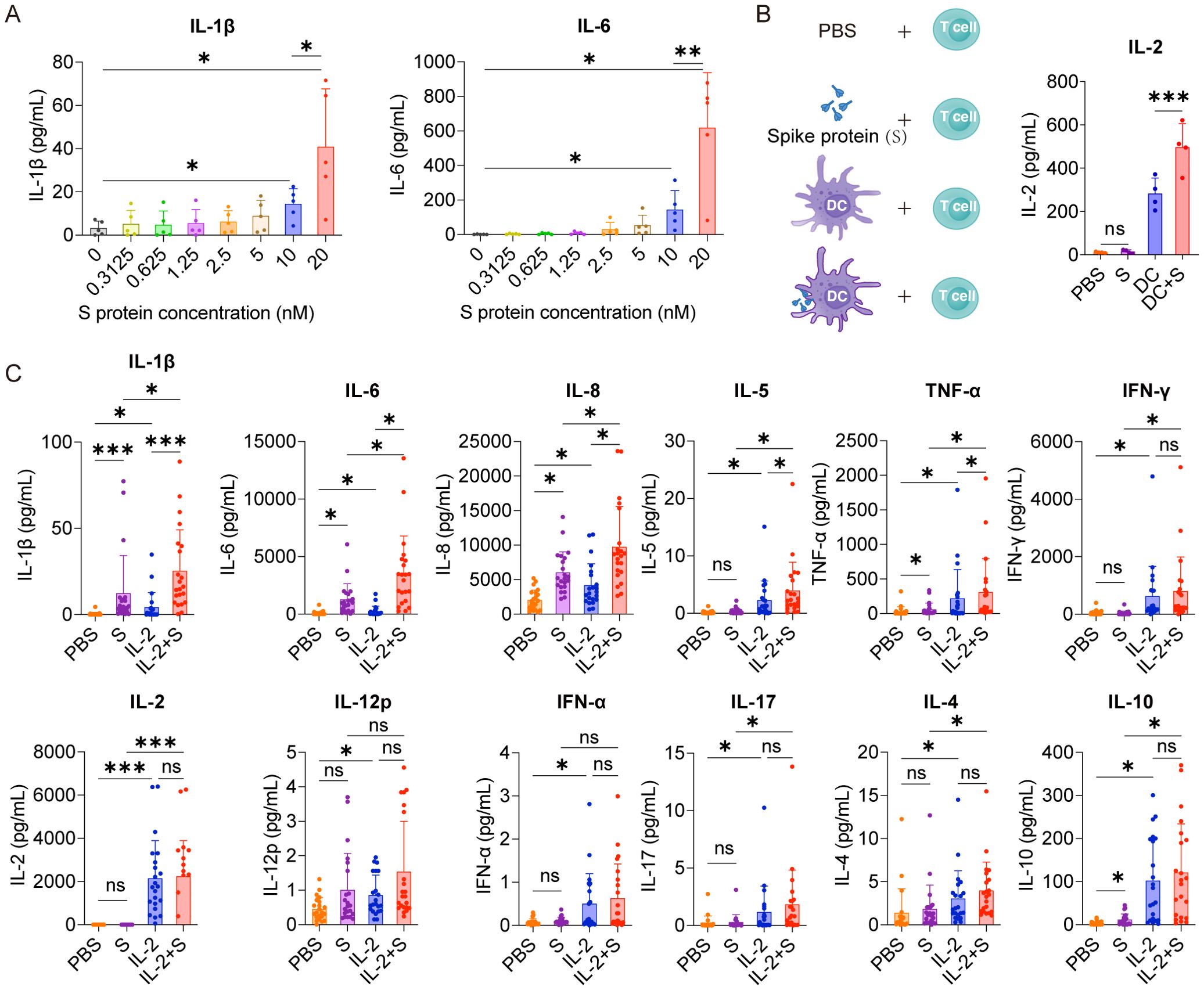
Figure 1. IL-2 cooperates with spike protein to stimulate PBMCs to secrete IL-6, IL-1β, and IL-8. (A) Quantifying concentrations of IL-1β and IL-6 in supernatants of PBMCs stimulated by different concentrations of spike protein for 16 h according to CBA (n = 5 biological replicates). (B) Bar graph (right) showing the concentration of IL-2 in the supernatants of T cells cocultured with DC, DC activated with spike protein, spike protein, or PBS as indicated by the schema (left). (n = 4 biological replicates). (C) Quantifying concentrations of cytokines by CBA in the supernatants of PBMCs treated with PBS, spike protein, IL-2, or IL-2 combined with spike protein for 16 h (n = 22 biological replicates). S, spike protein. Data are presented as mean ± SD. ns, not significant, *p < 0.05, **p < 0.01, and ***p < 0.001 as analyzed by one-way ANOVA (A, B) or Kruskal–Wallis test (C).
Among various cytokines involved in the CRS of patients with COVID-19, IL-2 has drawn our particular attention. This is due to the noticeable elevation of IL-2 in the plasma of many patients with severe COVID-19 (6, 19, 20), and high-dose IL-2 administration causes capillary leak syndrome (21, 22), a severe clinical manifestations of CRS (23). Although the source of IL-2 remains elusive, these studies hint at the crucial role of IL-2 in developing CRS in patients with severe COVID-19. Spike protein is a virus antigen which can be recognized by antigen-producing cells, such as DCs. We examined the effect of DCs loaded with spike protein on T-cell activation and found that DCs loaded with spike protein can stimulate T cells to secrete higher levels of IL-2 than did control DCs (Figure 1B). Along this line, we observed an increased secretion of IL-1β, IL-6, IL-8, IL-5, and TNF-α from PBMCs treated with the combination of IL-2 and spike protein, as compared with spike protein or IL-2 alone (Figure 1C). In contrast, IL-2 alone stimulated PBMCs to secrete IFN-γ, IL-12p, IFN-α, IL-17, IL-4 and IL-10, whereas the combination of spike protein and IL-2 did not show any synergistic effect on the secretion of these cytokines (Figures 1C, D). Furthermore, we found that the hemagglutinin protein of influenza virus and IL-2 did not synergistically stimulate PBMCs to secrete CRS-related cytokines (Supplementary Figure S2). Together, these data suggest that IL-2 released by T cells activated by DCs stimulated with spike protein may serve as an amplifier in inducing CRS in patients with COVID-19 in a manner of cooperation with spike protein.
3.2 Monocytes are the critical cells for the cooperation between IL-2 and spike protein in stimulating PBMCs to secrete inflammatory cytokines
IL-6 is a critical cytokine involved in the pathogenesis of CRS in patients with COVID-19. In severe cases of COVID-19, elevated levels of IL-6 have been observed and are pointed out to contribute to the systemic inflammatory response and organ damage seen in some patients (24, 25). Therefore, as a readout, IL-6 was used to analyze which cells in PBMCs are responders or effectors of the synergistic effect of IL-2 and spike protein to release cytokines. Intracellular staining and flow cytometry analysis revealed that spike protein, rather than IL-2, significantly increased the production of IL-6 in monocytes in PBMCs, and the combination of spike protein and IL-2 further enhanced this effect (Figure 2A). However, no increase in the expression level of IL-6 was observed in NK cells, B cells, or T cells (Figure 2B, Supplementary Figure S3A). These findings indicate that IL-2 can stimulate monocytes in PBMCs to produce more IL-6 in cooperation with spike protein. These results indicate that the spike protein and IL-2 work together to stimulate PBMC to release inflammatory factors that are associated with CRS, possibly through monocytes. To further determine the involvement of monocytes, they were removed from PBMCs (the clearance rate of monocytes is equal to or greater than 95%; Supplementary Figure S3B) and then stimulated with spike protein, IL-2, or spike protein combined with IL-2. Compared with PBMCs, the levels of various cytokines, including IL-1β, IL-6, IL-8, IL-4, IL-2, and IL-17 decreased significantly in the supernatant of PBMCs without monocytes after treatment with spike protein combined with IL-2. Of note, IL-1β, IL-6, and IL-8 levels also decreased significantly in the supernatant of PBMCs treated with spike protein after monocytes removal (Figures 2C, D). These results indicate that monocytes play an essential role in the cytokine secretion by PBMCs stimulated by spike protein or IL-2 in cooperation with spike protein.
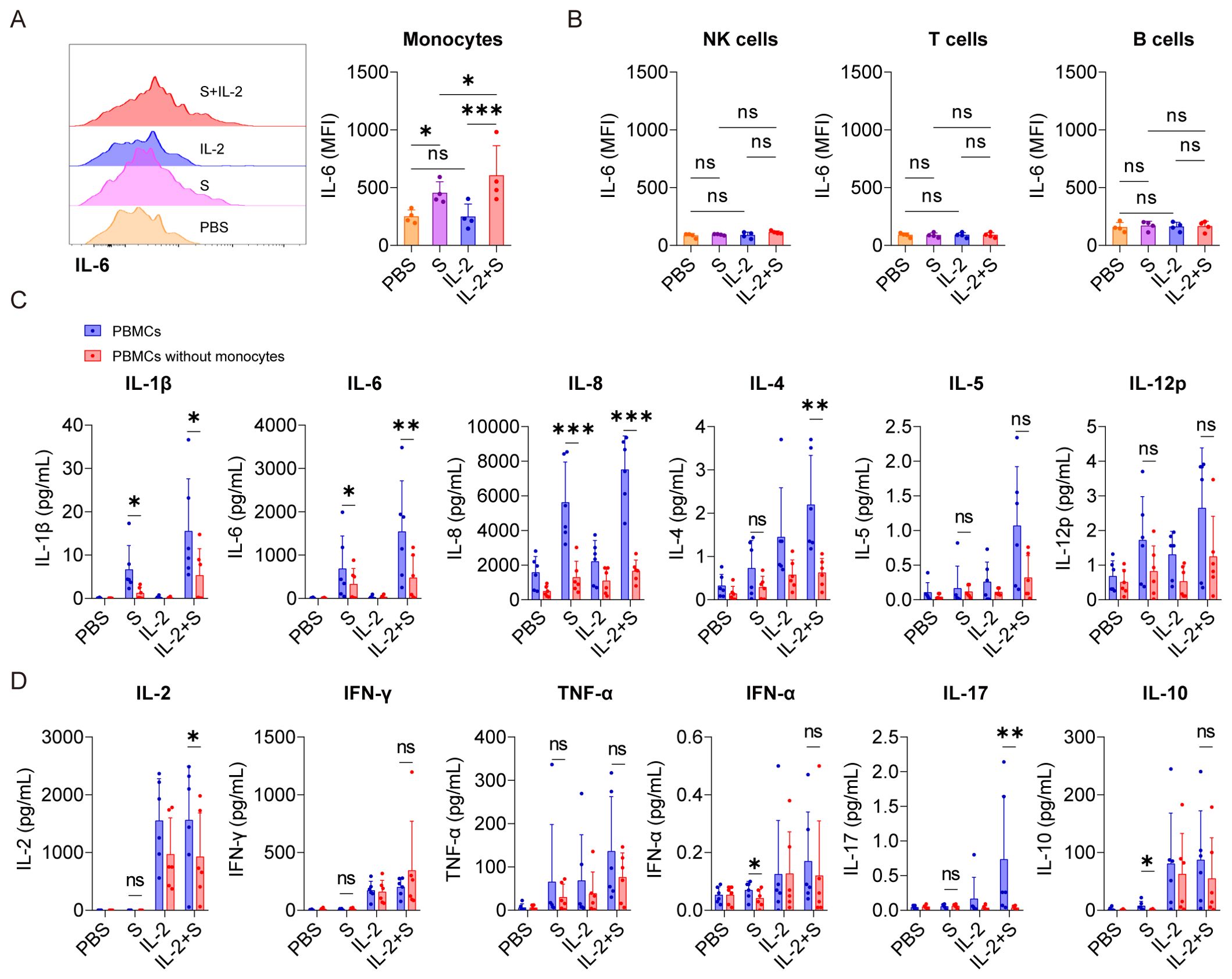
Figure 2. Monocytes are pivotal cells in the secretion of CRS-related cytokines in PBMCs co-stimulated with IL-2 and spike protein. (A) Representative intracellular staining analysis (left) and quantification (right) of the expression of IL-6 by flow cytometry in monocytes of PBMCs stimulated with PBS, spike protein, IL-2, or spike protein combined with IL-2 for 16 h (n = 4 biological replicates). MFI, mean fluorescence intensity. (B) Quantification of the expression of IL-6 in NK cells, T cells, and B cells of PBMCs described in (A). (C, D) Quantifying the concentrations of cytokines by CBA in the supernatants of PBMCs and PBMCs without monocytes stimulated with PBS, spike protein, IL-2, or spike protein combined with IL-2 for 16 h (n = 6 biological replicates). S, spike protein. Data are presented as mean ± SD. ns, not significant, *p < 0.05, **p < 0.01, and ***p < 0.001 as analyzed by one-way ANOVA (A, B), paired Student’s t-test (C), or Mann–Whitney U-test (D).
3.3 IL-2 activates NF-κB of monocytes via stimulating PBMCs to release TNF-α and IFN-γ
To investigate the mechanism underlying the synergistic effect of IL-2 and spike protein in inducing CRS, transcriptomic analysis was conducted on PBMCs treated with PBS, spike protein, IL-2, or a combination of spike protein and IL-2. Our analysis revealed that stimulation with spike protein activated multiple signaling pathways, including NF-κB, TNF, and Janus kinase-signal transducer and activator of transcription (JAK-STAT) in PBMCs (Supplementary Figures S4A–C). Notably, the combination of IL-2 and spike protein further amplified the activation of NF-κB, TNF, Toll-like receptor, and JAK-STAT signaling pathways compared to spike protein alone (Figures 3A–C). Flow cytometry analysis showed that spike protein facilitated the phosphorylation of p65, an indicator of activation of the NF-κB signaling pathway, in monocytes within PBMCs, and IL-2 combined with spike protein further enhanced this effect (Figure 3D).
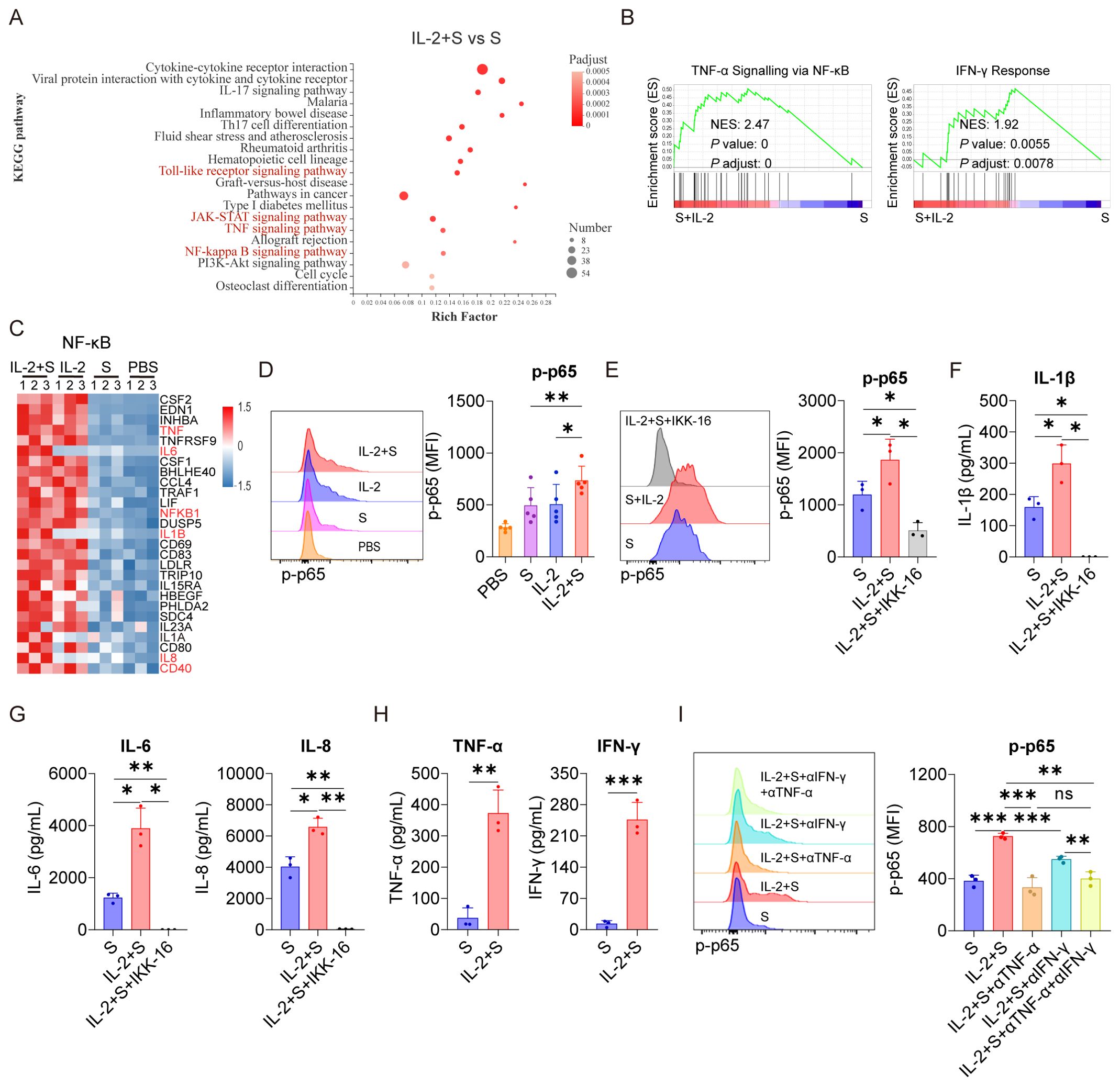
Figure 3. TNF-α and IFN-γ mediate the activation of NF-κB in PBMCs upon the synergistic stimulation by spike protein and IL-2. (A) Bubble plot showing the enrichment of Kyoto Encyclopedia of Genes and Genome (KEGG) pathways based on the transcriptomic analysis of PBMCs treated with IL-2 combined with spike protein or spike protein alone for 16 h (n = 3 biological replicates). (B) Gene set enrichment analysis (GSEA) shows the TNF-α signaling via NF-κB and the IFN-γ signatures in the transcriptomic analysis described in (A) NES, normalized enrichment score; FDR, false discovery rate. (C) The heat map illustrates the differentially expressed genes in the NF-κB pathway in PBMCs treated with IL-2 combined with spike protein, IL-2, spike protein, or PBS for 16 h. (D) Representative intracellular staining analysis (left) and quantification (right) of the expression of p-p65 by flow cytometry in monocytes of PBMCs stimulated with IL-2 combined with spike protein, IL-2, spike protein, or PBS for 16 h (n = 5 biological replicates). (E) Representative intracellular staining analysis (left) and quantification (right) of the expression of p-p65 in monocytes of PBMCs stimulated with spike protein, spike protein combined with IL-2, or spike protein combined with IL-2 and IKK-16 for 16 h (n = 3 biological replicates). (F, G) Quantifying concentrations of IL-1β, IL-6, and IL-8 by CBA in the supernatants of PBMCs in (E). (H) Quantifying concentrations of TNF-α and IFN-γ in the supernatants of PBMCs stimulated with spike protein or spike protein combined with IL-2 for 16 h (n = 3 biological replicates). (I) Representative intracellular staining analysis (left) and quantification (right) of the expression of p-p65 in monocytes of PBMCs stimulated with spike protein, spike protein combined with IL-2 and control IgG, or spike protein combined with IL-2 and TNF-α or/and IFN-γ blocking antibodies for 16 h (n = 3 biological replicates). S, spike protein. Data are presented as mean ± SD. ns, not significant, *p < 0.05, **p < 0.01, and ***p < 0.001 as analyzed by paired Student’s t-test (D, H) or one-way ANOVA (E, F, G, I).
Significantly, inhibiting the activation of NF-κB with IKK Inhibitor VII (IKK-16) not only significantly reduced the secretion of IL-6, IL-1β, and IL-8 by PBMCs stimulated by spike protein (Supplementary Figures S4D, E) but also blocked the secretion of these cytokines by PBMCs stimulated by IL-2 in cooperation with spike protein (Figures 3E–G). As TNF and JAK-STAT signaling pathways were significantly enriched in PBMCs treated with IL-2 combined with spike protein compared to spike protein alone (Figures 3A, B), it appears that IL-2 may regulate these pathways in PBMCs. The JAK-STAT signaling pathway is tightly regulated by IFN and plays an important role in various biological processes (26–28). Interestingly, in the IL-2 combined with spike protein group, TNF-α and IFN-γ levels were significantly increased compared to the spike protein group (Figure 3H). IL-2 alone elevated TNF-α and IFN-γ levels in PBMCs, too (Figure 1C). Importantly, blocking TNF-α or/and IFN-γ with antibodies during PBMCs stimulation with IL-2 combined with spike protein inhibited the activation of the NF-κB signaling pathway (Figure 3I).
Based on these findings, it can be inferred that IL-2 plays a crucial role in activating the NF-κB signaling pathway in PBMCs by promoting the secretion of TNF-α and IFN-γ. Previous studies have reported that spike protein also activates the NF-κB signaling pathway (29). Therefore, the interplay between IL-2 and TNF-α/IFN-γ might be crucial in mediating the synergistic effect of IL-2 and spike protein in inducing CRS in patients with COVID-19.
3.4 TNF-α and IFN-γ cooperate with spike protein to stimulate monocytes to secrete IL-1β, IL-6, and IL-8
As a critical transcription factor, NF-κB is involved in producing of many inflammatory cytokines, such as IL-1β and IL-6 (30). Because the activation of NF-κB in PBMCs induced by IL-2 relies on TNF-α and IFN-γ (Figure 3I), we sought to explore whether the secretion of CRS-related cytokines by PBMCs stimulated with IL-2 and spike protein also depends on these cytokines. Our results demonstrated that the TNF-α blocking antibodies effectively suppressed the secretion of IL-1β, IL-6, and IL-8 in PBMCs stimulated by IL-2 and spike protein. Similarly, the IFN-γ blocking antibodies inhibited the secretion of IL-1β and IL-6 in PBMCs stimulated under the same condition. Interestingly, the combined blocking effect of TNF-α and IFN-γ was not significantly different from that of single blocking antibodies for TNF-α (Figure 4A), suggesting that TNF-α plays a more important role in mediating IL-2-induced cytokines secretion, especially IL-8. Moreover, TNF-α or IFN-γ, when combined with spike protein, stimulated the secretion of CRS-related cytokines in PBMCs, with the effect of their combination being the most potent (Figure 4B). Finally, we observed that IKK-16, an NF-κB inhibitor, completely inhibited the secretion of IL-1β, IL-6, and IL-8 by PBMCs stimulated with TNF-α or IFN-γ that cooperate with spike protein (Supplementary Figure S5). Collectively, TNF-α and IFN-γ are indispensable in activating NF-κB and inducing CRS-related cytokines in PBMCs via cooperating with spike protein.
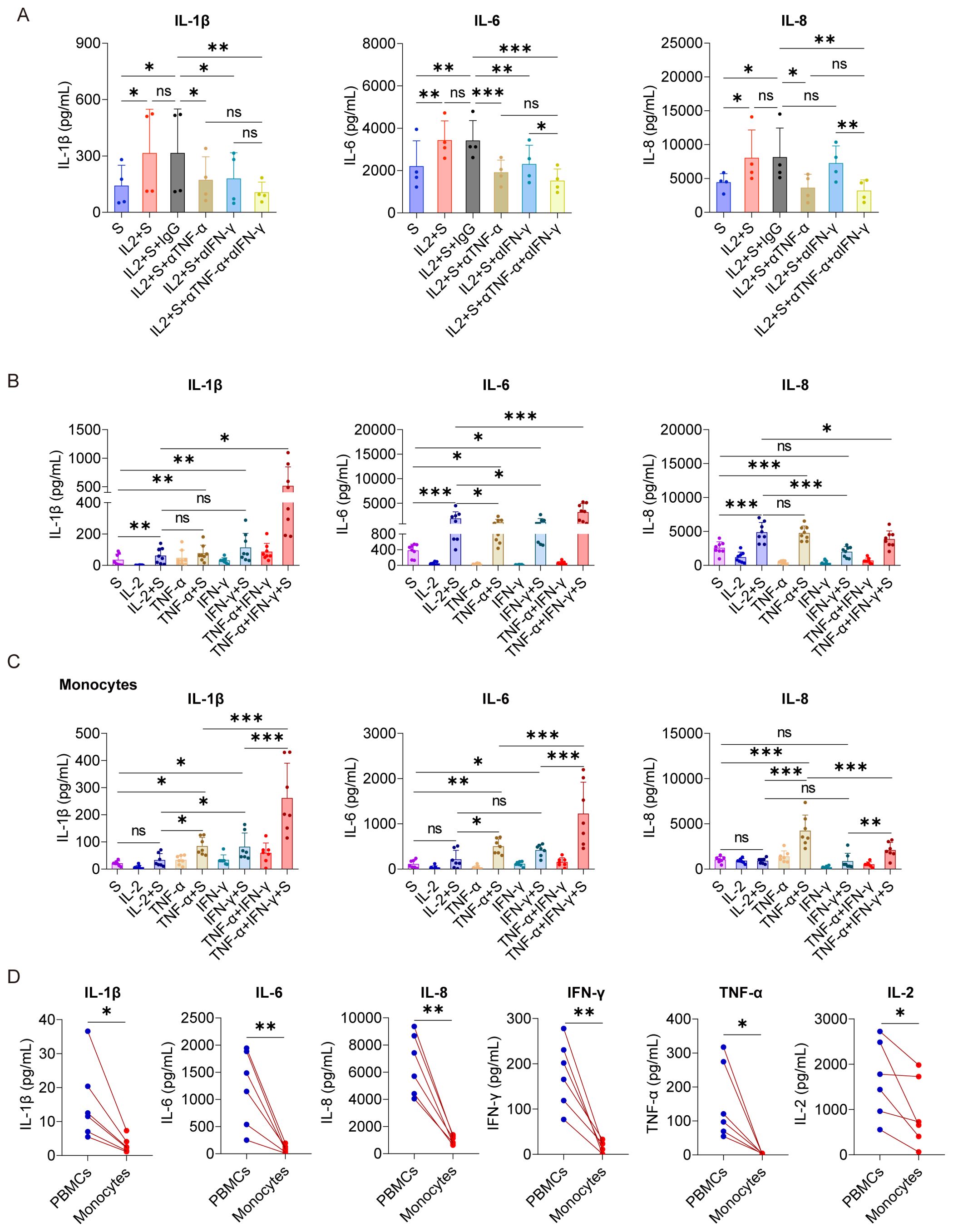
Figure 4. TNF-α and IFN-γ cooperate with spike protein to stimulate monocytes to release IL-1β, IL-6, and IL-8. (A) Quantifying concentrations of IL-1β, IL-6, and IL-8 by CBA in the supernatants of PBMCs stimulated with spike protein and spike protein combined with IL-2 together with/without blocking antibodies against TNF-α or IFN-γ for 16 h (n = 4 biological replicates). (B) Quantifying concentrations of IL-1β, IL-6, and IL-8 by ELISA in the supernatants of PBMCs stimulated with spike protein, IL-2, TNF-α, IFN-γ, or spike protein combined with these cytokines for 16 h (n = 8 biological replicates). (C) Quantifying concentrations of IL-1β, IL-6, and IL-8 by CBA in the supernatants of monocytes stimulated with spike protein, IL-2, TNF-α, IFN-γ, or spike protein combined with these cytokines for 16 h (n = 7 biological replicates). (D) Quantifying concentrations of IL-1β, IL-6, IL-8, IFN-γ, TNF-α, and IL-2 by CBA in the supernatants of PBMCs and monocytes stimulated with spike protein combined with IL-2 for 16 h. Each red line connects PBMCs and monocytes from the same donor (n = 6 biological replicates). S, spike protein. Data are presented as mean ± SD. ns, not significant, *p < 0.05, **p < 0.01, and ***p < 0.001 as analyzed by one-way ANOVA (A–C) or paired Student’s t-test (D).
As we demonstrated that monocytes are the critical cells for the cooperation between IL-2 and spike protein in stimulating PBMCs to secrete inflammatory cytokines (Figure 2), we further tested the synergistic effect of TNF-α/IFN-γ and spike protein in purified monocytes. As expected, TNF-α or IFN-γ, when combined with spike protein, stimulated monocytes to secrete IL-1β, IL-6, and IL-8, with the most potent effect observed when spike protein was combined with TNF-α and IFN-γ (Figure 4C). However, surprisingly, no synergism was observed when IL-2 was combined with spike protein in purified monocytes (Figure 4C). These observations suggest that other immune cells are involved in mediating the synergistic effect of IL-2 and spike protein in PBMCs. Therefore, to test this hypothesis, we treated PBMCs and monocytes from the same volunteer with IL-2 and spike protein and then measured cytokine levels in the supernatant. The results showed that the expressions of IL-1β, IL-6, and L-8 in the supernatant of the monocyte group were remarkably lower than those in the PBMCs group from the same volunteer (Figure 4D). Of note, the induction of TNF-α and IFN-γ was also strikingly lower in monocytes than in the PBMCs under the same stimulatory conditions, raising the possibility that the less potent effect of IL-2 and spike protein in monocytes as compared to PBMCs is due to the lower induction of TNF-α and IFN-γ (Figures 4C, D). Overall, these results indicate that IL-2 and spike protein work together to stimulate other immune cells to release TNF-α and IFN-γ, thereby facilitating monocytes to secrete IL-1β, IL-6, and IL-8.
3.5 NK cells and T cells play essential roles in the synergistic stimulation of CRS-related cytokines by IL-2 and spike protein via IFN-γ and TNF-α
To identify the immune cells responsible for secreting TNF-α and IFN-γ in PBMCs stimulated by IL-2, we conducted intracellular staining to analyze the secretion of these cytokines in NK cells, T cells, B cells, and monocytes within PBMCs. Our results indicated that, compared with the PBS control group, IL-2 could increase the secretion of TNF-α in NK cells and IFN-γ in NK cells and T cells. Compared with the spike protein group, IL-2 combined with spike protein can promote the secretion of TNF-α in NK cells and monocytes and IFN-γ in NK cells and T cells (Figures 5A–C). To further investigate the role of NK cells and T cells in the secretion of inflammatory factors by PBMCs stimulated by IL-2 and spike protein, we cocultured monocytes with NK cells or T cells, respectively, and then stimulated them with IL-2 and spike protein. Our finding showed that combining IL-2 and spike protein significantly elevated IL-1β, IL-6, and IL-8 in monocytes cocultured with T cells and those cocultured with NK cells (Figures 5D–F). However, stimulating monocytes alone did not have the same effect (Figures 5D, E). Blocking TNF-α or IFN-γ antibodies can inhibit the release of IL-1β, IL-6, and IL-8 from monocytes and NK cells or T cells, which have been stimulated by spike protein combined with IL-2 (Figure 5G). These data further support the notion that NK cells and T cells play essential roles in the synergistic stimulation of CRS-related cytokines by IL-2 and spike protein via IFN-γ and TNF-α.
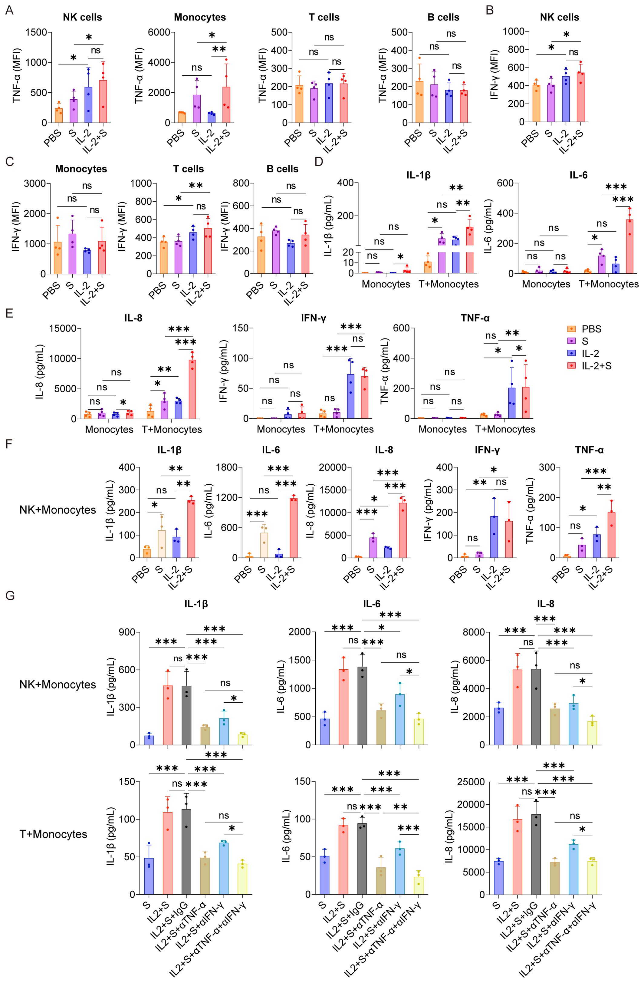
Figure 5. T cells and NK cells play essential roles in the synergistic stimulation of CRS-related cytokines by IL-2 and spike protein via IFN-γ and TNF-α. (A–C) Quantification of the expression of TNF-α and IFN-γ by flow cytometry in NK cells, monocytes, T cells, and B cells of PBMCs stimulated with PBS, spike protein, IL-2, or spike protein combined with IL-2 for 16 h (n = 4 biological replicates). (D, E) Quantifying concentrations of IL-1β, IL-6, IL-8, IFN-γ, and TNF-α by CBA in the supernatants of monocytes with/without cocultivation of T cells from the respective donor under stimulation with PBS, spike protein, IL-2, or spike protein combined with IL-2 for 16 h (n = 4 biological replicates). (F) Quantifying concentrations of IL-1β, IL-6, IL-8, IFN-γ, and TNF-α by CBA in the supernatants of monocytes cocultured with NK cells from the respective donor under stimulation with PBS, spike protein, IL-2, or spike protein combined with IL-2 for 16 h (n = 3 biological replicates). (G) Quantifying concentrations of IL-1β, IL-6, and IL-8 by CBA in the supernatants of monocytes and NK cells or T cells stimulated with spike protein, spike protein combined with IL-2 together with/without blocking antibodies against TNF-α or IFN-γ for 16 h (n = 3 biological replicates). S, spike protein. Data are presented as mean ± SD. ns, not significant, *p < 0.05, **p < 0.01, and ***p < 0.001 as analyzed by one-way ANOVA.
3.6 IL-2 induces an increase in the expression of CD40 and, in turn, facilitates the surface localization of TLR4 in monocytes via TNF-α and IFN-γ
As we have demonstrated that NF-κB plays a critical role in mediating the synergistic effect of spike protein with IL-2, TNF-α, or IFN-γ (Figures 3E, F and Supplementary Figure S4), we sought to explore its upstream responder to spike protein and these cytokines. Through high-throughput sequencing technology, we discovered significant activation of the Toll-like receptor signaling pathway in PBMCs in the present IL-2 combined with spike protein when compared to spike protein alone (Figures 3A, 6A). It has been reported that TLR4 is a receptor of spike protein (11, 12). As IL-1β and IL-6 are classic downstream of the TLR4–NF-κB signaling cascade, IL-2, TNF-α, and IFN-γ may exert their effects via this pathway as well. We found that spike protein reduces the membrane surface expression of TLR4 in monocytes of PBMCs (Figure 6B) without affecting its transcription (Figure 6C). Spike protein binding to TLR4 leads to the internalization of TLR4, which is consistent with the effect of LPS on TLR4. Interestingly, IL-2 combined with spike protein was found to reverse the effect of spike protein on decreasing TLR4 membrane expression without affecting its transcription (Figures 6B, C).
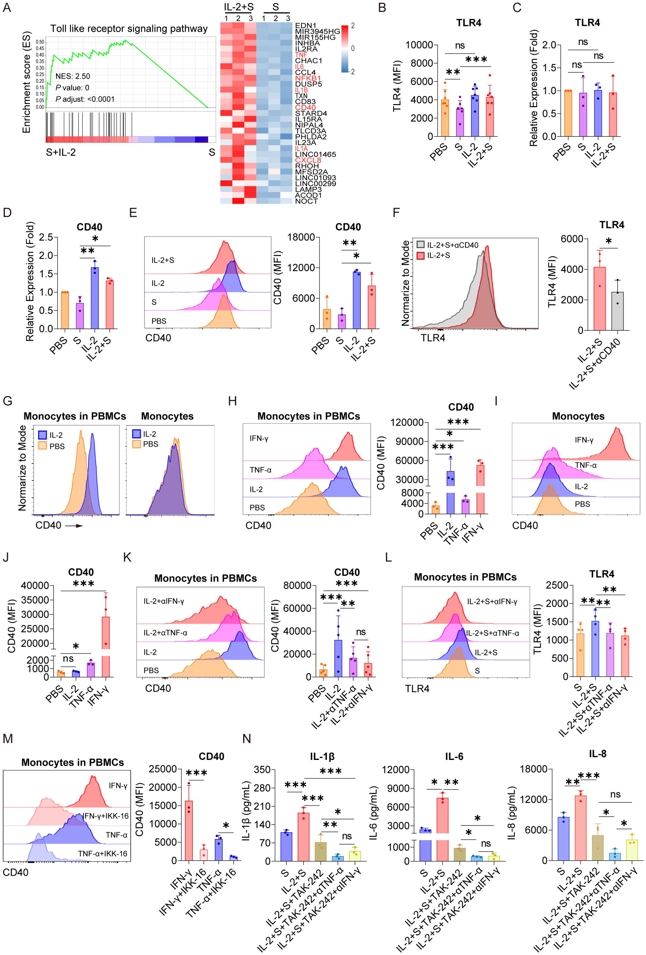
Figure 6. IL-2 induces an increase in the expression of CD40 and, in turn, facilitates the surface localization of TLR4 in monocytes via TNF-α and IFN-γ. (A) GSEA and heat map showing the differentially expressed genes of TLR signaling pathway based on transcriptomic analysis in PBMCs treated with spike protein combined with IL-2 and spike protein alone for 16 h (n = 3 biological replicates). (B) Quantification of the expression of TLR4 by flow cytometry on monocytes in PBMCs treated with PBS, spike protein, IL-2, or IL-2 combined with spike protein for 16 h (n = 7 biological replicates). (C, D) Quantification of the expression of TLR4 and CD40 by qPCR analysis in purified monocytes treated with PBS, spike protein, IL-2, or IL-2 combined with spike protein for 16 h (n = 3 biological replicates). (E) Representative staining analysis (left) and quantification (right) of the expression of CD40 by flow cytometry on monocytes in PBMCs stimulated with PBS, spike protein, IL-2, or spike protein combined with IL-2 for 16 h (n = 3 biological replicates). (F) Representative staining analysis (left) and quantification (right) of the expression of TLR4 on monocytes of PBMCs stimulated with spike protein combined with IL-2, along with the addition of CD40 blocking antibody or control IgG (n = 3 biological replicates). (G) Representative staining analysis of the expression of CD40 by flow cytometry on monocytes in PBMCs (left) and purified monocytes (right) treated with PBS or IL-2 for 16 h. (H) Representative staining analysis (left) and quantification (right) of the expression of CD40 by flow cytometry on monocytes in PBMCs stimulated by IL-2, TNF-α, or IFN-γ for 16 h (n = 3 biological replicates). (I, J) Representative staining analysis (left) and quantification (right) of the expression of CD40 by flow cytometry on purified monocytes stimulated by IL-2, TNF-α, or IFN-γ for 16 h (n = 3 biological replicates). (K) Representative staining analysis (left) and quantification (right) of the expression of CD40 by flow cytometry on monocytes in PBMCs stimulated with IL-2 along with the addition of blocking antibodies against TNF-α or IFN-γ or control IgG for 16 h (n = 5 biological replicates). (L) Representative staining analysis (left) and quantification (right) of the expression of TLR4 by flow cytometry on monocytes in PBMCs stimulated with IL-2 combined with spike protein, along with the addition of blocking antibodies against TNF-α or IFN-γ or control IgG for 16 h (n = 4 biological replicates). (M) Representative staining analysis (left) and quantification (right) of the expression of CD40 by flow cytometry on monocytes in PBMCs stimulated with TNF-α or IFN-γ along with the addition of NF-kB inhibitor IKK-16 for 16 h (n = 3 biological replicates). (N) Quantifying concentrations of IL-1β, IL-6, and IL-8 by CBA in the supernatants of PBMCs stimulated with spike protein combined with IL-2, along with the addition of TLR4 inhibitor and blocking antibodies against TNF-α or IFN-γ or control IgG for 16 h (n = 3 biological replicates). S, spike protein. Data are presented as mean ± SD. ns, not significant, *p < 0.05, **p < 0.01, and ***p < 0.001 as analyzed by one-way ANOVA (B–E, H, J–M) or paired Student’s t-test (F).
In recent studies, several proteins that regulate the stabilization, internalization, intracellular trafficking, and recycling of TLR4 have been identified (31–36). We noticed a significant upregulation of CD40, which has been reported to be able to upregulate membrane expression of TLR4 binding LPS (37), by IL-2 combined with spike protein compared to spike protein alone in PBMCs (Figure 6A). qPCR analysis confirmed that IL-2 significantly increased the transcription of CD40 in purified monocytes (Figure 6D) and also elevated its surface expression of monocytes in PBMCs (Figure 6E). Importantly, blocking antibodies against CD40 strongly reduced the expression of TLR4 on the membrane surface of monocyte of PBMCs stimulated with IL-2 and spike protein (Figure 6F). The secretion of TLR4 downstream inflammatory cytokines such as IL-6, IL-1β, and IL-8 was also significantly inhibited upon CD40 blocking (Supplementary Figure S6), underscoring the vital role of CD40 in mediating the TLR4–NF-κB signaling pathway in response to synergistic stimulation of spike protein and IL-2.
Although IL-2 increased the expression of CD40 on monocytes in PBMCs, purified monocytes directly stimulated with IL-2 did not exhibit any significant increase in the expression of CD40 at all (Figure 6G). This finding suggests the involvement of other immune cells in CD40 regulation. Based on the data in Figures 3–5, we hypothesized that TNF-α or IFN-γ might play a role in CD40 regulation. Along this line, we found that TNF-α or IFN-γ stimulated PBMCs markedly upregulated the expression of CD40 on the surface of monocytes in PBMCs, with IFN-γ having a potency similar to IL-2 (Figure 6H). Notably, the expression of CD40 in monocytes stimulated by TNF-α or IFN-γ significantly increased, whereas IL-2 had no effect (Figures 6I, J), suggesting that TNF-α and IFN-γ may mediate the role of IL-2 in upregulating the expression of CD40 in PBMCs, as they are upregulated by IL-2 (Figure 3H). Furthermore, blocking antibodies against TNF-α or IFN-γ effectively attenuated the upregulation of CD40 on the surface of monocytes within PBMCs treated with IL-2 (Figure 6K) and reversed the upregulation of TLR4 on the surface of monocytes in PBMCs treated with spike protein and IL-2 (Figure 6L). These results suggest that the regulation of membrane expression CD40 and TLR4 in response to synergistic stimulation of spike protein and IL-2 is mediated by TNF-α and IFN-γ. Moreover, we found that IKK-16, an inhibitor of NF-κB, could inhibit the promotion of TNF-α and IFN-γ on CD40 expression (Figure 6M).
Considering the vital role of the TLR4–NF-κB signaling pathway in mediating the synergistic stimulation of spike protein and IL-2 in inducing inflammatory cytokines, this pathway presents a promising target for preventing or reducing CRS in patients with COVID-19. Consistent with the effect of IKK-16, we found that TAK242, a TLR4 inhibitor, significantly inhibited the release of IL-1β, IL-6, and IL-8 in PBMCs co-treated with IL-2 and spike protein (Figure 6N). Notably, this inhibitory effect was further enhanced when combined with blocking antibodies against TNF-α or IFN-γ (Figure 6N).
Our findings suggested that IL-2 stimulation of PBMCs leads to a significant secretion of IFN-γ and TNF-α, resulting in the upregulation of CD40 on the surface of monocytes. The increased interaction of CD40-CD40L then maintains the stable membrane localization of TLR4 and prolongs its interaction with spike protein, subsequently hyper-activating NF-κB in monocytes. These findings shed light on the mechanism underlying the synergistic effect of spike protein and IL-2 in inducing inflammatory cytokine release. They may provide important insights for developing effective therapeutic strategies to prevent or reduce CRS in patients with COVID-19.
4 Discussion
CRS is a common complication observed in patients with severe COVID-19 (38). IL-6 and IL-1 are considered to be essential cytokines in CRS. We observed that spike protein can stimulate PBMCs to produce IL-1β and IL-6 in a dose-dependent manner. Therefore, we speculated that an increase in viral load could lead to elevated spike protein levels and trigger CRS. One prominent feature of CRS in patients with severe COVID-19 is the apparent increase in levels of cytokines. Many patients with severe COVID-19 exhibit a noticeable elevation of IL-2 in their plasma (6, 19, 20). It has been reported that high-dose IL-2 administration causes capillary leak syndrome (21, 22), and, coincidentally, capillary leak syndrome is a severe clinical manifestation of CRS (23). These studies hint at the crucial role of IL-2 in CRS in patients with severe COVID-19. Along this line, our study revealed that DCs stimulated by spike protein activate T cells to release more IL-2 than those stimulated with DCs alone. Therefore, we speculate that spike protein may promote DCs to activate T cells to release a large amount of IL-2, leading to capillary leak syndrome in patients with severe COVID-19. IL-2 plays a critical role in the immune system, and we found that IL-2 alone can stimulate PBMCs to produce multiple cytokines (IFN-α, IFN-γ, and TNF-α), which may lead to the persist inflammation. Additionally, IL-2 and spike protein of SARS-CoV-2 synergistically stimulated PBMCs to produce a large amount of IL-1β, IL-6, and IL-8. This synergistic effect was dose-dependent with spike protein (Supplementary Figure S7). These results further reveal that the continuous accumulation of virus antigens and persistent inflammatory reactions are the primary causes of CRS, which is consistent with the clinical symptoms of severe COVID-19 cases. Clinically, patients with severe COVID-19 have persistent virus and inflammation (39, 40). Thus, our findings indicate that IL-2 is essential in CRS in patients with severe COVID-19.
Patients with severe COVID-19 with CRS often exhibit activation of NF-κB, as well as upregulation of TNF-α and IFN-γ (6, 41, 42). However, the underlying mechanism is vague. In this study, we found that IL-2 activated the NF-κB pathway of monocytes by stimulating PBMCs to release TNF-α and IFN-γ. It has been reported that spike protein of SARS-CoV-2 activates monocytes through binding to TLR4, which is an important upstream regulator of NF-κB (12, 43–45). We confirmed that spike protein activated the NF-κB, but, when combined with IL-2, the NF-κB pathway becomes even more strongly activated. NF-κB inhibitor not only represses the NF-κB activation induced by spike protein or IL-2 combined with spike protein but also reduces the release of IL-1β, IL-6, and IL-8 from PBMCs stimulated by IL-2 combined with spike protein. These indicate that the NF-κB is vital in the interplay between IL-2 and spike protein in inducing CRS-related inflammatory factors in PBMCs. Targeting the NF-κB pathway could, therefore, prove helpful in preventing and treating CRS in patients with COVID-19. Aspirin may have lung-protective effects and reduce the need for mechanical ventilation, ICU admission, and in-hospital mortality in hospitalized patients with COVID-19. However, some studies state that aspirin has no improvement effect on critically ill patients. The mechanism of how aspirin plays a role in COVID-19 infection is unclear. Because aspirin can inhibit NF-κB activity (46, 47), we speculate that aspirin may reduce the risk of severe CRS in patients with mild COVID-19 by inhibiting the activity of NF-κB. However, this effect is not expected to be significant in severe patients who have already developed various complications, including CRS. Overall, our study clarified the interplay between IL-2, TNF-α, IFN-γ, and CRS in patients with COVID-19 and highlighted potential avenues for therapeutic intervention.
We found that spike protein of SARS-CoV-2 reduces the membrane expression of TLR4 on monocytes, which may be due to the internalization of TLR4 after binding to its ligand (48). The internalized TLR4 locates at the endosome and involves in TRIF-dependent signaling pathways, leading to a decreased and delayed activation of NF-κB compared to the TLR4-MyD88 signaling axis (49). In addition, the internalized TLR4 would eventually be degraded by lysosomes, further weakening the NF-κB signaling activated by membrane TLR4 (50). Of note, after adding IL-2, the decrease in TLR4 membrane expression caused by spike protein was reversed, and the TLR4–NF-κB pathway was further activated. This occurred because IL-2 maintained the stable localization of TLR4 on the surface of monocytes treated with spike protein by upregulating the expression of CD40. The interaction between CD40 and CD40L helped stabilize TLR4 on the surface of monocytes treated with spike protein, thus avoiding its rapid internalization and degradation. Our research is consistent with previous reports in which they state that the binding of CD40 and CD40L can improve the stability of TLR4 on the surface of DCs (37). Although IL-2 could not directly stimulate the expression of CD40 on monocytes, it achieved this effect through IFN-γ and TNF-α. It should be noted that the impact of TNF-α was weaker than that of IFN-γ, consistent with literature reports (51). These results indicate that TNF-α and IFN-γ are crucial in enhancing the TLR4–NF-κB pathway via transcriptional regulation of CD40. Our study further showed that TNF-α neutralizing antibody combined with a TLR4 inhibitor inhibits the synergistic stimulation of IL-2 and spike protein in monocytes releasing CRS-related inflammatory factors such as IL-1β, IL-6, and IL-8. Therefore, the combination of TLR4 inhibitor and TNF-α neutralizing antibody may represent a promising therapeutic strategy in treating CRS in patients of COVID-19 and other syndromes, such as CAR-T cell anti-tumor therapy, where CRS is a common side effect.
It has been reported that three individuals developed capillary leak syndrome after receiving the SARS-CoV-2 RNA vaccine (52). The exact cause is unclear. The research revealed that one person had a history of capillary leak syndrome, another had a history of sepsis, and the third person had a history of epilepsy. We found that the three diseases have one thing in common: increased inflammatory factors such as TNF-α in the body (53–56). Therefore, we point out that the spike protein produced by SARS-CoV-2 RNA vaccine may interact with these increased inflammatory factors, particularly TNF-α, which can lead to severe CRS. It is also shown in our research that spike protein combined with IL-2 can still stimulate PBMCs of 10 vaccine donors to secrete CRS-related IL-1β, IL-6, and IL-8. These results indicate that the body’s persistence of spike protein and inflammation (such as IL-2, TNF-α, and IFN-γ) may induce CRS. Therefore, our research suggests that inflammatory individuals with inflammation may not be suitable for the SARS-CoV-2 RNA vaccine.
Several clinical studies have shown that monocytes are involved in the developing of CRS in patients with severe COVID-19 (57–60), but, currently, no effective in vitro model exists for verification. We found that, when monocytes were removed from PBMCs, the synergistic effect of IL-2 and spike protein in stimulating PBMCs to release CRS-related cytokines significantly decreased. This indicated that our model could effectively certify that monocytes are the critical cells for releasing CRS-related cytokines. However, direct stimulation of monocytes with IL-2 combined with spike protein did not enhance the release of inflammatory factors, suggesting that IL-2 is not the primary factor directly cooperating with spike protein to stimulate monocytes to release inflammatory factors. Our study also found that PBMCs stimulated with IL-2 exhibited increased secretion of TNF-α and IFN-γ from NK cells and IFN-γ from T cells. Coculturing NK cells or T cells with monocytes in the medium containing IL-2 and spike protein significantly increased the release of inflammatory factors, including IL-1β, IL-6, and IL-8. These findings further suggest that NK cells, T cells, and monocytes play an essential role in CRS induced by SARS-CoV-2. Our research provides novel insights into the interplay between immune cells and cytokines produced by CRS, laying the foundation for a better understanding of this severe complication related to COVID-19.
The study has a limitation in that it only examines the spike protein of SARS-CoV-2 instead of the virus itself. The spike protein is just a single protein and cannot fully represent the actual condition of SARS-CoV-2 infection. However, the spike protein has been found in some patients with long COVID-19 plasma, and its role in the body is still unclear. This study can help explain the possible mechanism by which a single spike protein functions in patients with COVID-19. Our research suggests that, if there is a significant increase in IL-2, IFN-γ, or TNF-α, then the long-lasting presence of the spike protein in the body may trigger an excessive release of inflammatory factors related to CRS, causing severe damage to the body.
5 Conclusion
Our research suggests that, after SARS-CoV-2 enters the human body, spike protein can activate DCs, which, in turn, stimulate T cells producing a large amount of IL-2. IL-2 can stimulate NK cells to release TNF-α and IFN-γ and T cells to release IFN-γ. The increased level of IFN-γ enhances the membrane-located TLR4 via upregulating the transcription of its partner CD40 in monocytes, resulting in prolonged interaction between spike protein and TLR4 and subsequent activation of NF-κB. Simultaneously, the elevated level of TNF-α activates the NF-κB signaling pathway of monocytes. In such a condition, TNF-α and IFN-γ cooperate to enhance the activation of NF-κB–dependent transcription of CRS-related inflammatory cytokines such as IL-1β, IL-6, and IL-8 (Figure 7). Overall, our study shows that the development of CRS requires the involvement of multiple immune cells, and IL-2, TNF-α, and IFN-γ may act as the primary factors in triggering CRS, providing promising avenues for clinical interventions for patients with COVID-19.
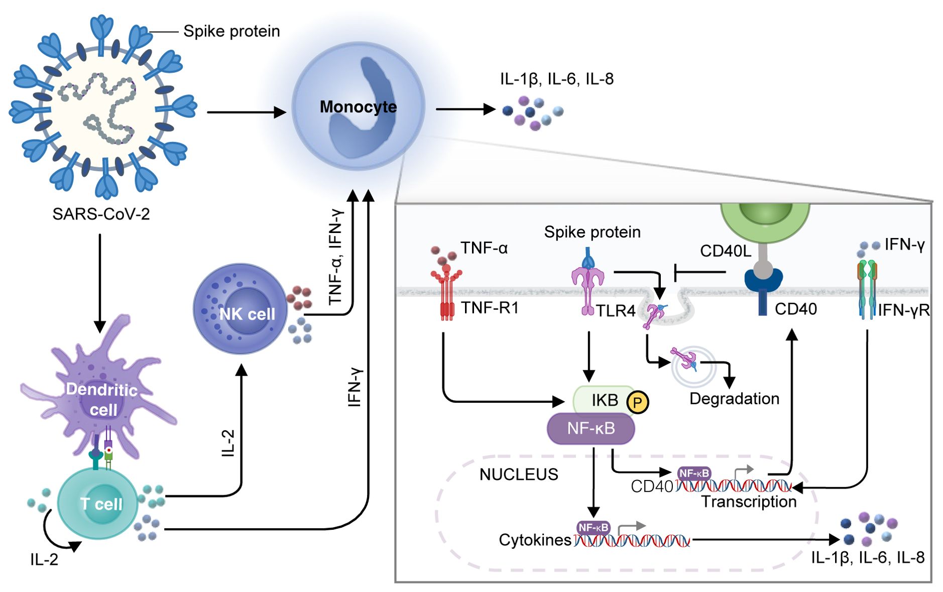
Figure 7. A diagram of the mechanism of spike protein cooperating with IL-2 to stimulate immune cells to produce CRS-related inflammatory factors. DCs loaded with spike protein stimulate T cells to secrete IL-2, which subsequently facilitates the production of TNF-α and IFN-γ by NK cells and IFN-γ by T cells. IFN-γ increases the transcription of CD40, which promotes the stable localization of TLR4 on the membrane surface of monocytes, leading to a constant interaction between spike protein and TLR4 and activation of NF-κB. TNF-α also activates NF-κB signaling in monocytes, which cooperates with IFN-γ to modulate NF-κB–dependent transcription of CRS-related inflammatory cytokines such as IL-1β, IL-6, and IL-8.
Data availability statement
The datasets presented in this study can be found in online repositories. The names of the repository/repositories and accession number(s) can be found below: GSE241843 (GEO).
Ethics statement
The studies involving humans were approved by the ethics committee of the First Hospital of Jilin University. The studies were conducted in accordance with the local legislation and institutional requirements. The participants provided their written informed consent to participate in this study.
Author contributions
CN: Data curation, Supervision, Writing – original draft, Writing – review & editing. TL: Funding acquisition, Writing – review & editing. YC: Methodology, Project administration, Writing – review & editing. SZ: Methodology, Writing – review & editing. LZ: Methodology, Writing – review & editing. NC: Methodology, Writing – review & editing. LQ: Methodology, Writing – review & editing. YW: Methodology, Writing – review & editing. ML: Data curation, Methodology, Writing – review & editing. XZ: Supervision, Writing – original draft, Writing – review & editing. JC: Supervision, Writing – original draft, Writing – review & editing.
Funding
The author(s) declare that financial support was received for the research, authorship, and/or publication of this article. This work was supported by the National Key Research and Development Program of China under Grant number 2020YFA0707704, the Innovative Program of National Natural Science Foundation of China under Grant number 82050003, National Natural Science Foundation of China under Grant numbers 82371838, 82273186, 82002962, and 81874052, Jilin Provincial Science and Technology Department under Grant numbers YDZJ202401438ZYTS, 20240304037SF, 20200602032ZP, 20210401076YY, and YDZJ202202CXJD004, Jilin Province Labor Resources and Social Security Department under Grant number 2023RY03, Jilin Provincial Development and Reform Commission under Grant number 2021C010, and Changchun Science and Technology Bureau under Grant number 23YQ03.
Conflict of interest
The authors declare that the research was conducted in the absence of any commercial or financial relationships that could be construed as a potential conflict of interest.
Publisher’s note
All claims expressed in this article are solely those of the authors and do not necessarily represent those of their affiliated organizations, or those of the publisher, the editors and the reviewers. Any product that may be evaluated in this article, or claim that may be made by its manufacturer, is not guaranteed or endorsed by the publisher.
Supplementary material
The Supplementary Material for this article can be found online at: https://www.frontiersin.org/articles/10.3389/fimmu.2024.1444643/full#supplementary-material
Supplementary Figure S1 | Spike protein stimulates PBMCs to secrete IL-1β, IL-6, and IL-8. Quantifying concentrations of cytokines by CBA in the supernatants of PBMCs stimulated by spike protein for 16 hrs (n = 5 biological replicates). S, spike protein. Data are presented as mean ± SD. ns, not significant, *p < 0.05, and **p < 0.01 as analyzed by paired Student’s t-test (A) or Mann-Whitney U test (B).
Supplementary Figure S2 | IL-2 cannot cooperate with hemagglutinin protein to stimulate PBMCs secreting IL-1β and IL-6. Quantifying concentrations of cytokines by CBA in the supernatants of PBMCs treated with PBS, spike protein, IL-2, IL-2 combined with spike protein (S), hemagglutinin protein (HA), or IL-2 combined with HA for 16 hrs (n = 5 biological replicates). Data are presented as mean ± SD. ns, not significant and ***p < 0.001 as analyzed by one-way ANOVA.
Supplementary Figure S3 | Analysis of IL-6 expression in different immune cells in PBMCs. (A) Representative intracellular staining analysis of the expression of IL-6 using flow cytometry in monocytes, NK cells, T cells, and B cells. The analysis was carried out on PBMCs that were stimulated with PBS, spike protein, IL-2, or spike protein combined with IL-2 for 16 hours. (B) Compare PBMCs before and after removing monocytes. (C) Compare the purity of monocytes before and after sorting from PBMCs. (D) Compare the purity of T cells before and after sorting from PBMCs. (E) Compare the purity of NK cells before and after sorting from PBMCs.
Supplementary Figure S4 | Spike protein activates NF-κB to facilitate monocyte transcription of IL-1β, IL-6, and IL-8. (A) Bubble plot showing the enrichment of KEGG pathways based on the transcriptomic analysis of PBMCs treated with spike protein or PBS for 16 hrs. (B, C) Heat maps showing the differentially expressed genes in the NF-κB signaling pathway, TNF-α signaling pathway, and JAK-STAT signaling pathway in PBMCs treated with spike protein or PBS for 16 hrs (n = 3 biological replicates). (D) Representative intracellular staining analysis (left) and quantification (right) of the expression of p-p65 by flow cytometry in monocytes of PBMCs stimulated with PBS, spike protein, or spike protein combined with IKK-16 (n = 3 biological replicates). (E) Quantifying concentrations of IL-1β, IL-6, and IL-8 by CBA in the supernatants of PBMCs stimulated with PBS, spike protein, or spike protein combined with IKK-16 for 16 hrs (n = 3 biological replicates). S, spike protein. Data are presented as mean ± SD. ns, not significant, *p < 0.05 and **p < 0.01 as analyzed by one-way ANOVA (D, E).
Supplementary Figure S5 | Inhibition of NF-κB reduces the secretion of IL-1β, IL-6, and IL-8 in PBMCs stimulated by spike protein together with TNF-α or IFN-γ. Quantifying concentrations of IL-1β, IL-6, and IL-8 by CBA in the supernatants of PBMCs stimulated with spike protein, spike protein combined with TNF-α or IFN-γ, and spike protein combined with TNF-α or IFN-γ together with IKK-16 for 16 hrs (n = 3 biological replicates). S, spike protein. Data are presented as mean ± SD. ns, not significant, *p < 0.05, **p < 0.01, and ***p < 0.001 as analyzed by one-way ANOVA.
Supplementary Figure S6 | Reduction in the secretion of IL-1β, IL-6, and IL-8 in PBMCs stimulated with IL-2 and spike protein upon blocking CD40. Quantifying concentrations of IL-1β, IL-6, and IL-8 by CBA in the supernatants of PBMCs stimulated with spike protein combined with IL-2 with/without CD40 blocking antibody for 16 hrs (n = 4 biological replicates). S, spike protein. Data are presented as mean ± SD. *p < 0.05, and **p < 0.01 as analyzed by paired Student’s t-test.
Supplementary Figure S7 | The synergistic effects of spike protein with IL-2, TNF-α and IFN-γ were spike protein dose-dependent. Quantifying concentrations of IL-1β and IL-6 in supernatants of PBMCs stimulated by different concentrations of spike protein combining with IL-2, TNF-α and IFN-γ for 16 hrs according to CBA (n = 3 biological replicates). S, spike protein. Data are presented as mean ± SD. *p < 0.05, **p < 0.01, and ***p < 0.001 as analyzed by two-way ANOVA.
Abbreviations
CRS, cytokine release syndrome; TNF, tumor necrosis factor; IL, interleukin; IFN, interferon; ICU, intensive care unit; DC, dendritic cells; NK, natural killer; PBMCs, peripheral blood mononuclear cells.
References
1. Hu B, Guo H, Zhou P, Shi ZL. Characteristics of SARS-coV-2 and COVID-19. Nat Rev Microbiol. (2021) 19:141–54. doi: 10.1038/s41579-020-00459-7
2. Wang Y, Perlman S. COVID-19: inflammatory profile. Annu Rev Med. (2022) 73:65–80. doi: 10.1146/annurev-med-042220-012417
3. Delorey TM, Ziegler CGK, Heimberg G, Normand R, Yang Y, Segerstolpe A, et al. COVID-19 tissue atlases reveal SARS-CoV-2 pathology and cellular targets. Nature. (2021) 595:107–13. doi: 10.1038/s41586-021-03570-8
4. Xiao N, Nie M, Pang H, Wang B, Hu J, Meng X, et al. Integrated cytokine and metabolite analysis reveals immunometabolic reprogramming in COVID-19 patients with therapeutic implications. Nat Commun. (2021) 12:1618. doi: 10.1038/s41467-021-21907-9
5. Que Y, Hu C, Wan K, Hu P, Wang R, Luo J, et al. Cytokine release syndrome in COVID-19: a major mechanism of morbidity and mortality. Int Rev Immunol. (2022) 41:217–30. doi: 10.1080/08830185.2021.1884248
6. Huang C, Wang Y, Li X, Ren L, Zhao J, Hu Y, et al. Clinical features of patients infected with 2019 novel coronavirus in Wuhan, China. Lancet. (2020) 395:497–506. doi: 10.1016/S0140-6736(20)30183-5
7. Mehta P, McAuley DF, Brown M, Sanchez E, Tattersall RS, Manson JJ, et al. COVID-19: consider cytokine storm syndromes and immunosuppression. Lancet. (2020) 395:1033–4. doi: 10.1016/S0140-6736(20)30628-0
8. Giamarellos-Bourboulis EJ, Netea MG, Rovina N, Akinosoglou K, Antoniadou A, Antonakos N, et al. Complex immune dysregulation in COVID-19 patients with severe respiratory failure. Cell Host Microbe. (2020) 27:992–1000 e3. doi: 10.1016/j.chom.2020.04.009
9. Sen S, Dey A, Bandhyopadhyay S, Uversky VN, Maulik U. Understanding structural malleability of the SARS-CoV-2 proteins and relation to the comorbidities. Brief Bioinform. (2021) 22:bbab232. doi: 10.1093/bib/bbab232
10. Jackson CB, Farzan M, Chen B, Choe H. Mechanisms of SARS-CoV-2 entry into cells. Nat Rev Mol Cell Biol. (2022) 23:3–20. doi: 10.1038/s41580-021-00418-x
11. Zhao Y, Kuang M, Li J, Zhu L, Jia Z, Guo X, et al. Publisher Correction: SARS-CoV-2 spike protein interacts with and activates TLR4. Cell Res. (2021) 31:825. doi: 10.1038/s41422-021-00501-0
12. Zhao Y, Kuang M, Li J, Zhu L, Jia Z, Guo X, et al. SARS-CoV-2 spike protein interacts with and activates TLR41. Cell Res. (2021) 31:818–20. doi: 10.1038/s41422-021-00495-9
13. To KK, Tsang OT, Leung WS, Tam AR, Wu TC, Lung DC, et al. Temporal profiles of viral load in posterior oropharyngeal saliva samples and serum antibody responses during infection by SARS-CoV-2: an observational cohort study. Lancet Infect Dis. (2020) 20:565–74. doi: 10.1016/S1473-3099(20)30196-1
14. Walsh KA, Jordan K, Clyne B, Rohde D, Drummond L, Byrne P, et al. SARS-CoV-2 detection, viral load and infectivity over the course of an infection. J Infect. (2020) 81:357–71. doi: 10.1016/j.jinf.2020.06.067
15. Pan Y, Zhang D, Yang P, Poon LLM, Wang Q. Viral load of SARS-CoV-2 in clinical samples. Lancet Infect Dis. (2020) 20:411–2. doi: 10.1016/S1473-3099(20)30113-4
16. Gajjela BK, Zhou MM. Calming the cytokine storm of COVID-19 through inhibition of JAK2/STAT3 signaling. Drug Discovery Today. (2022) 27:390–400. doi: 10.1016/j.drudis.2021.10.016
17. Liao M, Liu Y, Yuan J, Wen Y, Xu G, Zhao J, et al. Single-cell landscape of bronchoalveolar immune cells in patients with COVID-19. Nat Med. (2020) 26:842–4. doi: 10.1038/s41591-020-0901-9
18. Tan Y, Tang F. SARS-CoV-2-mediated immune system activation and potential application in immunotherapy. Med Res Rev. (2021) 41:1167–94. doi: 10.1002/med.21756
19. Liu J, Li S, Liu J, Liang B, Wang X, Wang H, et al. Longitudinal characteristics of lymphocyte responses and cytokine profiles in the peripheral blood of SARS-CoV-2 infected patients. EBioMedicine. (2020) 55:102763. doi: 10.1016/j.ebiom.2020.102763
20. Akbari H, Tabrizi R, Lankarani KB, Aria H, Vakili S, Asadian F, et al. The role of cytokine profile and lymphocyte subsets in the severity of coronavirus disease 2019 (COVID-19): A systematic review and meta-analysis. Life Sci. (2020) 258:118167. doi: 10.1016/j.lfs.2020.118167
21. Boyman O, Sprent J. The role of interleukin-2 during homeostasis and activation of the immune system. Nat Rev Immunol. (2012) 12:180–90. doi: 10.1038/nri3156
22. van Haelst Pisani C, Kovach JS, Kita H, Leiferman KM, Gleich GJ, Silver JE, et al. Administration of interleukin-2 (IL-2) results in increased plasma concentrations of IL-5 and eosinophilia in patients with cancer. Blood. (1991) 78:1538–44. doi: 10.1182/blood.V78.6.1538.1538
23. Case R, Ramaniuk A, Martin P, Simpson PJ, Harden C, Ataya A. Systemic capillary leak syndrome secondary to coronavirus disease 2019. Chest. (2020) 158:e267–e8. doi: 10.1016/j.chest.2020.06.049
24. Liu B, Li M, Zhou Z, Guan X, Xiang Y. Can we use interleukin-6 (IL-6) blockade for coronavirus disease 2019 (COVID-19)-induced cytokine release syndrome (CRS)? J Autoimmun. (2020) 111:102452. doi: 10.1016/j.jaut.2020.102452
25. Smoke SM, Raja K, Hilden P, Daniel NM. Early clinical outcomes with tocilizumab for severe COVID-19: a two-centre retrospective study. Int J Antimicrob Agents. (2021) 57:106265. doi: 10.1016/j.ijantimicag.2020.106265
26. Di Bona D, Cippitelli M, Fionda C, Camma C, Licata A, Santoni A, et al. Oxidative stress inhibits IFN-alpha-induced antiviral gene expression by blocking the JAK-STAT pathway. J Hepatol. (2006) 45:271–9. doi: 10.1016/j.jhep.2006.01.037
27. O’Connell D, Bouazza B, Kokalari B, Amrani Y, Khatib A, Ganther JD, et al. IFN-gamma-induced JAK/STAT, but not NF-kappaB, signaling pathway is insensitive to glucocorticoid in airway epithelial cells. Am J Physiol Lung Cell Mol Physiol. (2015) 309:L348–59. doi: 10.1152/ajplung.00099.2015
28. Li Y, Cui H, Li S, Li X, Guo H, Nandakumar KS, et al. Kaempferol modulates IFN-gamma induced JAK-STAT signaling pathway and ameliorates imiquimod-induced psoriasis-like skin lesions. Int Immunopharmacol. (2023) 114:109585. doi: 10.1016/j.intimp.2022.109585
29. Forsyth CB, Zhang L, Bhushan A, Swanson B, Zhang L, Mamede JI, et al. The SARS-coV-2 S1 spike protein promotes MAPK and NF-kB activation in human lung cells and inflammatory cytokine production in human lung and intestinal epithelial cells. Microorganisms. (2022) 10:1996. doi: 10.3390/microorganisms10101996
30. Al-Griw MA, Salter MG, Wood IC. Blocking of NF−kB/p38 MAPK pathways mitigates oligodendrocyte pathology in a model of neonatal white matter injury. Acta Neurobiol Exp (Wars). (2022) 82:52–64. doi: 10.55782/ane-2022-005
31. Kim D, Kim JY. Anti-CD14 antibody reduces LPS responsiveness via TLR4 internalization in human monocytes. Mol Immunol. (2014) 57:210–5. doi: 10.1016/j.molimm.2013.09.009
32. Tatematsu M, Yoshida R, Morioka Y, Ishii N, Funami K, Watanabe A, et al. Raftlin controls lipopolysaccharide-induced TLR4 internalization and TICAM-1 signaling in a cell type-specific manner. J Immunol. (2016) 196:3865–76. doi: 10.4049/jimmunol.1501734
33. Cao D, Luo J, Chen D, Xu H, Shi H, Jing X, et al. CD36 regulates lipopolysaccharide-induced signaling pathways and mediates the internalization of Escherichia coli in cooperation with TLR4 in goat mammary gland epithelial cells. Sci Rep. (2016) 6:23132. doi: 10.1038/srep23132
34. Liu Y, Zhang M, Zhong H, Xie N, Wang Y, Ding S, et al. LncRNA SNHG16 regulates RAS and NF-kappaB pathway-mediated NLRP3 inflammasome activation to aggravate diabetes nephropathy through stabilizing TLR4. Acta Diabetol. (2023) 60:563–77. doi: 10.1007/s00592-022-02021-8
35. Ciesielska A, Matyjek M, Kwiatkowska K. TLR4 and CD14 trafficking and its influence on LPS-induced pro-inflammatory signaling. Cell Mol Life Sci. (2021) 78:1233–61. doi: 10.1007/s00018-020-03656-y
36. Aerbajinai W, Lee K, Chin K, Rodgers GP. Glia maturation factor-gamma negatively modulates TLR4 signaling by facilitating TLR4 endocytic trafficking in macrophages. J Immunol. (2013) 190:6093–103. doi: 10.4049/jimmunol.1203048
37. Frleta D, Noelle RJ, Wade WF. CD40-mediated up-regulation of Toll-like receptor 4-MD2 complex on the surface of murine dendritic cells. J Leukoc Biol. (2003) 74:1064–73. doi: 10.1189/jlb.0203062
38. Yongzhi X. COVID-19-associated cytokine storm syndrome and diagnostic principles: an old and new Issue. Emerg Microbes Infect. (2021) 10:266–76. doi: 10.1080/22221751.2021.1884503
39. Moss P. The T cell immune response against SARS-CoV-2. Nat Immunol. (2022) 23:186–93. doi: 10.1038/s41590-021-01122-w
40. Zheng S, Fan J, Yu F, Feng B, Lou B, Zou Q, et al. Viral load dynamics and disease severity in patients infected with SARS-CoV-2 in Zhejiang province, China, January-March 2020: retrospective cohort study. BMJ. (2020) 369:m1443. doi: 10.1136/bmj.m1443
41. Hariharan A, Hakeem AR, Radhakrishnan S, Reddy MS, Rela M. The role and therapeutic potential of NF-kappa-B pathway in severe COVID-19 patients. Inflammopharmacology. (2021) 29:91–100. doi: 10.1007/s10787-020-00773-9
42. Kircheis R, Haasbach E, Lueftenegger D, Heyken WT, Ocker M, Planz O. NF-kappaB pathway as a potential target for treatment of critical stage COVID-19 patients. Front Immunol. (2020) 11:598444. doi: 10.3389/fimmu.2020.598444
43. Manik M, Singh RK. Role of toll-like receptors in modulation of cytokine storm signaling in SARS-CoV-2-induced COVID-19. J Med Virol. (2022) 94:869–77. doi: 10.1002/jmv.27405
44. Brandao SCS, Ramos JOX, Dompieri LT, Godoi E, Figueiredo JL, Sarinho ESC, et al. Is Toll-like receptor 4 involved in the severity of COVID-19 pathology in patients with cardiometabolic comorbidities? Cytokine Growth Factor Rev. (2021) 58:102–10. doi: 10.1016/j.cytogfr.2020.09.002
45. Conte C. Possible link between SARS-coV-2 infection and parkinson’s disease: the role of toll-like receptor 4. Int J Mol Sci. (2021) 22:7135. doi: 10.3390/ijms22137135
46. Huo X, Zhang X, Yu C, Cheng E, Zhang Q, Dunbar KB, et al. Aspirin prevents NF-kappaB activation and CDX2 expression stimulated by acid and bile salts in oesophageal squamous cells of patients with Barrett’s oesophagus. Gut. (2018) 67:606–15. doi: 10.1136/gutjnl-2016-313584
47. Liao D, Zhong L, Duan T, Zhang RH, Wang X, Wang G, et al. Aspirin suppresses the growth and metastasis of osteosarcoma through the NF-kappaB pathway. Clin Cancer Res. (2015) 21:5349–59. doi: 10.1158/1078-0432.CCR-15-0198
48. Kagan JC, Su T, Horng T, Chow A, Akira S, Medzhitov R. TRAM couples endocytosis of Toll-like receptor 4 to the induction of interferon-beta. Nat Immunol. (2008) 9:361–8. doi: 10.1038/ni1569
49. Lin C, Wang H, Zhang M, Mustafa S, Wang Y, Li H, et al. TLR4 biased small molecule modulators. Pharmacol Ther. (2021) 228:107918. doi: 10.1016/j.pharmthera.2021.107918
50. Gangloff M. Different dimerisation mode for TLR4 upon endosomal acidification? Trends Biochem Sci. (2012) 37:92–8. doi: 10.1016/j.tibs.2011.11.003
51. Lee SJ, Qin H, Benveniste EN. Simvastatin inhibits IFN-gamma-induced CD40 gene expression by suppressing STAT-1alpha. J Leukoc Biol. (2007) 82:436–47. doi: 10.1189/jlb.1206739
52. Matheny M, Maleque N, Channell N, Eisch AR, Auld SC, Banerji A, et al. Severe exacerbations of systemic capillary leak syndrome after COVID-19 vaccination: A case series. Ann Intern Med. (2021) 174:1476–8. doi: 10.7326/L21-0250
53. Wang B, Li Q, Wang H, Du X, Lai Q, Li X, et al. TNF-alpha: A serological marker for evaluating the severity of hippocampal sclerosis in medial temporal lobe epilepsy? J Clin Neurosci. (2024) 123:123–9. doi: 10.1016/j.jocn.2024.03.030
54. Wang J, He L, Jin Z, Lu G, Yu S, Hu L, et al. Immune dysfunction-associated elevated RDW, APACHE-II, and SOFA scores were a possible cause of 28-day mortality in sepsis patients. Infect Drug Resist. (2024) 17:1199–213. doi: 10.2147/IDR.S442169
55. Xie Z, Chan E, Yin Y, Ghosh CC, Wisch L, Nelson C, et al. Inflammatory markers of the systemic capillary leak syndrome (Clarkson disease). J Clin Cell Immunol. (2014) 5:1000213. doi: 10.4172/2155-9899.1000213
56. Zhai GH, Zhang W, Xiang Z, He LZ, Wang WW, Wu J, et al. Diagnostic value of sIL-2R, TNF-alpha and PCT for sepsis infection in patients with closed abdominal injury complicated with severe multiple abdominal injuries. Front Immunol. (2021) 12:741268. doi: 10.3389/fimmu.2021.741268
57. Falck-Jones S, Osterberg B, Smed-Sorensen A. Respiratory and systemic monocytes, dendritic cells, and myeloid-derived suppressor cells in COVID-19: Implications for disease severity. J Intern Med. (2022) 293:130–43. doi: 10.1111/joim.13559
58. Ma Y, Qiu F, Deng C, Li J, Huang Y, Wu Z, et al. Integrating single-cell sequencing data with GWAS summary statistics reveals CD16+monocytes and memory CD8+T cells involved in severe COVID-19. Genome Med. (2022) 14:16. doi: 10.1186/s13073-022-01021-1
59. Merad M, Martin JC. Pathological inflammation in patients with COVID-19: a key role for monocytes and macrophages. Nat Rev Immunol. (2020) 20:355–62. doi: 10.1038/s41577-020-0331-4
Keywords: SARS-CoV-2, spike protein, monocyte, cytokine release syndrome, NF-κB, CD40
Citation: Niu C, Liang T, Chen Y, Zhu S, Zhou L, Chen N, Qian L, Wang Y, Li M, Zhou X and Cui J (2024) SARS-CoV-2 spike protein induces the cytokine release syndrome by stimulating T cells to produce more IL-2. Front. Immunol. 15:1444643. doi: 10.3389/fimmu.2024.1444643
Received: 06 June 2024; Accepted: 13 August 2024;
Published: 18 September 2024.
Edited by:
Remo Castro Russo, Federal University of Minas Gerais, BrazilReviewed by:
Sandhya Bansal, St. Joseph’s Hospital and Medical Center, United StatesLucero Ramón-Luing, National Institute of Respiratory Diseases-Mexico (INER), Mexico
Copyright © 2024 Niu, Liang, Chen, Zhu, Zhou, Chen, Qian, Wang, Li, Zhou and Cui. This is an open-access article distributed under the terms of the Creative Commons Attribution License (CC BY). The use, distribution or reproduction in other forums is permitted, provided the original author(s) and the copyright owner(s) are credited and that the original publication in this journal is cited, in accordance with accepted academic practice. No use, distribution or reproduction is permitted which does not comply with these terms.
*Correspondence: Jiuwei Cui, Y3VpandAamx1LmVkdS5jbg==; Xin Zhou, eGluemhvdUBqbHUuZWR1LmNu; Min Li, bGltaW4xOTg0QGpsdS5lZHUuY24=
†These authors share senior authorship
 Chao Niu
 Tingting Liang
 Yongchong Chen1
 Shan Zhu
 Lei Zhou
 Naifei Chen
 Yufeng Wang
 Xin Zhou
 Jiuwei Cui