 Tao Li1
 Xinlong Lin1
 Binhai Shen1Wujian Zhang2Yangyang Liu3Hongbin Liu1
 Ye Wang3Lijun Zheng3
 Fachao Zhi1*
- 1Guangdong Provincial Key Laboratory of Gastroenterology, Department of Gastroenterology, Institute of Gastroenterology of Guangdong Province, Nanfang Hospital, Southern Medical University, Guangzhou, China
- 2Department of General Surgery of the First Affiliated Hospital of Heilongjiang University of Traditional Chinese Medicine, Haerbin, China
- 3Guangzhou ZhiYi Biotechnology Co. Ltd., Guangzhou, China
A Corrigendum on
Akkermansia muciniphila suppressing nonalcoholic steatohepatitis associated tumorigenesis through CXCR6+ natural killer T cells
by Li T, Lin X, Shen B, Zhang W, Liu Y, Liu H, Wang Y, Zheng L and Zhi F (2022) Front. Immunol. 13:1047570. doi: 10.3389/fimmu.2022.1047570
In the published article, there was an error in Figure 1F as published. According to the experimental design of this paper, the original purpose of Figure 1F was to confirm the decreased abundance of A. muciniphila in the colon of STAM mice (20 weeks old) compared to control mice. However, in Figure 1F, we included FISH results of STAM mice at 4, 10, 16 weeks of age by mistake, which was not consistent with the description in the Results part. According to the description in the Results part, the comparison of the FISH results of STAM mice (20 weeks old) with control mice should be include in Figure 1F, which makes the results more readable and rigorous. The corrected Figure 1F and its corrected figure legend appear below.
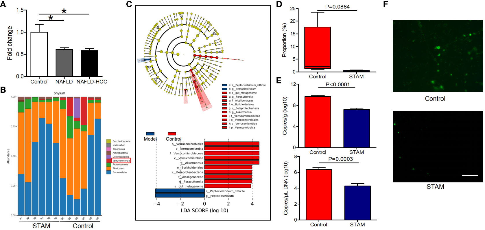
Figure 1 The intestinal abundance of A muciniphila was decreased in patients and mice with NAFLD-HCC (STAM at 20 weeks) (A) The relative abundance of A muciniphila in patients with NAFLD or NAFLD-HCC and healthy controls by qPCR (n=6). *P<0.05 by unpaired Student’s t test. (B) Comparison of the faecal microbiota between STAM at 20 weeks and controls at the species level by 16S rRNA sequencing. (C) LEfSe analysis of the faecal microbiota between the STAM at 20 weeks and healthy controls. Cladogram displays the taxonomic tree of differentially abundant taxa. Histogram represents the LDA scores of bacteria with significant differential abundance between the compared groups, identified by different colors. (D) The proportion of Akkermansia in the faecal microbiota was compared. (E) qPCR validation of the abundance of A muciniphila in STAM at 20 weeks and control. (F) FISH detection of A. muciniphila on the surface of the colon from STAM mice at 20 weeks of age and control mice. Data are presented as the mean ± SEM and were analysed by unpaired Student’s t test. NAFLD, non-alcoholic fatty liver disease; HCC, hepatocellular carcinoma; STAM, streptozotocin+high fat diet-treated mice.
The authors apologize for this error and state that this does not change the scientific conclusions of the article in any way. The original article has been updated.
Publisher’s note
All claims expressed in this article are solely those of the authors and do not necessarily represent those of their affiliated organizations, or those of the publisher, the editors and the reviewers. Any product that may be evaluated in this article, or claim that may be made by its manufacturer, is not guaranteed or endorsed by the publisher.
Keywords: cancer progression, Akkermansia muciniphila, tumor immune surveillance, nonalcoholic fatty liver disease, hepatocellular carcinoma - metabolic syndrome - non-alcoholic fatty liver disease (NAFLD) - non-alcoholic steatohepatitis - NASH-HCC
Citation: Li T, Lin X, Shen B, Zhang W, Liu Y, Liu H, Wang Y, Zheng L and Zhi F (2023) Corrigendum: Akkermansia muciniphila suppressing nonalcoholic steatohepatitis associated tumorigenesis through CXCR6+ natural killer T cells. Front. Immunol. 14:1297103. doi: 10.3389/fimmu.2023.1297103
Received: 19 September 2023; Accepted: 08 November 2023;
Published: 27 November 2023.
Edited and Reviewed by:
Lan Wu, Vanderbilt University Medical Center, United StatesCopyright © 2023 Li, Lin, Shen, Zhang, Liu, Liu, Wang, Zheng and Zhi. This is an open-access article distributed under the terms of the Creative Commons Attribution License (CC BY). The use, distribution or reproduction in other forums is permitted, provided the original author(s) and the copyright owner(s) are credited and that the original publication in this journal is cited, in accordance with accepted academic practice. No use, distribution or reproduction is permitted which does not comply with these terms.
*Correspondence: Fachao Zhi, emhpZmM0MTUzMkAxNjMuY29t