- 1Division of Rheumatology, Allergy and Immunology, Stony Brook University Renaissance School of Medicine, Stony Brook, NY, United States
- 2Biodata Mining and Discovery Section, National Institute of Arthritis and Musculoskeletal and Skin Diseases, Bethesda, MD, United States
- 3Department of Family, Population and Preventive Medicine, Stony Brook University Renaissance School of Medicine, Stony Brook, NY, United States
- 4Division of Gastroenterology and Hepatology, Stony Brook University Renaissance School of Medicine, Stony Brook, NY, United States
- 5Inflammatory Disease Section, National Human Genome Research Institute, National Institutes of Health, Bethesda, MD, United States
NOD-like receptors (NLRs) are intracellular sensors associated with systemic autoinflammatory diseases (SAIDs). We investigated the largest monocentric cohort of patients with adult-onset SAIDs for coinheritance of low frequency and rare mutations in NOD2 and other autoinflammatory genes. Sixty-three patients underwent molecular testing for SAID gene panels after extensive clinical workups. Whole exome sequencing data from the large Atherosclerosis Risk in Communities (ARIC) study of individuals of European-American ancestry were used as control. Of 63 patients, 44 (69.8%) were found to carry combined gene variants in NOD2 and another gene (Group 1), and 19 (30.2%) were carriers only for NOD2 variants (Group 2). The genetic variant combinations in SAID patients were digenic in 66% (NOD2/MEFV, NOD2/NLRP12, NOD2/NLRP3, and NOD2/TNFRSF1A) and oligogenic in 34% of cases. These variant combinations were either absent or significantly less frequent in the control population. By phenotype-genotype correlation, approximately 40% of patients met diagnostic criteria for a specific SAID, and 60% had mixed diagnoses. There were no statistically significant differences in clinical manifestations between the two patient groups except for chest pain. Due to overlapping phenotypes and mixed genotypes, we have suggested a new term, “Mixed NLR-associated Autoinflammatory Disease “, to describe this disease scenario. Gene variant combinations are significant in patients with SAIDs primarily presenting with mixed clinical phenotypes. Our data support the proposition that immunological disease expression is modified by genetic background and environmental exposure. We provide a preliminary framework in diagnosis, management, and interpretation of the clinical scenario.
Introduction
Systemic autoinflammatory diseases (SAIDs) are characterized by abnormal innate immune responses. Nucleotide-binding oligomerization domain (NOD)-like receptors (NLRs) are intracellular sensors that modulate innate immunity, and include NOD2, Pyrin, Cryopyrin, and NLRP12, NLRP1 and NLRC4 among others (1). Germline and somatic variants of NLRs are linked to polygenic and monogenic diseases, such as NOD2-associated diseases (2), and periodic fever syndromes (3). Blau syndrome or early-onset sarcoidosis (at age 4 and younger) is an autosomal dominant granulomatous disease and is caused by NOD2 mutations of high penetrance (4, 5). Another NOD2-associated disease is Yao syndrome (YAOS, OMIM #617321), formerly designated NOD2-associated autoinflammatory disease. This disease is characterized by recurrent episodes of fever, dermatitis, arthralgias, distal leg swelling, gastrointestinal complaints, sicca-like symptoms, and eyelid swelling. The specific NOD2 mutations increase susceptibility to inflammation and serve as diagnostic markers for the disease (6–8). Classical hereditary periodic fever syndromes encompass a recessively inherited Familial Mediterranean Fever (FMF; OMIM 249100), a dominantly inherited Cryopyrin-associated Periodic Syndromes (CAPS), and Tumor Necrosis Factor Receptor-associated Periodic Syndrome (TRAPS; OMIM 142680). These diseases are linked to novel and rare pathogenic missense variants that yield mutated proteins with a gain of function in various inflammatory pathways. Depending on the mutation’s impact on protein function, patients present with a spectrum of disease severity and manifestations. Patients with CAPS typically have cold-induced conjunctivitis, urticaria and arthralgia, known as Familial Cold Autoinflammatory Syndrome type 1 (FCAS1, OMIM #120100) (9), or they can present with a severe early-onset disease caused by monoallelic high-penetrance NLRP3 mutations (NOMID; OMIM 607115). However, an intermediate disease phenotype has been associated with a low-penetrance variant, Gln705Lys (Q705K), in NLRP3 (7, 8). Similar to FCAS1 in clinical phenotype, Familial Cold Autoinflammatory Syndrome type 2 (FCAS2, OMIM #611762), also called NLRP12-AID, is associated with heterozygous loss-of-function mutations in NLRP12; nearly half of patients reported to date harbor the low -penetrance NLRP12 variant, Phe402Leu (F402L) (10). TRAPS is an autosomal dominant disease characterized by recurrent fever, centrifugal rash, migratory myalgias underlying the rash, and periorbital swelling/pain; it is caused by monoallelic missense variants in the extracellular domain of TNFR1. The low-penetrance variant, Arg121Gln (R121Q; aka R92Q), has been reported in patients with a milder non-specific inflammatory disease (11, 12). Although these SAIDs share overlapping clinical phenotypes, they are genetically distinct and follow classical recessive or dominant mode of inheritance (13, 14).
Molecular technologies in genomic medicine, especially next-generation sequencing, are increasingly being used clinically to identify related genetic markers for an accurate diagnosis of SAIDs and other immunological diseases (15). Digenic or oligogenic inheritance of low-frequency and low-penetrance gene variants has been reported in individual patients, leading to challenges for clear interpretation of their clinical significance. We and others have previously reported cases and case series of gene variant combinations in SAID patients (14, 16). Herein, we provide detailed clinical and genetic information for the largest single-site cohort of adult patients who carry two or more variant combinations of NOD2 and other SAID-linked genes. In conjunction with the literature, we provide the most up-to-date information on these SAIDs and genetics, and our experience in diagnosis and management.
Materials and methods
Electronic medical records of a cohort of patients with SAIDs were retrospectively reviewed. These patients presented with a constellation of recurrent fever, rash, arthralgia, abdominal pain/diarrhea and/or chest pain among others. Patients were referred and managed by subspecialists in the Center of Autoinflammatory Disease at Stony Brook University Hospital between 2016 and April 2023. These patients were encountered after multidisciplinary care and had undergone frequently repetitive diagnostic testing. Systemic autoimmune diseases such as classic connective tissue diseases and vasculitis were ruled out; in addition, most had complete evaluations by gastroenterologists, with negative evaluations for inflammatory bowel disease (IBD). Magnetic resonance imaging of the head, echocardiography, and computerized tomography of the chest, abdomen, and pelvis were conducted if indicated. Malignant diseases were excluded. Due to unclear diagnoses and the presence of autoinflammatory clinical features, all patients underwent molecular testing including a 6-gene panel, i.e., MEFV, TNFRSF1A, NLRP3, MVK, NLRP12, and NOD2 (DDC, Middlefield, Ohio, USA). A total of 44 patients were identified in our entire cohort of patients with SAIDs to carry both NOD2 and other SAID-associated gene variants (NOD2+other gene variants, Group 1). An individual SAID was diagnosed based on characteristic phenotype and specific genotype, as well as the classification criteria for periodic fever syndromes (17). In order to examine potential differences between patients with NOD2 ± other gene variants, we selected typical cases of YAOS with NOD2 variants alone (Group 2) for phenotypic comparison between the two groups. YAOS was diagnosed according to our published criteria, i.e., characteristic phenotype and specific NOD2 variants with the exclusion of relevant diseases, such as early onset sarcoidosis or Blau syndrome (BS) and IBD (7, 18, 19). YAOS-associated NOD2 variants are often compound heterozygous for IVS8 + 158(rs5743289, Minor Allele Frequency, MAF=0.10 in gnomAD) and another one or more NOD2 variants, such as Arg702Trp (R702W/SNP8; rs2066804; MAF=0.025),1007fs (SNP13; rs2066847; MAF=0.015), Val955Ile (V955I; rs5743291; MAF=0.06) or rare NOD2 variants (20). A single heterozygous NOD2 variant, such as IVS8 + 158, V955I or rare variants are also seen.
To estimate the distribution and frequency of the combined NOD2+ other variant alleles identified in Group 1 patients in a control population, our collaborators at the National Institutes of Health (NIH) used the dbGaP database, the Atherosclerosis Risk in Communities (ARIC) study with dbGaP accession number phs000280.v8. p2. The ARIC study includes a cohort population and several community surveillance populations in the US. ARIC initiated community-based surveillance in 1987 for myocardial infarction and coronary heart disease incidence and mortality and created a prospective cohort of 15,792 Black and White adults ages 45 to 64 years (21). There were 2,952 unrelated individuals selected based on European-American ancestry and the availability of high-quality whole exome sequencing (WES) data. Combined gene alleles identified in the SAID patients were analyzed in the ARIC control population (Group 3). The study was approved by the Stony Brook University Institutional Research Board.
Statistical analysis
Two-sample t-test test was used to compare continuous variables such as age between two patient groups. The Chi-square test with exact p value from Monte Carlo simulation was used to compare categorical variables such as sex. In addition, mean+/-SD were reported for continuous variables; column percentages were reported for categorical variables. Fisher’s exact test was used to compare prevalence of different genotypes between Group 1 patients and the control population, Group 3. The significance level is set at p<0.05 and all analysis was performed using SAS 9.4 (SAS Institute Inc., Cary, NC).
Results
The demographic, clinical, and laboratory data of patients in both groups
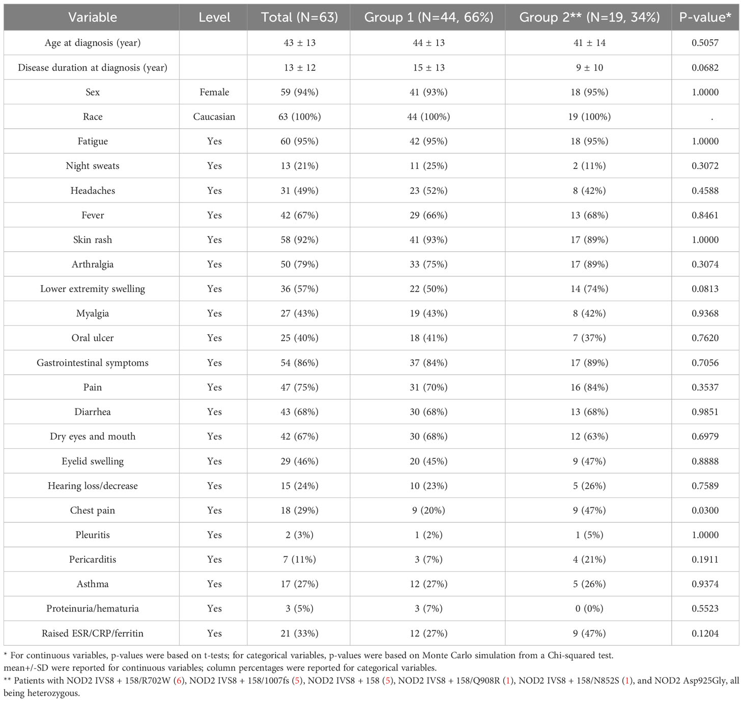
A total of 63 adult patients with SAIDs were included in this study, among whom 44 (69.8%) carried combined variants in two or more genes, and there were 19 (30.2%) YAOS patients with characteristic clinical features and the specific NOD2 variants. All 44 patients in Group 1 were Caucasian and 93% were female; mean age was 44 ± 13 years, and disease duration 15 ± 13 years at the time of diagnosis. The latter underscores a prolonged diagnostic delay due to lack of recognition. The demographic, clinical, and laboratory data of patients in both groups are listed (Table 1), and these parameters were compared between the two groups. There were no statistically significant differences between the two groups except for a higher rate of chest pain in Group 2. All patients presented with a constellation of inflammatory symptoms, including recurrent fever, rash, arthralgias/distal leg swelling, gastrointestinal complaints, sicca-like symptoms, and eyelid swelling. Other notable symptoms were myalgias, oral ulcers, chest pain/pleuritis/pericarditis, asthma, and hearing loss. Most patients (80%) reported no family history of periodic fever syndromes.
Genotyping results of patients in both groups and control population
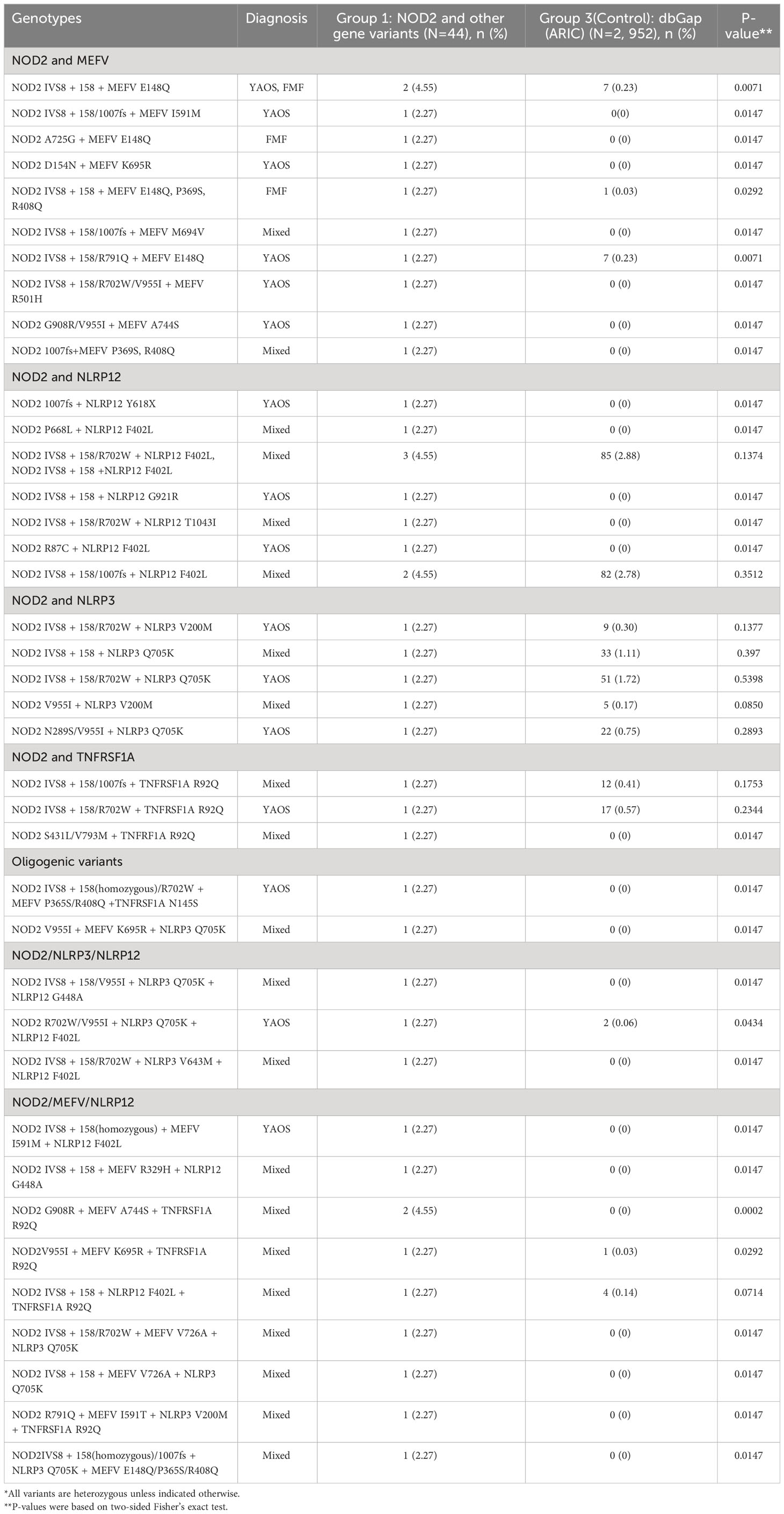
Of the 44 SAID patients in Group 1, all patients carried NOD2 monoallelic or biallelic variants, as well as other gene variants. Most patients carried digenic variants, while oligogenic variants were found in a minority of patients. Among the digenic variants were NOD2/MEFV, NOD2/NLRP12, NOD2/NLRP3, and NOD2/TNFRSF1A in descending order of frequency. The oligogenic variants were NOD2/NLRP3/NLRP12, NOD2/MEFV/NLRP12, NOD2/MEFV/TNFRSF1A, NOD2/NLRP12/TNFRSF1A, and NOD2/MEFV/NLRP3 (Table 2). Most gene combinations were composed of low frequency/penetrance and rare variants, with the digenic variants in NOD2 and MEFV being the most common. To compare the distribution and frequency of these combined SAID-associated genetic variants in patients (Group 1) with those in the control population (Group 3), we used and analyzed the WES data of 2,952 subjects with European-American ancestry (Table 2). The combined gene variants identified in patients (Group 1) were either absent (23/44) or significantly lower in the ARIC control population. All 19 patients in Group 2 carried NOD2 variants, compound heterozygote mostly.
Diagnostic challenge
Clinical phenotypes with features suggestive of autoinflammatory disease were found for all patients at presentation. Following detailed phenotypic evaluations and phenotype-genotype correlations, 16/44(36%) of patients in Group 1 were diagnosed as YAOS, 3/44(6.8%) as atypical FMF, and the remaining with mixed diagnoses of two or more SAIDs, such as YAOS, FMF, NLRP3-AID, NLRP12-AID, and TRAPS. Having NOD2 as the denominator in all patients with combined variants, we asked if there were similarities between these patients with NOD2 ± other gene variants. Our results demonstrated no statistically significant differences in demographics, clinical phenotypes and laboratory results between the two groups except for lower frequency of chest pain in Group 1 (Table 1), suggesting similar but mixed clinical phenotypes among patients with NOD2 ± other gene variants. No significant internal solid organ damage or dysfunction was found for either group. However, some patients in both groups experienced frequent disease flares over a prolonged period of time, resulting in impaired ability to function physically and mentally. Nearly half of the patients in either group received IL-1 inhibitors (Canakinumab or Anakinra), many after trials of colchicine or sulfasalazine, with good response.
Discussion
Potential significance of combined gene variants
SAIDs are generally associated with variants in a single gene locus, though variant combinations in two or more relevant genes can be identified in individual patients as in our study. As a result, challenges arise for diagnosis and management. In the current study, genetic variants classified as variants of uncertain significance in NOD2 and MEFV genes based on the Infevers database were found to be the most frequently inherited https://infevers.umai-montpellier.fr/web/index.php.
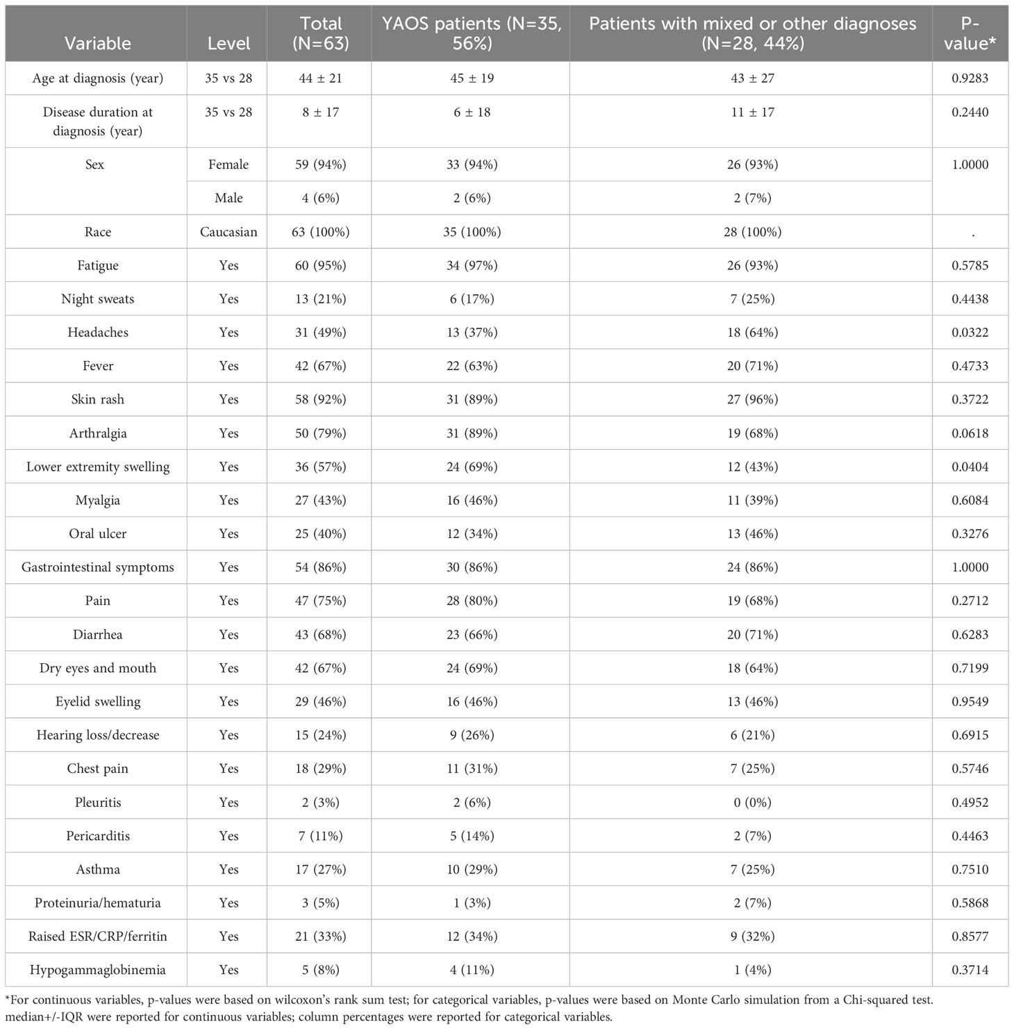
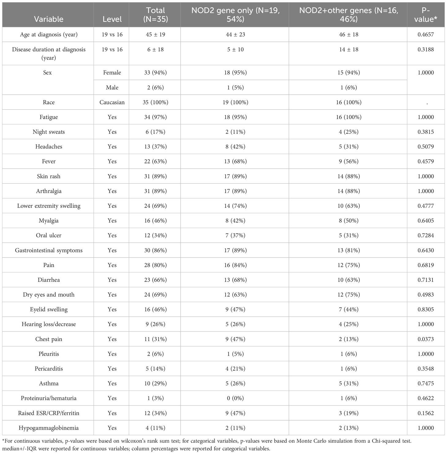
What could be the clinical significance of combined variants in individual SAID patients who present with adult-onset disease? We did not observe significant differences between the groups in clinical phenotypes, which likely extends to disease course and therapy. As seen in Table 1, YAOS was diagnosed more frequently among patients with combined NOD2 and other SAID gene variants, and the majority of patents with YAOS carried compound NOD2 variants or rare variants. This might indicate more influences of the NOD2 variants on phenotypic expression in the genetic background containing multiple SAID genes. We stratified patients in both groups into subgroups: YAOS patients with NOD2 variants only (n=19), patients diagnosed with YAOS and NOD2/other gene variants (n=16), and patients with mixed or other diagnoses (n=28). Further analyses showed statistically significant differences between YAOS patients (n=35) vs patients with mixed or other diagnoses (n=28). Lower extremity swelling as a characteristic finding for YAOS was significantly higher in YAOS patients (69%) than patients with mixed or other diagnoses (43%, P value=0.0404), whereas headache was significantly higher in patients with mixed or other diagnoses (Table 3, Supplemental). In addition, chest pain was significantly higher in YAOS patients with NOD2 variants only than those with NOD2+other gene variants (Table 4, Supplemental). These data indicate that patients with NOD2+other gene variants may not have more severe diseases and poorer outcomes than those with variants in a single gene locus. Another possibility is that combined variants could be synergistic, antagonistic, or perhaps both, depending on the type of gene combinations.
Molecular pathways underlying combined gene expression
To understand the role of these intracellular sensors in autoinflammatory diseases, individual genes(NOD2, NLRP3, NLRP12, and MEFV) and their downstream pathways are schematically depicted in Figure 1 based on literature review (1, 2). NLR interactions are complex, and NOD2 may function in concert with other immune sensors to regulate immune and inflammatory responses in different tissues. NOD2 shares a similar molecular structure with NLRP3 and NLRP12, and has an important role with regard to innate immune responses in gut. NOD2 recognizes a bacterial wall component, Muramyl Dipeptide (MDP), and functions in defense against microbial infection, in the regulation of the inflammatory process, and apoptosis (2). NOD2 mutations are linked to Crohn’s disease (40% patients), Blau syndrome, and YAOS. NOD2 signaling pathway involves receptor interacting protein kinase 2 (RIP2) that can be activated dependently on or independently of NOD2 (22). There is a cross-talk between NOD2 and Toll-like receptors (TLRs). NOD2 activation causes interferon regulatory factor 4(IRF4) expression, which in turn binds to tumor necrosis factor receptor associated factor 6(TRAF6) and RIP2, leading to NF-kB activation (23, 24). NOD2 mutations have been recently shown to cause loss of NOD2 cross-regulatory function involving IRF4 (25). In murine models, NOD2, together with NLRP3, caspase-1, Apoptosis-associated Speck-like protein containing a Caspase activation and recruitment domain (ASC), and RIP2, is required for MDP-induced IL-1 release (26). The NLRP3 inflammasome mediates intestinal inflammation in NOD2-deficient mice (27). NLRP3 also interacts with IRF4 (28).The function of NLRP12 is yet unclear in humans. In mice, NLRP12 has been shown to play a role in the proteasomal degradation of NOD2 and to promote bacterial tolerance and colonization by enteropathogens. NLRP12 suppresses MDP-induced NF-κB activation by targeting the NOD2/RIP2 complex. MDP tolerance is lost in murine monocytes deficient for NLRP12 (29, 30). There may be indirect interactions between NLRP12 and IRF4 (31). MEFV gene is an immediate early gene for interferon-gamma (32). These data suggest cooperative and interrelated roles of some NLRs in disease, and that IRF4 could be a key transcription factor for the orchestration of NLR functions in immune cells. Further study of the role of IRF4 within the context of NLR interactions in autoinflammatory disease is needed. In addition, we previously reported a combination of NOD2 and UBA1 mutations in a patient with an autoinflammatory disease and VEXAS syndrome, noting that both genes are involved in, or regulated by, the ubiquitin pathway (33). Taken together, our study has extended the understanding of the enormous complexity of the genetic influences that underlie autoinflammatory diseases whose clinical characteristics are often superimposable. In other words, similar clinical phenotypes in SAIDs may be caused in different genetic background, suggesting the biological complexity behind the disease phenotypes. Based on our study results and the literature data, it would be reasonable to perform higher-order molecular testing, as first-level testing may not suffice to reveal the underlying genetic mechanisms.
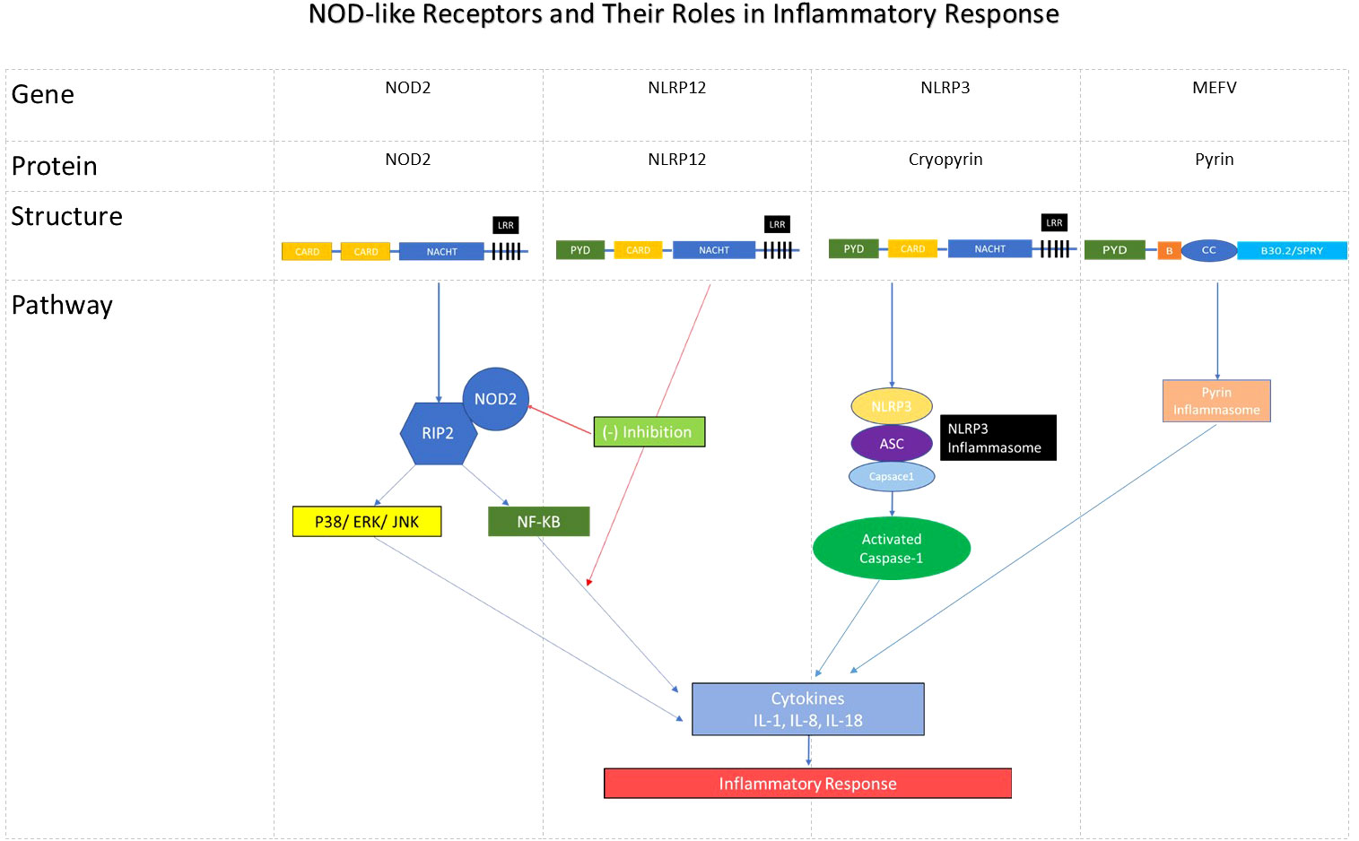
Figure 1 NOD-like receptors and their roles in inflammatory response. The diagram shows the names of respective NLR gene/encoded protein and its structure. These proteins give rise to inflammatory response via NOD2/RIPK2/NF-kB and other pathways involving the NLRP3- and Pyrin inflammasomes. NLRP12 can inhibit RIPK2/NOD2 complex and NF-kB. Note: CARD, Caspase-recruiting domain; LRR, Leucine rich repeat; RIPK2, Receptor interacting protein kinase; P38, 3 Mitogen activated protein kinases (MAPKs); ERK, extracellular signal-regulated kinase (ERK); JNK, c-JUN N terminal kinase (JNK); ASC, Apoptosis-associated speck-like protein containing a CARD.
Implication of low frequency and low penetrance variants in SAIDs
The majority of patients with combined gene variants carried both low-frequency/low- penetrance and rare variants. These variants are MEFV Glu148Gln (E148Q; rs3743930; MAF=0.07 in gnomAD, as high as 0.30 in Asian populations), NLRP3 Gln705Lys (Q705K; rs35829419; MAF=0.038), NLRP12 Phe402Leu (F402L; rs199985574; MAF=0.05), and TNFRSF1A Arg121Gln (R92Q; rs4149584; MAF=0.012), and selected NOD2 variants. These previously reported risk alleles have been associated with susceptibility to SAIDs or they play a role in modifying disease expression (10, 12, 30, 34–37). Notably, these low-frequency variants were found to be concurrent with rare ones in most patients. Furthermore, the distribution and frequency of these variant combinations in patients were either absent or significantly lower in the control population. These data suggest that these variant combinations are clinically significant. Genetic variants contributing to disease lie on a spectrum from rare alleles with large effect sizes to more common alleles with small effect sizes. There is a “gray zone” between these two extremes, as in this study, which is poorly defined with regard to terminology, classification and clinical reportability (38). These autoinflammatory diseases with low penetrance variants have been recently classified as the new category of “Genetically Transitional Disease” (GTD). GTD straddles the old distinction between monogenic and polygenic, where a large-effect mutation is necessary, but not sufficient, to cause disease (39). GTD emphasizes the key role of genetic background in modifying both penetrance and expressivity of a mutation or variant. This concept may also be important for the genetic counseling of these patients, supplementing the traditional interpretation of monogenic autosomal dominant or recessive diseases.
Genome-Wide Association Studies (GWAS) have revealed thousands of genetic variants associated with hundreds of human diseases. However, those that reach genome-wide statistical significance explain only a small fraction of heritability. Common variants may explain more than 50% heritability of many common diseases, including Crohn’s disease, type I diabetes, and multiple sclerosis (40). Overall, the composite effect of variants of small effect may have equal or greater impact than a rare pathogenic mutation with a large effect for more common human phenotypes (41). This concept and theory may in part help explain the implication of our findings, i. e., the coexistence of low penetrance variants in individual patients. Our data also suggest that a combination of genetic defects in different genes, which converge to a common pathogenic pathway, may have a synergistic effect and predispose individuals to SAIDs. For example, CAPS, NLRP12-AID and TRAPS have been classified as IL-1 mediated autoinflammatory diseases based on the patients’ response to IL-1 inhibitors (42, 43). The variant combinations in our study also support the conventional wisdom that genetic background influences both penetrance and phenotypic expressivity of gene mutations (39). Genetic background refers to all other related genes that may interact with the gene of interest to potentially influence specific phenotype in concert with environment (44). Our data may also serve as an example to understand the interactions between genes of interest, genetic background, and environmental or other factors in genetic diseases. Typical monogenic diseases are autosomal dominant disease (Huntington’s disease, HD) and recessive disease (Cystic fibrosis, CF). In these Mendelian diseases, there are slight but not predominant differences in female: male ratio, 54.5%/45.5% for HD (45), and 47.1%/52.9% for CF (46). One explanation is that primary mutations are highly penetrant and major players in these diseases. The candidate gene interactions with genetic background contribute to disease. For example, primary CFTR gene interacts with its modifier gene in the background contribute to CF (47). Unlike monogenic diseases, diseases like YAOS are mostly associated with reduced penetrance variants and are considered as GTD. Based on the GTD model of necessity and insufficiency, the candidate gene, for example, NOD2, genetic background containing certain SAID genes, and other factors such as sex hormones could play more important roles in the disease. Hormonal imbalance is known to cause inflammation or immune response (48), whereas androgen acts via its receptor on macrophages to suppress inflammation or immune response (24). Consequently, this could skew towards female predominance in the disease. Concretely, NOD2 variants are the denominator in all combinations of variants in our study; if NOD2 is considered as the candidate gene, other gene variants such as MEFV, NLRP3, NLRP12 andTNFRSF1A may be entertained as modifying alleles within genetic background. As noted in Table 3, among the overwhelming majority of 44 patients with combined variants, each carried a different combination of a limited number of the SAID genes to constitute a different but related genetic background, as analogous to the ten Arabic numerals from 0 to 9 for writing numbers or codes. Although most NOD2 variants are low-penetrant, their effect could be upwardly influenced by additional germline genetic alleles (39). As most of these gene variants are gain-of-function, they might have been under a positive evolutionary selection as they could provide better immune responses against various pathogens (49).Future genome-based studies in large cohorts of patients may help identify more SAIDs-associated modifying alleles. Acquired somatic mutations in the same genes may further contribute to disease expressivity in adult-onset autoinflammatory and autoimmune diseases.
Digenic/oligogenic disease: conceptual utility in SAIDs
The term “Digenic disease” refers to combinations of variants in two genes (50), and encompasses disorders in which both genes are required for expression or situations where a modifier gene significantly influences phenotype. The term “Oligogenic disease” refers to variant combinations in multiple genes. Based on these concepts, variant combinations in our study may be classified as “Digenic” in the majority of cases and “Oligogenic” in the minority. These variant combinations in our patients may be significant for the following reason. FMF is generally classified as a monogenic recessive disease, caused by biallelic missense gain-of-function mutations in MEFV. However, approximately 25% of FMF patients only carry monoallelic MEFV mutations (51). In fact, several studies have shown the monoallelic association with FMF (52–55), suggesting it is a dominant disease in some cases, where GTD model may apply. In our study, most patients with NOD2/MEFV variants were heterozygous for MEFV E148Q. Several studies of its pathogenic role in classic FMF were conducted with mixed results (35), but most literature data have favored its contributory role in autoinflammatory phenotypes that may or may not be classified as typical FMF as is the case with two recent independent Turkish studies (36, 56). An Israeli study showed that MEFV E148Q is likely a contributory genetic factor when coinherited with M694V (35). Based on our current study, we would assume that patients with carriage of both NOD2 and heterozygous MEFV mutations could mimic biallelic compound heterozygotes of MEFV mutations leading to autoinflammatory diseases with mixed phenotypic expressivity. This could explain a proportion of FMF patients with monoallelic MEFV mutations.
In our study, approximately 20% of patients reported a family history of similar symptoms. A co-segregation analysis of three families in our cohort found that symptomatic relatives shared digenic or oligogenic variants with the probands. Functional studies of some individual low penetrance variants involved in our study were previously conducted by others with ambigous results, specifically in regard to the MEFV E148Q variant. Cells expressing NLRP3 Q705K have mildly increased caspase 1 activity and cleavage, and such patients responded to an IL-1 inhibitor therapy (34). In another study, human monocytic cell lines transduced with Q705K produced significantly higher level of IL-1β and IL-18 than wild type, indicating a gain-of-function (57). In addition, we previously demonstrated abnormal NOD2 expression, NOD2 pathway activation, and a cytokine profile in patients harboring NOD2 variants, IVS8 + 158 and R702W (58).
Mixed NLR-associated autoinflammatory disease
Genotype-phenotype correlation may be readily apparent in patients with monoallelic or biallelic variants in a single gene, but can be challenging concerning combined variants from different genes. Additionally, NOD2 was the denominator in all the combined variants, and our study indicates that these NOD2 variants together with other relevant SAID-associated gene variants contribute to disease pathogenesis. We, therefore, have suggested a new term at the American College of Rheumatology Annual Meeting in 2022, mixed NLR-associated Autoinflammatory Disease (NLR-AID) to denote NLR involvement in the mixed diagnosis, which could be assigned an ICD10 code in the future for insurance billing and research purposes. Biologic therapy with IL-1 inhibitors are generally effective for mixed NLR-AID, YAOS, as well as hereditary periodic fever syndromes (59, 60). One would ask whether mixed NLR-AID could represent an independent entity that might be caused by a hidden pathogenic mutation. To clarify about it, whole exome or genome sequencing would be needed.
In conclusion, this is the largest single site-cohort of autoinflammatory disease patients with NOD2+ other gene variants. Most patients underwent a diagnostic odyssey during a prolonged “mysterious” illness. Unlike many common diseases for which there are readily available guidelines or consensus, SAIDs are rare diseases, and best evidence may come from case reports and case series (61). We provide rational interpretations and our experience with regard to diagnosis, classification, and management.
Limitations
This is a single center study with benefit of uniformity and standardization of the study population. Relative to studies of common diseases, the sample size of our current study is small because the disease entity and associated clinical scenario is rare. We hope that more studies using similar cohorts of patients with these combined gene variants should be performed to replicate our findings.
Data availability statement
The datasets presented in this study can be found in online repositories. The names of the repository/repositories and accession number(s) can be found in the article/supplementary material.
Ethics statement
The studies involving humans were approved by the Institutional Review Board of Stony Brook University. The studies were conducted in accordance with the local legislation and institutional requirements. The ethics committee/institutional review board waived the requirement of written informed consent for participation from the participants or the participants’ legal guardians/next of kin because of retrospective review of electronic medical records.
Author contributions
HN: Data curation, Formal Analysis, Investigation, Resources, Writing – review and editing. ZD: Data curation, Investigation, Resources, Writing – review and editing, Methodology. BN: Investigation, Resources, Writing – review and editing. JY: Resources, Writing – review and editing, Data curation. MY: Resources, Writing – review and editing, Investigation. OA: Resources, Writing – review and editing. PG: Resources, Writing – review and editing. IA: Writing – review and editing, Data curation, Formal Analysis, Investigation. QY: Data curation, Formal Analysis, Investigation, Writing – review and editing, Conceptualization, Methodology, Project administration, Resources, Supervision, Validation, Writing – original draft.
Funding
The author(s) declare that no financial support was received for the research, authorship, and/or publication of this article.
Acknowledgments
The authors are grateful to Baozhong Xin, PhD and Heng Wang, MD for genetic testing at DDC, Middlefield, OH, USA. The authors are also grateful to Mrs. Lyn Hastings, Communication Specialist, the Department of Medicine, Stony Brook University Renaissance School of Medicine for making the figure.
Conflict of interest
The authors declare that the research was conducted in the absence of any commercial or financial relationships that could be construed as a potential conflict of interest.
Publisher’s note
All claims expressed in this article are solely those of the authors and do not necessarily represent those of their affiliated organizations, or those of the publisher, the editors and the reviewers. Any product that may be evaluated in this article, or claim that may be made by its manufacturer, is not guaranteed or endorsed by the publisher.
References
1. Mason DR, Beck PL, Muruve DA. Nucleotide-binding oligomerization domain-like receptors and inflammasomes in the pathogenesis of non-microbial inflammation and diseases. J Innate Immun (2012) 4:16–30. doi: 10.1159/000334247
2. Yao Q. Nucleotide-binding oligomerization domain containing 2: Structure, function, and diseases. Semin Arthritis Rheumatol (2013) 43:125–30. doi: 10.1016/j.semarthrit.2012.12.005
3. Aksentijevich I, Schnappauf O. Molecular mechanisms of phenotypic variability in monogenic autoinflammatory diseases. Nat Rev Rheumatol (2021) 17:405–25. doi: 10.1038/s41584-021-00614-1
4. Matsuda T, Kambe N, Takimoto-Ito R, Ueki Y, Nakamizo S, Saito MK, et al. Potential benefits of TNF targeting therapy in Blau syndrome, a NOD2-associated systemic autoinflammatory granulomatosis. Front Immunol (2022) 13:895765. doi: 10.3389/fimmu.2022.895765
5. Tromp G, Kuivaniemi H, Raphael S, Ala-Kokko L, Christiano A, Considine E, et al. Genetic linkage of familial granulomatous inflammatory arthritis, skin rash, and uveitis to chromosome 16. Am J Hum Genet (1996) 59:1097–107.
6. Yao Q, Kontzias A. Expansion of phenotypic and genotypic spectrum in Yao syndrome: A case series. J Clin Rheumatol (2022) 28:e156–60. doi: 10.1097/RHU.0000000000001655
7. Yao Q, Shen M, McDonald C, Lacbawan F, Moran R, Shen B. NOD2-associated autoinflammatory disease: a large cohort study. Rheumatol (Oxford) (2015) 54:1904–12. doi: 10.1093/rheumatology/kev207
8. Yao Q, Schils J. Distal lower extremity swelling as a prominent phenotype of NOD2-associated autoinflammatory disease. Rheumatol (Oxford) (2013) 52:2095–7. doi: 10.1093/rheumatology/ket143
9. Yao Q, Furst DE. Autoinflammatory diseases: an update of clinical and genetic aspects. Rheumatol (Oxford) (2008) 47:946–51. doi: 10.1093/rheumatology/ken118
10. Shen M, Tang L, Shi X, Zeng X, Yao Q. NLRP12 autoinflammatory disease: a Chinese case series and literature review. Clin Rheumatol (2017) 36:1661–7. doi: 10.1007/s10067-016-3410-y
11. Yao Q, Englund KA, Hayden SP, Tomecki KJ. Tumor necrosis factor receptor associated periodic fever syndrome with photographic evidence of various skin disease and unusual phenotypes: case report and literature review. Semin Arthritis Rheumatol (2012) 41:611–7. doi: 10.1016/j.semarthrit.2011.07.008
12. Aksentijevich I, Galon J, Soares M, Mansfield E, Hull K, Oh HH, et al. The tumor-necrosis-factor receptor-associated periodic syndrome: new mutations in TNFRSF1A, ancestral origins, genotype-phenotype studies, and evidence for further genetic heterogeneity of periodic fevers. Am J Hum Genet (2001) 69:301–14. doi: 10.1086/321976
13. Yao Q, Su LC, Tomecki KJ, Zhou L, Jayakar B, Shen B. Dermatitis as a characteristic phenotype of a new autoinflammatory disease associated with NOD2 mutations. J Am Acad Dermatol (2013) 68:624–31. doi: 10.1016/j.jaad.2012.09.025
14. Yao Q, Li E, Shen B. Autoinflammatory disease with focus on NOD2-associated disease in the era of genomic medicine. Autoimmunity (2019) 52:48–56. doi: 10.1080/08916934.2019.1613382
15. Simon A, van der Meer JW, Vesely R, Myrdal U, Yoshimura K, Duys P, et al. Approach to genetic analysis in the diagnosis of hereditary autoinflammatory syndromes. Rheumatol (Oxford) (2006) 45:269–73. doi: 10.1093/rheumatology/kei138
16. Karamanakos A, Tektonidou M, Vougiouka O, Gerodimos C, Katsiari C, Pikazis D, et al. Autoinflammatory syndromes with coexisting variants in Mediterranean FeVer and other genes: Utility of multiple gene screening and the possible impact of gene dosage. Semin Arthritis Rheumatol (2022) 56:152055. doi: 10.1016/j.semarthrit.2022.152055
17. Gattorno M, Hofer M, Federici S, Vanoni F, Bovis F, Aksentijevich I, et al. Classification criteria for autoinflammatory recurrent fevers. Ann Rheum Dis (2019) 78:1025–32. doi: 10.1136/annrheumdis-2019-215048
19. Yao Q, Shen B. A systematic analysis of treatment and outcomes of NOD2-associated autoinflammatory disease. Am J Med (2017) 130:365 e13–e18. doi: 10.1016/j.amjmed.2016.09.028
20. Navetta-Modrov B, Nomani H, Yun M, Yang J, Salvemini J, Aroniadis O, et al. A novel nucleotide-binding oligomerization domain 2 genetic marker for Yao syndrome. J Am Acad Dermatol (2023) 89:166–8. doi: 10.1016/j.jaad.2023.02.029
21. Wright JD, Folsom AR, Coresh J, Sharrett AR, Couper D, Wagenknecht LE, et al. (Atherosclerosis risk in communities) study: JACC focus seminar 3/8. J Am Coll Cardiol (2021) 77:2939–59. doi: 10.1016/j.jacc.2021.04.035
22. Watanabe T, Minaga K, Kamata K, Sakurai T, Komeda Y, Nagai T, et al. RICK/RIP2 is a NOD2-independent nodal point of gut inflammation. Int Immunol (2019) 31:669–83. doi: 10.1093/intimm/dxz045
23. Watanabe T, Asano N, Meng G, Yamashita K, Arai Y, Sakurai T, et al. NOD2 downregulates colonic inflammation by IRF4-mediated inhibition of K63-linked polyubiquitination of RICK and TRAF6. Mucosal Immunol (2014) 7:1312–25. doi: 10.1038/mi.2014.19
24. Williamson KA, Yun M, Koster MJ, Arment C, Patnaik A, Chang TW, et al. Susceptibility of nucleotide-binding oligomerization domain 2 mutations to Whipple's disease. Rheumatol (Oxford) (2023) 19:kead372. doi: 10.1093/rheumatology/kead372
25. Mao L, Dhar A, Meng G, Fuss I, Montgomery-Recht K, Yang Z, et al. Blau syndrome NOD2 mutations result in loss of NOD2 cross-regulatory function. Front Immunol (2022) 13:988862. doi: 10.3389/fimmu.2022.988862
26. Pan Q, Mathison J, Fearns C, Kravchenko VV, Da Silva Correia J, Hoffman HM, et al. MDP-induced interleukin-1beta processing requires Nod2 and CIAS1/NALP3. J Leukoc Biol (2007) 82:177–83. doi: 10.1189/jlb.1006627
27. Umiker B, Lee HH, Cope J, Ajami NJ, Laine JP, Fregeau C, et al. The NLRP3 inflammasome mediates DSS-induced intestinal inflammation in Nod2 knockout mice. Innate Immun (2019) 25:132–43. doi: 10.1177/1753425919826367
28. Hsu ML, Zouali M. Inflammasome is a central player in B cell development and homing. Life Sci Alliance (2022) 6(2):e202201700. doi: 10.26508/lsa.202201700
29. Normand S, Waldschmitt N, Neerincx A, Martinez-Torres RJ, Chauvin C, Couturier-Maillard A, et al. Proteasomal degradation of NOD2 by NLRP12 in monocytes promotes bacterial tolerance and colonization by enteropathogens. Nat Commun (2018) 9:5338. doi: 10.1038/s41467-018-07750-5
30. Jeru I, Duquesnoy P, Fernandes-Alnemri T, Cochet E, Yu JW, Lackmy-Port-Lis M, et al. Mutations in NALP12 cause hereditary periodic fever syndromes. Proc Natl Acad Sci USA (2008) 105:1614–9. doi: 10.1073/pnas.0708616105
31. Wang S, Hu D, Wang C, Tang X, Du M, Gu X, et al. Transcriptional profiling of innate immune responses in sheep PBMCs induced by Haemonchus contortus soluble extracts. Parasit Vectors (2019) 12:182. doi: 10.1186/s13071-019-3441-8
32. Centola M, Wood G, Frucht DM, Galon J, Aringer M, Farrell C, et al. The gene for familial Mediterranean fever, MEFV, is expressed in early leukocyte development and is regulated in response to inflammatory mediators. Blood (2000) 95:3223–31. doi: 10.1182/blood.V95.10.3223
33. Rivera EG, Patnaik A, Salvemini J, Jain S, Lee K, Lozeau D, et al. SARS-CoV-2/COVID-19 and its relationship with NOD2 and ubiquitination. Clin Immunol (2022) 238:109027. doi: 10.1016/j.clim.2022.109027
34. Kuemmerle-Deschner JB, Verma D, Endres T, Broderick L, de Jesus AA, Hofer F, et al. Clinical and molecular phenotypes of low-penetrance variants of NLRP3: diagnostic and therapeutic challenges. Arthritis Rheumatol (2017) 69:2233–40. doi: 10.1002/art.40208
35. Eyal O, Shinar Y, Pras M, Pras E. Familial Mediterranean fever: Penetrance of the p.[Met694Val];[Glu148Gln] and p.[Met694Val];[=] genotypes. Hum Mutat (2020) 41:1866–70. doi: 10.1002/humu.24090
36. Tanatar A, Karadag SG, Sonmez HE, Cakan M, Ayaz NA. Comparison of pediatric familial mediterranean fever patients carrying only E148Q variant with the ones carrying homozygous pathogenic mutations. J Clin Rheumatol (2021) 27:182–6. doi: 10.1097/RHU.0000000000001261
37. Yao Q. Systemic autoinflammatory disease and genetic testing. Rheumatol Immunol Res (2021) 2:209–11. doi: 10.2478/rir-2021-0028
38. Senol-Cosar O, Schmidt RJ, Qian E, Hoskinson D, Mason-Suares H, Funke B, et al. Considerations for clinical curation, classification, and reporting of low-penetrance and low effect size variants associated with disease risk. Genet Med (2019) 21:2765–73. doi: 10.1038/s41436-019-0560-8
39. Yao Q, Gorevic P, Shen B, Gibson G. Genetically transitional disease: a new concept in genomic medicine. Trends Genet (2023) 39(2):98–108. doi: 10.1016/j.tig.2022.11.002
40. Golan D, Lander ES, Rosset S. Measuring missing heritability: inferring the contribution of common variants. Proc Natl Acad Sci USA (2014) 111:E5272–81. doi: 10.1073/pnas.1419064111
41. Gibson G. Rare and common variants: twenty arguments. Nat Rev Genet (2012) 13:135–45. doi: 10.1038/nrg3118
42. Romano M, Arici ZS, Piskin D, Alehashemi S, Aletaha D, Barron K, et al. The 2021 EULAR/American college of rheumatology points to consider for diagnosis, management and monitoring of the interleukin-1 mediated autoinflammatory diseases: cryopyrin-associated periodic syndromes, tumour necrosis factor receptor-associated periodic syndrome, mevalonate kinase deficiency, and deficiency of the interleukin-1 receptor antagonist. Arthritis Rheumatol (2022) 74:1102–21. doi: 10.1002/art.42139
43. Nigrovic PA, Lee PY, Hoffman HM. Monogenic autoinflammatory disorders: Conceptual overview, phenotype, and clinical approach. J Allergy Clin Immunol (2020) 146:925–37. doi: 10.1016/j.jaci.2020.08.017
44. Mullis MN, Matsui T, Schell R, Foree R, Ehrenreich IM. The complex underpinnings of genetic background effects. Nat Commun (2018) 9:3548. doi: 10.1038/s41467-018-06023-5
45. Pham Nguyen TP, Bravo L, Gonzalez-Alegre P, Willis AW. Geographic barriers drive disparities in specialty center access for older adults with Huntington's disease. J Huntingtons Dis (2022) 11:81–9. doi: 10.3233/JHD-210489
46. Keogh RH, Szczesniak R, Taylor-Robinson D, Bilton D. Up-to-date and projected estimates of survival for people with cystic fibrosis using baseline characteristics: A longitudinal study using UK patient registry data. J Cyst Fibros (2018) 17:218–27. doi: 10.1016/j.jcf.2017.11.019
47. Becker T, Pich A, Tamm S, Hedtfeld S, Ibrahim M, Altmuller J, et al. Genetic information from discordant sibling pairs points to ESRP2 as a candidate trans-acting regulator of the CF modifier gene SCNN1B. Sci Rep (2020) 10:22447. doi: 10.1038/s41598-020-79804-y
48. Sharmeen S, Nomani H, Taub E, Carlson H, Yao Q. Polycystic ovary syndrome: epidemiologic assessment of prevalence of systemic rheumatic and autoimmune diseases. Clin Rheumatol (2021) 40(12):4837–43. doi: 10.1007/s10067-021-05850-0
49. Park YH, Remmers EF, Lee W, Ombrello AK, Chung LK, Shilei Z, et al. Ancient familial Mediterranean fever mutations in human pyrin and resistance to Yersinia pestis. Nat Immunol (2020) 21:857–67. doi: 10.1038/s41590-020-0705-6
50. Nachtegael C, Gravel B, Dillen A, Smits G, Nowe A, Papadimitriou S, et al. Scaling up oligogenic diseases research with OLIDA: the Oligogenic Diseases Database. Database J Biol Database Curation (2022) 2022:baac023. doi: 10.1093/database/baac023
51. Marek-Yagel D, Berkun Y, Padeh S, Abu A, Reznik-Wolf H, Livneh A, et al. Clinical disease among patients heterozygous for familial Mediterranean fever. Arthritis Rheumatol (2009) 60:1862–6. doi: 10.1002/art.24570
52. Kocabey M, Cankaya T, Bayram MT, Ulgenalp A, Caglayan AO, Giray Bozkaya O. Investigation of different genomic variants in familial Mediterranean fever cases with monoallelic MEFV mutation. Clin Exp Rheumatol (2023) 28. doi: 10.55563/clinexprheumatol/2z3l1u
53. Federici L, Rittore-Domingo C, Kone-Paut I, Jorgensen C, Rodiere M, Le Quellec A, et al. A decision tree for genetic diagnosis of hereditary periodic fever in unselected patients. Ann Rheum Dis (2006) 65:1427–32. doi: 10.1136/ard.2006.054304
54. Padeh S, Shinar Y, Pras E, Zemer D, Langevitz P, Pras M, et al. Clinical and diagnostic value of genetic testing in 216 Israeli children with Familial Mediterranean fever. J Rheumatol (2003) 30:185–90.
55. Tchernitchko D, Moutereau S, Legendre M, Delahaye A, Cazeneuve C, Lacombe C, et al. MEFV analysis is of particularly weak diagnostic value for recurrent fevers in Western European Caucasian patients. Arthritis Rheumatol (2005) 52:3603–5. doi: 10.1002/art.21408
56. Aydin F, Cakar N, Ozcakar ZB, Uncu N, Basaran O, Ozdel S, et al. Clinical features and disease severity of Turkish FMF children carrying E148Q mutation. J Clin Lab Anal (2019) 33:e22852. doi: 10.1002/jcla.22852
57. Verma D, Sarndahl E, Andersson H, Eriksson P, Fredrikson M, Jonsson JI, et al. The Q705K polymorphism in NLRP3 is a gain-of-function alteration leading to excessive interleukin-1beta and IL-18 production. PLoS One (2012) 7:e34977. doi: 10.1371/journal.pone.0034977
58. McDonald C, Shen M, Johnson EE, Kabi A, Yao Q. Alterations in nucleotide-binding oligomerization domain-2 expression, pathway activation, and cytokine production in Yao syndrome. Autoimmunity (2018) 51:53–61. doi: 10.1080/08916934.2018.1442442
59. De Benedetti F, Gattorno M, Anton J, Ben-Chetrit E, Frenkel J, Hoffman HM, et al. Canakinumab for the treatment of autoinflammatory recurrent fever syndromes. N Engl J Med (2018) 378:1908–19. doi: 10.1056/NEJMoa1706314
60. Yao Q. Research letter: Effectiveness of canakinumab for the treatment of Yao syndrome patients. J Am Acad Dermatol (2023) 88:653–4. doi: 10.1016/j.jaad.2019.09.020
Keywords: Nod2, NLRP3, NLRP12, autoinflammatory, familial Mediterranean fever, digenic, Yao syndrome
Citation: Nomani H, Deng Z, Navetta-Modrov B, Yang J, Yun M, Aroniadis O, Gorevic P, Aksentijevich I and Yao Q (2023) Implications of combined NOD2 and other gene mutations in autoinflammatory diseases. Front. Immunol. 14:1265404. doi: 10.3389/fimmu.2023.1265404
Received: 22 July 2023; Accepted: 09 October 2023;
Published: 19 October 2023.
Edited by:
Nobuo Kanazawa, Hyōgo College of Medicine Hospital, JapanReviewed by:
Katerina Laskari, University Hospital Zurich, SwitzerlandEmanuele Bizzi, ASST Fatebenefratelli Sacco, Italy
Hidenori Ohnishi, Gifu University, Japan
Copyright © 2023 Nomani, Deng, Navetta-Modrov, Yang, Yun, Aroniadis, Gorevic, Aksentijevich and Yao. This is an open-access article distributed under the terms of the Creative Commons Attribution License (CC BY). The use, distribution or reproduction in other forums is permitted, provided the original author(s) and the copyright owner(s) are credited and that the original publication in this journal is cited, in accordance with accepted academic practice. No use, distribution or reproduction is permitted which does not comply with these terms.
*Correspondence: Qingping Yao, cWluZ3BpbmcueWFvQHN0b255YnJvb2ttZWRpY2luZS5lZHU=
 Hafsa Nomani1
 Peter Gorevic
 Ivona Aksentijevich
 Qingping Yao