
94% of researchers rate our articles as excellent or good
Learn more about the work of our research integrity team to safeguard the quality of each article we publish.
Find out more
MINI REVIEW article
Front. Immunol., 11 January 2024
Sec. Alloimmunity and Transplantation
Volume 14 - 2023 | https://doi.org/10.3389/fimmu.2023.1242478
This article is part of the Research TopicCommunity series in Progress of Allo- and Xeno-transplantation Facilitating the Initial Xeno-Kidney and Islet Clinical Trials, volume IIView all 12 articles
 Haiyan Xu
 Xiaozhou He*
The search for kidney xenografts that are appropriate for patients with end-stage renal disease has been ongoing since the beginning of the last century. The major cause of xenograft loss is hyperacute and acute rejection, and this has almost been overcome via scientific progress. The success of two pre-clinical trials of α1,3-galactosyltransferase gene-knockout porcine kidneys in brain-dead patients in 2021 triggered research enthusiasm for kidney xenotransplantation. This minireview summarizes key issues from an immunological perspective: the discovery of key xenoantigens, investigations into key co-stimulatory signal inhibition, gene-editing technology, and immune tolerance induction. Further developments in immunology, particularly immunometabolism, might help promote the long-term outcomes of kidney xenografts.
Xenotransplantation can play key roles in reducing the kidney donor shortage. Since the first kidney xenotransplant in 1906 (1), great strides have led to achievements in xenotransplantation such that the risk of hyperacute and acute rejection is almost overcome (2, 3). Significant progress has been made in key issues in xenotransplantation (4–6). Important events in kidney xenotransplantation and the advancements of immunological theories and techniques in corresponding periods are listed in Figure 1.
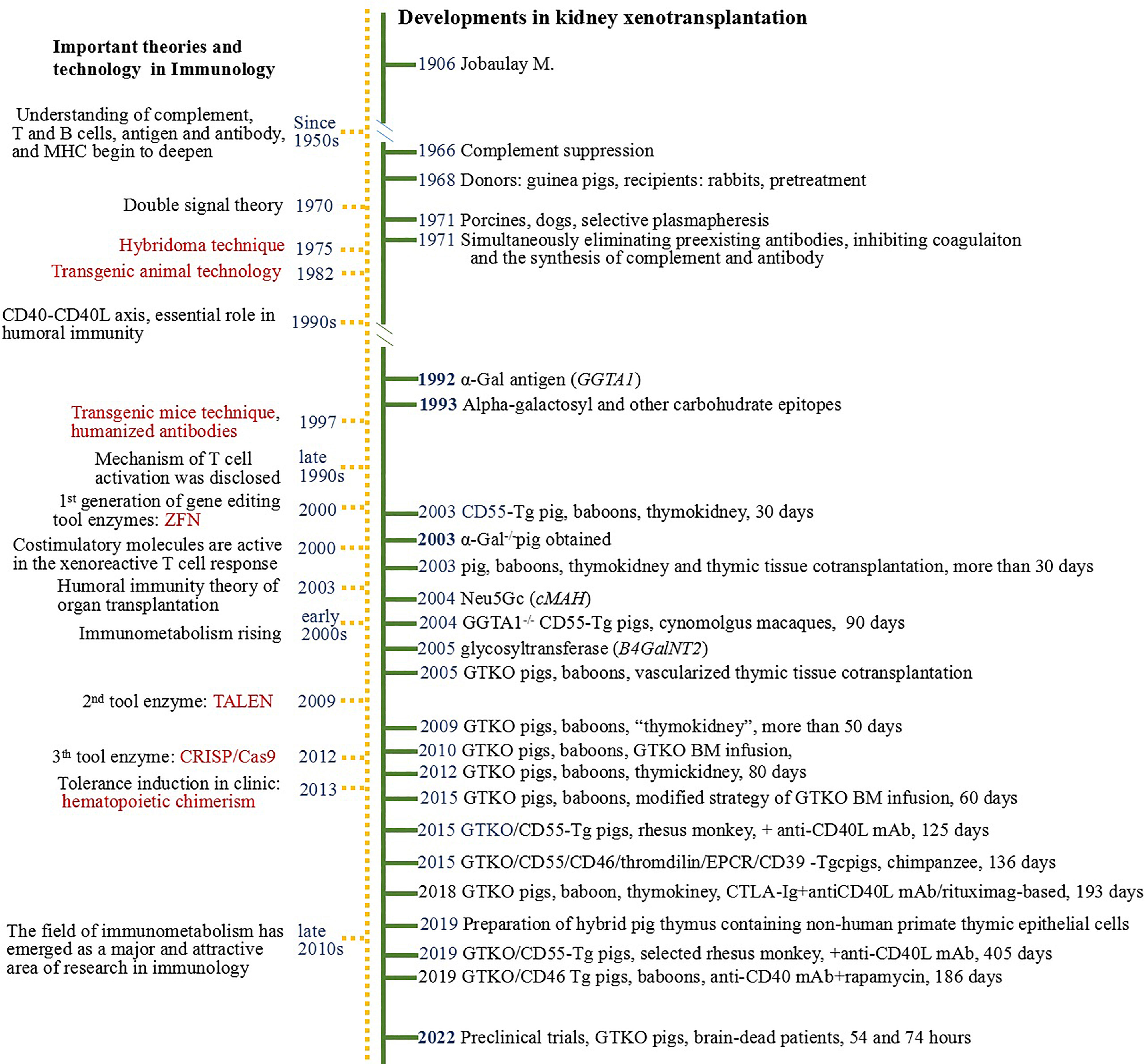
Figure 1 Timeline of developments in kidney xenotransplantation 1906-2022.
Here we especially discuss the pivotal developments of kidney xenotransplantation from an immunological perspective (Table 1).
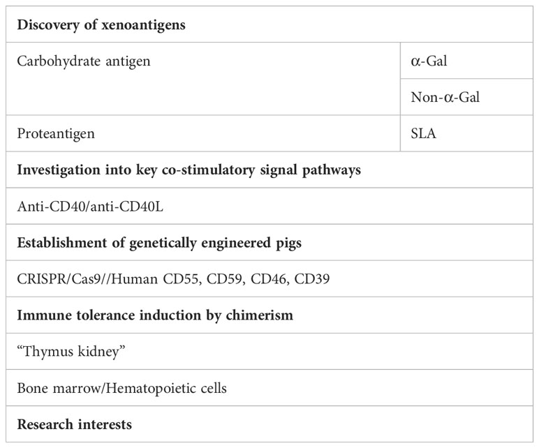
Table 1 Critical progress in promoting kidney xenograft survival.
The recognition of xenoantigens involved in hyperacute rejection has been a long and tortuous road. The first interzygotic twin transplantation in 1953 resulted in long recipient survival and revealed a new direction for organ transplantation. With the discovery and application of immunosuppressive agents, hyperacute rejection after allotransplantation could be controlled, and the survival of recipients gradually increased. However, hyperacute rejection after xenotransplantation cannot be controlled by the empirical application of immunosuppressive agents (7).
Recipient rabbits treated with homogenized guinea pig liver mixtures survived longer after guinea pig kidney grafts were transplanted (8). This inspired many attempts to reduce hyperacute rejection of xenografts, such as the selective removal of plasma components (9), elimination of extant antibodies, inhibition of coagulation, as well as the synthesis of complement and antibodies (10). The results suggested that hyperacute rejection of xenografts is strongly associated with donor antigens, plasma composition, and antibody synthesis, similar to hyperacute rejection during allotransplantation.
The red blood cell surface galactose antigen (DGalα1→3DGal) that induces hyperacute homotransplant rejection due to an ABO mismatch was identified in the late 1980s (11). During xenotransplantation, hyperacute rejection results in an abnormal increase in immunoglobulin (Ig)M serum levels rather than in IgG levels. This indicates that the recipient’s immune system first recognizes the specific antigens harbored in xenografts.
Due to the emergence of monoclonal antibodies (mAbs) using hybridomas, human anti-swine antibodies waere generated and used to identify significant carbohydrate structures for xenotransplantation (12). Then the α-galactosyltransferase (α-Gal) was found, which is encoded by the α-1,3-galactosyltransferase (GGTA1) gene (13). Other carbohydrate antigens, such as non-fucosylated chondroitin sulfate monolayers and linear antigens, are also found, locating on the surfaces of all porcine vascular endothelial cells. These antigens tightly bind to anti-Gal isogalectin β4 antibodies and specifically bind to natural, human anti-α-Gal antibodies. Gal epitopes are expressed abundantly in the brush margins of proximal convoluted tubules, moderately in distal convoluted tubules, and not at all in renal collecting tubules and glomeruli. A specific antigen-antibody reaction activates the complement system, leading to a powerful cytotoxic effect that leads to hyperacute grafts (14–18). The discovery of the α-Gal antigen was a major breakthrough in xenotransplantation.
Thereafter, considerable efforts were directed toward decreasing hyperacute rejection of kidney xenotransplants by removing anti-porcine antibodies in vitro, short-term infusions of specific carbohydrates (19), or the absorption of anti-xenoantigen antibodies produced in the spleen and kidneys (20). Soluble Gal proteins can partially inhibit human rejection of porcine kidneys. Intravenous infusions of bovine serum albumin-Gal in vivo can essentially maintain the depletion of circulating anti-Gal antibodies and prevent or delay antibody deposition and the acute humoral rejection of pig-to-baboon xenografts, but it might be associated with liver damage (21).
Transgenic technology was established in 1981 using microinjections; and a transgenic mouse model was created in 1982. The first generation of the gene-editing tool, zinc finger nuclease, was introduced during the late 1990s, and another, transcriptional activator-like effector nuclease, was identified in 2009. These gene-editing techniques had a positive global impact on life sciences.
Pigs with α-Gal knockout (α-Gal-/-, GTKO) are important xenotransplantation models (22–24). In the α-Gal-/- pigs to baboon kidney xenotransplantation models, most recipients did not develop hyperacute rejection; however, they succumbed to acute humoral rejection. The significantly increased abundance of peripheral anti-non-α-Gal antibodies in recipients suggested that non-α-Gal antigens in kidney xenografts might trigger the production of large amounts of corresponding antibodies. Thereafter, non-α-Gal antigens were recognized as obstacles to α-Gal-/- pig organ xenotransplantation (25). The α-Gal antigen is crucial for hyperacute rejection, and non-α-Gal antigens play important roles in humoral rejection of xenotransplants. In addition to α-Gal and non-α-Gal, other carbohydrate antigens have a complex spatial distribution in porcine kidneys and are strongly associated with the outcome of porcine kidney xenotransplantation (26).
Non-α-Gal antigens, such as N-glycolylneuraminic acid (Neu5Gc; HD antigen), encoded by the cytidine monophospho-N-acetylneuraminic acid hydroxylase (cMAH) gene have been identified (27, 28). Compared with GGTA1-/- pig xenotransplantation, humoral rejection is reduced in GGTA1-/-/CMAH-/- pigs xenotransplantation (29), implying that the immune heritability of the Neu5Gc antigen potentially plays an important role in pig-human xenotransplantation. The other carbohydrate non-α-Gal antigen, glycosyltransferase, (SD(a) antigen), is encoded by the β-1,4-N-acetyl-galactosaminyl transferase (B4GalNT2) gene (30, 31).
Clustered regularly interspaced short palindromic repeats (CRISPR)-associated protein (Cas9) is a third-generation gene-editing tool. Porcine embryonic fibroblasts with GGTA1-/-/Gal-/- were initially created using CRISPR/Cas9 in 2014 (32). Since then, CRISPR/Cas9 has become the preferred means of generating genetically engineered pigs. The serum of many waitlisted patients contained only a minimal number of antibodies that reacted with peripheral blood mononuclear cells from GGTA1-/-/CMAH-/-/B4GalNT2-/- pigs. However, anti-human leukocyte antigen antibodies in some sensitized patients cross-reacted with porcine major histocompatibility complex (MHC) I antibodies (33). Pigs with simultaneous MHC and three antigen (GGTA1/CMAH/B4GalNT2) inactivation have been generated using the CRISPR/Cas method (34). Natural and inducible anti-SDa plays important roles in GTKO pig-to-rhesus monkey xenotransplant rejection, thus providing further support for the notion that Gal and SDa antigens should be simultaneously targeted (35). Exploration of new key non-α-Gal antigens is currently underway (36).
SLAs are being discovered to play an important role in swine innate and adoptive immune responses. In some sensitized kidney transplant-waitlisted patients, some human leucocyte antigen (HLA) antibodies cross-react with SLA class I (37). SLA II is also a xenoantigen (38, 39). And triple (GGTA1, CMAH, B2M) genes modified pigs expressed the SLA Ilow phenotype, which effects on immune status and susceptibility to human immune responses (40). In vitro human TNF-α could increase SLA I expression, while human IL-17 could decrease TNF-α-mediated SLA-I upregulation (41), and downregulation of SLA expression decreases the strength of xenogeneic immune responses towards renal tubular epithelial cells (42). These data may support the SLA-silencing strategy application to prevent xenogeneic cellular immune responses.
Diversity and specificity of immunoglobulins suggests that cellular and humoral immune responses are not separate entities, but complementary components. T and B lymphocytes interact to activate and differentiate into effector cells under specific circumstances. During this process, co-stimulatory signals, such as cluster of differentiation (CD) 40 and its ligand CD40L, CD28-B7, and inducible T cell co-stimulator ligand (ICOS) and its ligand ICOSL, play indispensable roles, and the effects of CD40L-CD40 signaling on xenotransplantation have been extensively investigated.
The 35 kDa polypeptide CD40 is mainly expressed in B lymphocytes (43, 44). After CD40L was identified (44–46), numerous in vivo and in vitro findings showed that the CD40L-CD40 pathway is essential for T cell responses and specific antibody production by B lymphocytes (47–52). The biological effects of anti-CD40L mAb, as well as other related mAbs, including anti-CD80, anti-CD86 mAbs, and biologicals, such as hCTLA4-Ig, have been extensively studied in vitro and in vivo (53–55). Results suggest that blocking the CD40-CD40L pathway, or combined blocking of the CD28-B7 signal could effectively inhibit T cell activation and suppress the production of specific antibodies.
Data from pig to non-human primates (NHPs) organ xenotransplants reveal that anti-CD40L mAb suppresses CD40-CD40L co-stimulatory signals and decreases T cell-mediated immune responses, whereas natural anti-Gal antibodies are detectable at baseline (56). The application of anti-CD40L mAbs to NHPs is safe (57, 58) and blocking the CD40L-CD40 signal might induce immune nonresponse to a xenotransplant (59, 60); thus, prolonging xenograft survival (61–64). By comparison, co-stimulation blockades with an anti-CD40L agent is more successful than with an anti-CD40 agent (65–67).
Currently, the immunosuppressive regimen based on the blockade of the CD40-CD40L co-stimulation pathway is considered as an extremely important development in the xenotransplantation. As a biological agent, the affinity and effective doses of these mAbs for individuals, the mechanism of action, and the potential side effects, require further investigation.
Expression of the end-stage complement suppressor human CD59 seems to promote the survival of transplanted organs in vitro (68, 69). The complement protein CD55 (decay acceleration factor) regulates complements, whereas CD46 is an inhibitory regulator of the complement system. Knocking human CD55, CD59, and CD46 into the pig genomes resulted in their expression in vascular endothelial cells and suppressed damage caused by complement activation (70). Cynomolgus monkeys that received GGTA1-/-/CD55 transgene (Tg) pig kidneys survived for >90 days (71), which was surprising at the time. This also suggested that human CD55 knock-in promotes xenograft survival, in addition to preventing ureteral stenosis. Recipient rhesus monkeys with low levels of anti-pig antibodies were screened as recipients of GTKO/human CD55 Tg pigs’ kidneys, and the anti-CD40L mAbs applied after transplantation and conventional immunosuppressive protocol resulted in the recipients surviving for >125 days (72).
Thrombomodulin, endothelial protein C receptors, CD39, and other factors function in the regulation of human coagulation. Thrombomodulin and CD39 are involved in complement activation and the coagulation cascade during heterogeneous immune regulation (73–75). In the GTKO/human CD46, CD55, thrombomodulin, endothelial protein C receptors, and CD39 Tg porcine to baboon kidney xenotransplantation models, recipients who received anti-thymocyte globulin (ATG) and anti-CD20 mAb induction, along with anti-CD40 mAb-based immunosuppression therapy survived for up to 136 days (76). In the GTKO/human CD55 Tg porcine to rhesus monkey kidney xenotransplantation models, rhesus monkeys with low antibody titers were selected, some who received transient pan-T cell destruction and the anti-CD40L mAb-based immunotherapy protocol survived for 405 days (77).
The obtained experience in kidney xenotransplantation of genetically engineered pigs to NHPs has provided a solid foundation for pre-clinical trials. The surgeries, α-Gal knockout pigs to brain-dead patient kidney xenotransplantation, were conducted in the USA in 2021, and the survival of xenografts was 54 (2) and 74 (3) h.
Attempts to induce immune tolerance in xenografts by multiple low-dose xenoantigen inoculations have been unsuccessful. Transplanting fetal porcine thymus and liver tissues into mice to eliminate T and natural killer cells and removing the thymus induces specific tolerance to porcine antigens (78). The mouse CD4+ T cell repertoire developed in implanted pig thymus grafts indicated positive selection by porcine (xenogeneic) MHC antigens and negative selection by both mice (recipients) and porcine MHC; this suggested a high level of tolerant immunocompetence (79–81). Findings of kidney allotransplantation in large animals have indicated that the thymus is essential for rapid and stable immune tolerance (82, 83), implying the potential value of thymus transplants to induce tolerance.
The “thymus kidney” was invented by placing thymus tissues under a kidney quilt to facilitate autologous thymus transplantation. The results suggested that the abundance of peripheral CD4+CD45RA+ T cells increased steadily from 30 to 150 days after transplanting “thymus kidneys” into athymic micropigs, and recipient pigs had acquired immune tolerance. Vascularized donor thymus tissue can induce rapid and stable immune tolerance in recipients to MHC-unmatched allograft (84–86).
In “thymus kidneys” xenotransplantation models, recipient baboons transplanted with a “thymus kidney” graft from a human CD55 Tg pig survived for 30 days, and live thymic epithelial cells and thymic bodies, including a few baboon lymphocytes, were discovered under the renal capsule and omentum of the baboons. The “thymus kidney” can induce the production of non-responsive donor-specific cells and stable amounts of anti-α-Gal antibodies, thus inducing immune tolerance across the genetic immune barrier (87). Transplanting GTKO pig kidneys with the vascular thymus into baboons significantly extended recipients’ survival (88). Recipient baboons with or without cortisol transplanted with “thymus kidneys” from GTKO micropigs survived for >80 days with no signs of cellular rejection or IgG deposition in the transplants and no loss of the transplanted kidneys, suggesting establishment of donor-specific T cell tolerance (89).
Fetal porcine thymus grafts containing mice thymic epithelial cells implanted into mice improved the development of T cells in the thymus, increased the likelihood that they would develop tolerance to the grafts, and reconstructed the T cell population (90). The method for preparing donor thymus grafts enriched with recipient thymic epithelial cells in large animals (cynomolgus monkeys and micropigs) was established (91). This should induce the tolerance of transplanted solid organs, including the kidneys (92).
Mouse T cell receptor–transgenic T cells can be functionally educated using porcine MHC antigens (93). Human T cells develop normally in porcine thymus grafts and form specific tolerance to porcine MHC in immunodeficient mice (94). However, a mouse with a transplanted porcine thymus would develop analogous autoimmune diseases, in which mouse CD4+ T cells play a key role (95). Therefore, the differentiation of host T precursor cells in the porcine thymus should differ from the normal physiological state. The number of Tregs in the athymic mice that were grafted with porcine thymus was close to normal, but the regulatory function was not (96). Moreover, T cell differentiation in humanized mice after bone marrow (BM) transplantation revealed that the positive selection was inadequate (97).
These findings should be helpful for thymus transplantation in large animals. Autologous thymus tissues were co-transplanted with GTKO porcine kidneys in the clinical trial of transplantation in two brain-dead patients (2). The results exceeded expectations; however, the mechanisms of tolerance induction need to be further explored.
Transplanted BM or hematopoietic cells can establish chimera-induced tolerance (98). Long-term survival has been achieved using kidneys co-transplanted with BM (99). Moreover, the role of CD4+CD25+FoxP3+Treg cells in these results cannot be ignored (100–102).
Simon et al. (103) injected large doses of porcine spleen cells into baboons and found that low-level chimera status was maintained for almost 1.5 years, during which the baboons did not get sick. These results suggested that donor leukocyte infusion can be used to induce peripheral tolerance during xenotransplantation. Perhaps infusing BM cells with differentiation potential would be more advantageous for establishing chimera-induced immune tolerance.
Griesemer et al. found that baboons transplanted with GTKO BM alone in vivo developed peripheral chimeras within 28 days, and the abundance of anti-GTKO porcine antibody or porcine-specific cytotoxicity did not increase. However, anti-porcine and other specific antibodies appeared 14 days after transplantation in baboons that were co-transplanted with BM cells and kidneys, and relatively high levels of anti-Gal antibodies were detected when the porcine kidney was rejected (104). These data suggested that BM infusion is associated with a loss of anti-Gal antibodies. To improve chimerism, the infusion method was modified, and the results were successful, the donor pig kidneys in the two groups survived for 47 and 60 days, respectively (105).
The cell- and species-specific CD47/Signal regulatory protein α (Sirp-α) signaling pathway might be involved in clearing cells derived from porcine BM cells in recipients. Porcine BM transferred the human CD47 gene survived much longer in a recipient baboon, and the chimeras prolonged the survival of porcine skin grafts (106).
In the past decades, many solutions have been applied to solve the ethics and theoretical issues in kidney xenotransplantation, and the breakthrough achieved are encouraging. In addition to immunology-related issues, the transmission of porcine xenotransplantation-relevant viruses (such as porcine endogenous retroviruses, PERV) were well controlled (107). However, whether PERV remains inactivated depends on the stability of porcine genomes after modified by CRISPR/Cas 9 technique.
Comprehensive analysis suggested that, these following issues should be studied in deepth for a better survival of kidney xenografts.
Although CRISPR/Cas9 technology is widely applied, it has some limitations, such as off-target effects, low delivery efficiency, and the immune heritability of Cas9 protein. Any unexpected changes in the human (or xenograft) genome could result in serious and unintended consequences, including the activation of proto-oncogenes and production of new single nucleotide polymorphisms that can alter cellular behavior. In addition, >60% of the population harbors components of humoral and cellular immune responses to Cas9. Therefore, if sustained, Cas9 expression is required during treatment and the immune response induced by the Cas9 must be considered (108). Improvements in CRISPR/Cas9 technology will be conducive to the long-term outcome of clinical kidney xenotransplantation (109).
NHPs often serve as transplant recipients to determine the efficacy of xenotransplantation. However, the expression profile of α-Gal in NHPs differs from that in humans (110). Therefore, data from NHPs can only provide a reference for clinical xenotransplantation. Techniques have been developed to knock out multiple porcine genes (33, 111). However, recent data indicated that the loss of the non-Gal antigen, Neu5Gc, is associated with increased humoral rejection in pig-baboon kidney xenotransplants (112, 113). Therefore, an in-depth investigation of porcine carbohydrate antigens might provide a more comprehensive understanding of their roles in xenotransplantation.
In the most recent GTKO pig-baboon kidney xenotransplantation with an anti-CD40 mAb-based immunosuppressive regimen, results indicated that ATG and anti-CD20 mAb eliminated peripheral T and B lymphocytes and inhibited lymphocyte recovery; a decreased abundance of memory CD8+ T cells might determine long-term outcomes (114). The hCD47 expression in porcine endothelial cells and podocytes reduced the phagocytic effects of human and baboon macrophages on porcine endothelial cells and podocytes by rectifying the inter-species incompatibility of CD47/Sirp-α signaling (115). Results suggest that the expression of human CD47 in donor pig renal glomerular cells might be an important strategy for preventing proteinuria after xenotransplantation. The results of an in vivo study suggested that porcine podocytes expressing hCD47 inhibit the development of albuminuria in GTKO/hCD47 Tg pig-baboon kidney xenotransplantation (116). The underlying mechanism deserves more intensive investigation.
In addition to T and B lymphocyte, monocyte, macrophages, neutrophils, and natural killer (NK) cells should all involve in the initiation and advancements of rejection and outcome of xenografts. Nevertheless, we are just scratching the surface of the iceberg about the function and mechanisms of each type of cells. For instance, NK cells may play an effector role by releasing cytotoxicity granules against xenogeneic cells, or an affector role on other immune cells through cytokine secretion (117), and much work need to be carried out to promote xenograft acceptance by driving NK cells (118).
Pigs, NHPs, and humans significantly differ biologically and physiologically (119–121). All findings suggested that specific immune tolerance induction or immunosuppression regimen need to be developed, and immune mechanism of chronic rejection needs to be explored from multi-angle exploration.
Accumulating evidence suggests that various metabolites and metabolic networks intersect with the induction, regulation, and maintenance of trained immunity (122). Metabolism and the immunological state are inextricably linked, and immunometabolism is recognized as a major mechanism that is central to adaptive and innate immune regulation (123). Now, whether, which, and how metabolites are involved in immune regulation of kidney xenografts remains to be determined. Kidney xenografts grow abnormally in hosts like any other xenograft. The threshold for the ratio of transplanted kidney volume to host body weight is 25 cm2/kg; beyond this threshold, kidney xenografts become ischemic (124). This phenomenon reflects physiological differences between GTKO pigs and baboons and more importantly, a link between metabolism and the renal xenograft immune response. This is confirmed by the results that rituximab and CTLA4Ig might confer benefits in terms of symptomatic treatments (125–127).
Compared with the understanding of the alloimmune response, that of the heterologous immune mechanism is still in its infancy (128, 129). We believe that a deeper understanding of immunological theories and the development of techniques will continue to promote the progress of kidney xenotransplantation. Further studies of immunomechanisms in kidney xenotransplantation might help to promote the survival of kidney xenografts.
XH wrote the manuscript, and HX revised and reviewed the manuscript. All authors were involved in the creation of the manuscript and are responsible for the content of the work. All authors contributed to the article and approved the submitted version.
This study was funded by the National Natural Science Foundation of China (81272367).
We thank Editage (www.editage.cn) for English language editing.
The authors declare that the research was conducted in the absence of any commercial or financial relationships that could be construed as a potential conflict of interest.
All claims expressed in this article are solely those of the authors and do not necessarily represent those of their affiliated organizations, or those of the publisher, the editors and the reviewers. Any product that may be evaluated in this article, or claim that may be made by its manufacturer, is not guaranteed or endorsed by the publisher.
1. Rodger D, Hurst D. Mathieu Jaboulay's (1860-1913) contribution to xenotransplantation. Xenotransplantation (2022) 29:e12765. doi: 10.1111/xen.12765
2. Montgomery RA, Stem JM, Lonze BE, Tatapudi VS, Mangiola M, Wu M, et al. Results of two cases of pig-to-human kidney xenotransplantation. N Engl J Med (2022) 386:1889–98. doi: 10.1056/NEJMoa2120238
3. Porrett PM, Bj O, Kumar V, Houp J, Anderson D, AC K, et al. First clinical-grade porcine kidney xenotransplant using a human decedent model. Am J Transplant (2022) 22:1037–53. doi: 10.1111/ajt.16930
4. Cooper DKC. Advancing xenotransplantation to the clinic: how relevant is the pig-to-nonhuman primate kidney transplantation model today? Transplantation (2022) 106:1717–9. doi: 10.1097/TP.0000000000004097
5. Sykes M, Sachs DH. Progress in xenotransplantation: overcoming immune barriers. Nat Rev nephrol (2022) 18:745–61. doi: 10.1038/s41581-022-00624-6
6. Anderson DJ, Locke JE. Progress towards solving the donor organ shortage. Nat Rews Neph (2023) 19:83–4. doi: 10.1038/s41581-022-00664-y
7. Rowlands DT Jr., Kirkpatrick CH, Vatter AE, Wilson WE. Immunologic studies in human organ transplantation. IV. Serologic and pathologic studies following heterotransplantation of the kidney. Am J Pathol (1967) 50:605–22.
8. Owen ER. Prolonged survival in heterografted kidneys with transplantation antigen pretreatment. Nature (1968) 219:970–1.
9. Merkel FK, Bier M, Beavers CD, Merriman WG, Wilson C, Starzl TE. Modification of xenograft response by selective plasmapheresis. Transplant Proc (1971) 3:534–7.
10. Moberg AW, Shons AR, Gewurz H, Mozes M, Najarian JS. Prolongation of renal xenografts by the simultaneous sequestration of preformed antibody, inhibition of complement, coagulation and antibody synthesis. Transplant Proc (1971) 3:538–41.
11. Wood C, Kabat EA, Murphy LA, Goldstein IJ. Immunochimical studies of the combining sites of the two isolectins, A4 and B4, isolated from Bandeiraea simplicifolia. Arch Biochem Biophys (1979) 198:1–11. doi: 10.1016/0003-9861(79)90389-8
12. Good AH, Cooper DK, Malcolm AJ, Ippolito RM, Koren E, Neethling FA, et al. Identification of carbohydrate structures that bind human antiporcine antibodies: implications for discordant xenografting in humans. Transplant Proc (1992) 24:559–62.
13. Cooper DK, Good AH, Koren E, Orial R, Malcolm AJ, Ippolito RM, et al. Identification of alpha-galactosyl and other carbohydrate epitopes that are bound by human anti-pig antibodies: relevance to discordant xenografting in man. Transpl Immunol (1993) 1:198–205. doi: 10.1016/0966-3274(93)90047-C
14. Oriol R, Ye Y, Koren E, Cooper DK. Carbohydrate antigens of pig tissues reacting with human natural antiboies as potential targets for hyperacute vascular rejection in pig-to-man organ xenotransplantation. Transplantation (1993) 56:1433–42. doi: 10.1097/00007890-199312000-00031
15. Galili U. Interaction of the natural anti-Gal antibody with alpha-galactosyl epitopes: a major obstacle for xenotransplantation in humans. Immunol Today (1993) 14:480–2. doi: 10.1016/0617-5699(93)90261-i
16. Sandrin MS, Vaughan HA, Dabkowski PL.McKenzie IF. Anti-pig IgM antibodies in human serum react predominantly with Gal (alpha 1-3) Gal epitopes. Proc Natl Acad Sci USA (1993) 90:11391–5. doi: 10.1073/pnas.90.23.11391
17. McKenzie IF, Xing PX, Vaughan HA, Prenzoska J, Dabkowski PL, Sandrin MS. Distribution of the major xenoantigen (gal (alpha 1-3) gal) for pig to human xenografts. Transpl Immunol (1994) 2:81–6. doi: 10.1016/0966-3274(94)90032-9
18. Vaughan HA, Loveland BE, Sandrin MS. GALα(1,3)GAL is the major xenoepitope expressed on pig endothelial cells recognized by naturally occurring cytotoxic human antibodies. Transplantation (1994) 58:879–82. doi: 10.1097/000078-199410270-00003
19. Cairns T, Lee J, Goldberg L, Cook T, Simpson P, Sparckman D, et al. Inhibition of the pig to human xenograft reaction, using soluble Gal alpha 1-3Gal and Gal alpha 1-3Gal beta 1-4GleNAc. Transplantation (1995) 60:1202–7. doi: 10.1097/00007890-199512150-00004
20. Nitta K. Ex vivo spleen and kidney absorption of xenoreactive natural antibodies decreases severity of hyperacute rejection in pig-to-dog renal xenotransplantation. Hiroshima J Med Sci (1996) 45:119–25.
21. Gollackner B, Knosalla C, Houser S, Mauiyyed S, Buhler L, Kawai T, et al. Pig kidney transplantation in baboons treated intravenously with a bovine serum albumin-Galalpha1-3Gal conjugate. Xenotransplantation (2003) 10:606–14. doi: 10.1034/j.1399-3089.2003.00065.x
22. Lai L, Kolber-Simonds D, Park KW, Cheong HT, Greenstein JL, Im GS, et al. Production of alpha-1,3-galactosyltransferase knockout pigs by nuclear transfer cloning. Science (2002) 295:1089–92. doi: 10.1126/science.1068228
23. Phelps CJ, Koike C, Vaught TD, Boone J, Wells KD, Chen SH, et al. Production of alpha 1,3-galactosyltransferase-deficient pigs. Science (2003) 299:411–4. doi: 10.1126/science.1078942
24. Kolber-Simonds D, Lai L, Watt SR, Denaro M, Arn S, Augenstein ML, et al. Production of alpha-1,3-galactosyltransferase null pigs by means of nuclear transfer with fibroblasts bearing loss of heterozygosity mutations. Proc Natl Acad Sci USA (2004) 101:7335–40. doi: 10.1073/pnas.0307819101
25. Chen G, Qian H, Starzl T, Sun H, Garcia B, Wang X, et al. Acute rejection is associated with antibodies to non-Gal antigens in baboon using Gal-knockout pig kidneys. Nat Med (2005) 11:1295–8. doi: 10.1038/nm1330
26. Kirkeby S, Mikkelsen HB. Distribution of the alphaGal- and non-alphaGal T-antigens in the pig kidney: potential targets for rejection in pig-to-man xenotransplantation. Immunol Cell Biol (2008) 86:363–71. doi: 10.1038/icb.2008.1
27. Miwa Y, Kobayashi T, Nagasaka T, Liu D, yu M, Yokoyama I, et al. Are N-glycolylneuraminic acid (Hanganutziu-Deicher) antigens important in pig-to-human xenotransplantation? Xenotransplantation (2004) 11:247–53. doi: 10.1111/j.1399-3089.2004.00126.x
28. Martin MJ, Rayner JC, Gagneux P, Barnwell JB, Varki A. Evolution of human-chimpanzee differences in malaria susceptibility: relationship to human genetic loss of N-glycolylneuraminic acid. Proc Natl Acad Sci USA (2005) 102:12819–24. doi: 10.1073/pnas.0503819102
29. Burlak C, Paris LL, Lutz AJ, Sider RA, Estrada J, Li P, et al. Reduced binding of human antibodies to cells from GGTA1/CMAH KO pigs. Am J Transplant (2014) 14:1895–900. doi: 10.1111/ajt.12744
30. Byme GW, Du Z, Stalboerger P, Kogelberg H, McGregor CGA. Cloning and expression of porcine β1,4 N-acetylgalactosaminyl transferase encoding a new xenoreaction antigen. Xenotransplantation (2014) 21:543–54. doi: 10.1111/xen.12124
31. Byrne G, Ahmad-Villiers S, Du Z, McGregor C, et al. B4GALNT2 and xenotransplantation: a newly appreciated xenogeneic antigen. Xenotransplantation (2018) 25(5):e12394. doi: 10.1111/xen.12394
32. Sato M, Miyoshi K, Nagao Y, Nishi Y, Ohtsuka M, Nakamura S, et al. The combinational use of CRISPR/Cas9-based gene editing and targeted toxin technology enables efficient biallelic knockout of the α-1,3-galactosyltransferase gene in porcine embryonic fibroblasts. Xenotransplantation (2014) 21:291–300. doi: 10.1111/xen.12089
33. Martens GR, Teyes LM, Li P, Butler JR, Ladowski JM, Estrada JL, et al. Humoral reactivity of renal transplant- Waitlisted patients to cells from GGTA1/CMAH/B4GalNT2, and SLA class I knockout pigs. Transplantation (2017) 101:e86–92. doi: 10.1097/TP.0000000000001646
34. Fischer K, Rieblinger B, Hein R, Sfriso R, Zuber J, Fischer A, et al. Viable pigs after simultaneous inactivation of porcine MHC class I and three xenoreactive antigen genes GGTA1, CMAH and B4GALTN2. Xenotransplantation (2020) 27:e12560. doi: 10.1111/xen.12560
35. Feng H, Li T, Du J, Xia Q, Wang L, Chen S, et al. Both natural and induced anti-Sda antibodies play important roles in GTKO pig-to-rhesus monkey xenotransplantation. Front Immunol (2022) 13:849711. doi: 10.3389/fimmu.2022.849711
36. Bello-Gil D, Olivera-Ardid S, Tuzikov AB, Costa C, Bovin NV, Mañezl. Antibodies against hyaluronan oligosaccharides in xenotransplantation. Xenotransplantation (2023) 30:e12799. doi: 10.1111/xen.12799
37. Martens GR, Reyes L, Li P, Butler JR, Ladowski JM, Estrada JL, et al. Humoral reactivity of renal transplant-waitlisted patients to cells from GGTA1/CMAH/B4GalNT2, and SLA class I knockout pigs. Transplantation (2017) 101:e86–e92. doi: 10.1097/TP.0000000000001646
38. Ladowski JM, Reyes L, Martens GR, Butler JR, Wang Z, Eckhoff DE, et al. Swine leukocyte antigen class II is a xenoantigen. Transplantation (2018) 102:249–54. doi: 10.1097/TP.0000000000001924
39. Ladowski JM, Martens GR, Reyes LM, Wang Z, Eckhoff DE, Hauptfeld-Dolejsek V, et al. Examining the biosynthesis and xenoantigenicity of class II swine leukocyte antigen proteins. J Immunol (2018) 200:2957–64. doi: 10.4049/jimmunol.1800022
40. Hein R, Sake HJ, Pokoyski C, Hundrieser J, Brinkmann A, Baars W, et al. Triple (GGTA1, CMAH, B2M) modified pigs expressing ans SLA class I low phenotype- effects on immune status and susceptibility to human immune responses. Am J Transplant (2020) 20:988–98. doi: 10.1111/ajt.15710
41. Li W, Chen P, Zhao Y, Cao M, Hu W, Pan L, et al. Human IL-17 and TNF-α additively or synergistically regulate the expression of proinflammatory genes, coagulation-related genes, and tight junction genes in porcine aortic endothelial cells. Front Immunol (2022) 30:857311. doi: 10.3389/fimmu.2022.857311
42. Schmalkuche K, Schwinzer R, Wenzel N, Valdivia E, Petersen B, Blasczyk R, et al. Downregulation of swine leukocyte antigen expression decreases the strength of xenogeneic immune responses towards renal proximal tubular epithelial cells. Int J Mol Sci (2023) 24:12711. doi: 10.3390/ijms241612711
43. Clark EA. CD40: a cytokine receptor in search of a ligand. Tissue Antigens (1990) 36:33–6. doi: 10.1111/j.1399-0039.1990.tb01795.x
44. Armitage RJ, Fanslow WC, Strocckbine L, Sato TA, Clifford KN, Macduff BM, et al. Molecular and biological characterization of a murine ligand for CD40. Nature (1992) 357:80–2. doi: 10.1038/357080a0
45. Noelle RJ, Roy M, Shepherd DM, Stamenkovic I, Ledbetter JA, Aruffo A. A 39-kDa protein on activated helper T cells binds CD40 and transduces the signal for cognate activation of B cells. Proc Natl Acad Sci USA (1992) 89:6550–4. doi: 10.1073/pnas.89.14.6550
46. Hollenbaugh D, Grosmaire LS, Kullas CD, Chalupny NJ, Braesch-Andersen S, Noelle RJ, et al. The human T cell antigen gp39, a member of the TNF gene family, is a ligand for the CD40 receptor: expression of a soluble form of gp39 with B cell costimulatory activity. EMBO J (1992) 11:4313–21. doi: 10.1002/j.1460-2075.1992.tb05330.x
47. Spriggs MK, Armitage RJ, Strockbine L, Clifford KN, Macduff BM, Sato TA, et al. Recombinant human CD40 ligand stimulates B cell proliferation and immunoglobulin E secretion. J Exp Med (1992) 176:1543–50. doi: 10.1084/jem.176.6.1543
48. Nonoyama S, Hollenbaugh D, Aruffo A, Ledbetter JA, Ochs HD. B cell activation via CD40 is required for specific antibody production by antigen-stimulated human B cells. J Exp Med (1993) 178:1097–102. doi: 10.1084/jem.178.3.1097
49. Tsubata T, Wu J, Honjo T. B-cell apoptosis induced by antigen receptor crosslinking is blocked by a T-cell signal through CD40. Nature (1993) 364:645–8. doi: 10.1038/364645a0
50. Van den Eertwegh AJ, Noelle RJ, Roy M, Shepherd DM, Aruffo A, Ledbetter JA, et al. In vivo CD40-gp39 interactions are essential for thymus-dependent humoral immunity. I In vivo expression of CD40 ligand, cytokines, and antibody production delineates sites of cognate T-B cell interactions. J Exp Med (1993) 178:1555–65. doi: 10.1084/jem.178.5.1555
51. Foy TM, Shepherd DM, Durie FH, Aruffo A, Ledbetter JA, Noelle RJ. In vivo CD40-gp39 interactions are essential for thymus-dependent humoral immunity. II. Prolonged suppression of the humoral immune response by an antibody to the ligand for CD40, gp39. J Exp Med (1993) 178:1567–75. doi: 10.1084/jem.178.5.1567
52. Foy TM, Page DM, Waldschmidt TJ, Schoneveld A, Masters SR, Tygrett L, et al. An essential role for gp39, the ligand for CD40, in thymic selection. J Exp Med (1995) 182:1377–88. doi: 10.1084/jem.182.5.1377
53. Pearson TC, Trambley J, Odom K, Anderson DC, Cowan S, Bray R, et al. Anti-CD40 therapy extends renal allograft survival in rhesus macaques. Transplantation (2002) 74:933–40. doi: 10.1097/00007890-200210150-00006
54. Watanabe M, Kumagai-Braesch M, Yao M, Thunberg S, Berglund D, Sellberg F, et al. Ex vivo generation of donor antigen-specific immunomodulatory cells: a comparison study of anti-CD80/86 mAbs and CTLA4-Ig costimulatory blockade. Cell Transplant (2018) 27:1692–704. doi: 10.1177/0963689718794642
55. Lee RS, Yamada K, Womer KL, Pillsbury EP, Allison KS, Marolewski AE, et al. Blockade of CD28-B7, but not CD40-CD154, prevents costimulation of allogeneic porcine and xenogeneic human anti-porcine T cell responses. J Immunol (2000) 164:3434–44. doi: 10.4049/jimmunol.164.6.3434
56. BÜhler L, Yamada K, Kitamura H, Alwayn IP, Basker M, Appel 3JZ, et al. Pig kidney transplantation in baboons: anti-Gal(alphα)1-3Gal IgM alone is associated with acute humoral xenograft rejection and disseminated intravascular coagulation. Transplantation (2001) 72:1743–52. doi: 10.1097/00007890-200112150-00007
57. Knosalla C, Ryan D, Moran K, Gollackner B, Schuler W, DH S, et al. Initial experience with the human anti-human CD154 monoclonal antibodt, ABI793, in pig-to-baboon xenotransplantation. Xenotransplantation (2004) 11:353–60. doi: 10.1111/j.1399-3089.2004.00148.x
58. Bottino R, Knoll MF, Graeme-Wilson J, Klein EC, Ayares D, Trucco M, et al. Safe use of anti-CD154 monoclonal antibody in pig islet xenotransplantation in monkeys. Xenotransplantation (2017) 24:10. doi: 10.1111/xen.12283
59. Wu G, Pfeiffer S, Schröder C, Zhang T, Nguyen BN, Lea W, et al. Co-stimulation blockade targeting CD154 and CD28/B7 modulates the induced antibody response after a pig-to-baboon cardiac xenograft. Xenotransplantation (2005) 12:197–208. doi: 10.1111/j.1399-3089.2005.00221.x
60. BÜhler I, Alwayn IP, Basker M, Oravec G, Thall A, White-Scharf ME, et al. CD40-CD154 pathway blockade requires host macrophages to induce homural unresponsiveness to pig hematopoietic cells in baboons. Transplantation (2001) 72:1759–68. doi: 10.1097/00007890-200112150-00009
61. Thopmson P, Cardona K, Russell M, Badell IR, Shaffer V, Korbutt G, et al. CD40-specific costimulation blockade enhances neonatal porcine islet survival in nonhuman primates. Am J Transplant (2011) 11:947–57. doi: 10.1111/j.1600-6143.2011.03509.x
62. Thompson P, Badell IR, Lowe M, Turner A, Cano J, Avila J, et al. Alterative immunomodulatory strategies for xenotransplantation: CD40/154 pathway-sparing regimens promote xenograft survival. Am J Transplant (2012) 12:1765–75. doi: 10.1111/j.1600-6143.2012.04031.x
63. Cardona K, Korbutt GS, Milas Z, Lyon J, Cano J, Jiang W, et al. Long-term survival of neonatal porcine islets in nonhuman primates by targeting costimulation pathways. Nat Med (2006) 12:304–6. doi: 10.1038/nm1375
64. Mohiuddin MM, Singh AK, Corcoran PC, Iii MLT, Clark T, Lweis BG, et al. Chimeric 2C10R4 anti-CD40 antibody therapy is critical for long-term survival of GTKO.hCD46.h TBM pig-to-primate cardiac xenograft. Nat Commun (2016) 7:11138. doi: 10.1038/ncomms11138
65. Shin JS, Kim JM, Kim JS, Min BH, Kim YH, Kim HJ, et al. Long-term controls of diabetes in immunosuppressed nonhuman primates (NHP) by the transplantation of adult porcine islets. Am J Tranpsplant (2015) 15:2837–50. doi: 10.1111/ajt.13345
66. Yin D, Ma L, Shen J, Byrne GW, Logan JS, Chong ASF. CTLA-4Ig in combination with anti-CD40L prolongs xenograft survival and inhibits anti-gal ab production in GT-Ko mice. Am J Transplant (2002) 2:41–7. doi: 10.1034/j.1600-6143.2002.020108.x
67. Cooper DKC, Foote JB, Javed M, Nguyen HQ, Bikhet MH, Hansen-Estruch C, et al. Initial evidence that blockade of the CD40/CD154 costimulation pathway alone is sufficient as maintenance therapy in xenotransplantation. Xenotransplantation (2021) 28:e12721. doi: 10.1111/xen.12721
68. Kennedy SP, Rollins SA, Burton WV, Sims PJ, Bothwell AL, Squinto SP, et al. Protection of porcine aortic endothelial cells from complement-mediated cell lysis and activation by recombinant human CD59. Transplantation (1994) 57:1494–501. doi: 10.1097/00007890-199405270-00017
69. Kroshus TJ, Bolman 3RM, Dalmasso AP, Rollins SA, Guilmette ER, Williams BL, et al. Expression of human CD59 in transgenic pig organs enhances organ survival in an ex vivo xenogeneic perfusion model. Transplantation (1996) 61:1513–21. doi: 10.1097/00007890-199605270-00018
70. Neimann H, Rath D. Progress in reproductive biotechnology in swine. Theriogenology (2001) 56:1291–304. doi: 10.1016/s0093-691x(01)00630-6
71. Baldan N, Rigotti P, Calabrese F, Cadrobbi R, Dedja A, Iacooetti I, et al. Ureteral stenosis in HDAF pig-to primate renal xenotransplantation: a phenomenon related to immunological events? Am J Transplant (2004) 4:475–81. doi: 10.1111/j.1600-6143.2004.00407.x
72. Higginobotham L, Mathews D, Breeden CA, Song M, Farris 3AB, Larsen CP, et al. Pre-transplant antibody screening and anti-CD154 costimulation blockade promote long-term xenograft survival in a pig-to-primate kidney transplant model. Xenotransplantation (2015) 22:221–30. doi: 10.1111/xen.12166
73. Kim H, Hawthone WJ, Kang HJ, Lee YJ, Hwang J, Hurh S, et al. Human thrombomodulin regulates complement activation as well as the coagulation cascade in xeno-immune response. Xenotransplantation (2015) 22:260–72. doi: 10.1111/xen.12173
74. Hara H, Iwase H, Nguyen H, Miyagawa Y, Kuravi K, Foote JB, et al. Stable expression of the human thrombomodulin transgene in pig endothelial cells is associated with a reduction in the inflammatory response. Cytokine (2021) 148:155580. doi: 10.1016/j.cyto.2021.155580
75. Robson S, Wu Y, Sun X, Knosalla C, Dwyer K, Enjyoji K. Extonucleotidases of CD39 family modulate vascular inflammation and thrombosis in transplantation. Semin Thromb Hemost (2005) 31:217–33. doi: 10.1055/s-2005-869527
76. Iwase H, Liu H, Wijkstrom M, Zhou H, Singh J, Hara H, et al. Pig kidney graft survival in a baboon for 136 days: longest life-supporting organ graft survival to date. Xenotransplantation (2015) 22:302–9. doi: 10.1111/xen.12174
77. Kim SC, Mathews DV, Breeden CP, Higginbotham LB, Ladowski J, Martens G, et al. Long-term survival of pig-to-rhesus macaque renal xenogrfts is dependent on CD4 T cell depletion. Am J Transplant (2019) 19:2174–85. doi: 10.1111/ajt.15329
78. Lee LA, Gritsch HA, Sergio JJ, Arn JS, Glaser RM, Sablinski T, et al. Specific tolerance across a discordant xenogeneic transplantation barrier. Proc Natl Acad Sci USA (1994) 91:10864–7. doi: 10.1073/pnas.91.23.10864
79. Zhao Y, Sergio JJ, Swenson K, Arn JS, Sachs DH, Sykes M. Positive and negative selection of functional mouse CD4 cells by porcine MHC in pig thymus grafts. J Immunol (1997) 159:2100–7. doi: 10.4049/jimmunol.159.5.2100
80. Zhao Y, Fishman JA, Sergio JJ, Oliveros JL, Pearson DA, Szot GL, et al. Immune restoration by fetal pig thymus grafts in T cell-depleted, thymectomized mice. J Immunol (1997) 158:1641–9. doi: 10.4049/jimmunol.158.4.1641
81. Zhao Y, Swenson K, Sergio JJ, Sykes M. Pig MHC mediated positive selection of mouse CD4+ T cells with a mouse MHC-restricted TCR in pig thymus grafts. J Immunol (1998) 161:1320–6. doi: 10.4049/jimmunol.161.3.1320
82. Yamada K, Gianello PR, Ierino FL, Lorf T, Shimizu A, Meehan S, et al. Role of the thymus in transplantation tolerance in miniature swine. I. Requirement of the thymus for rapid and stable induction of tolerance to class I-mismatched renal allografts. J Exp Med (1997) 186:497–506. doi: 10.1084/jem.186.4.497
83. Yamada K, Ierino FL, Gianello PR, Shimizu A, Colvin RB, Sachs DH. Role of the thymus in transplantation tolerance in miniature swine. III. Surgical manipulation of the thymus interferes with stable induction of tolerance to class I-mismatched renal allografts. Transplantation (1999) 67:1112–9. doi: 10.1097/00007890-199904270-00005
84. Yamada K, Shimizu A, Inerino FL, Utsugi R, Barth RN, Esnaola N, et al. Thymic transplantation in miniature swine. I. Development and function of the “thymokidney”. Transplantation (1999) 68:1684–92. doi: 10.1097/00007890-199912150-00011
85. Yamada K, Gianello PR, Ierino FL, Fishbein J, Lorf T, Shimizu A, et al. Role of the thymus in transplantation tolerance in miniature swine. II. Effect of steroids and age on the induction of tolerance to class I mismatched renal allografts. Transplantation (1999) 67:458–67. doi: 10.1097/00007890-199902150-00020
86. Yamada K, Shimizu A, Utsugi R, Ierino FL, Gargollo P, Haller GW, et al. Thymic transplantation in miniature swine. II. Induction of tolerance by transplantation of composite thymokidneys to thymectomized recipients. J Immunol (2000) 164:3079–86. doi: 10.4049/jimmunol.164.6.3079
87. Barth RN, Yamamoto S, LaMattina JC, Kumagai N, Kitamura H, Vagefi PA, et al. Xenogeneic thymokidney and thymic tissue transplantation in a pig-to-baboon model: I. evidence for pig-specific T-cell unresponsiveness. Transplantation (2003) 75:1615–24. doi: 10.1097/01.TP.0000064335.50622.20
88. Yamada K, Yazawa K, Shimizu A, Iwanaga T, Hisashi Y, Nuhn M, et al. Marked prolongation of porcine renal xenograft survival in baboons through the use of alpha1,3-galactosyltransferase gene-knockout donors and the cotransplantation of vascularized thymic tissue. Nat Med (2005) 11:32–4. doi: 10.1038/nm1172
89. Griesemer AD, Hirakata A, Shimizu A, Moran S, Tena A, Iwaki H, et al. Results of gal-knockout porcine thymokidney xenografts. Am J Transplant (2009) 9:2669–78. doi: 10.1111/j.1600-6143.2009.02849.x
90. Fudaba Y, Onoe T, Chittenden M, Shimizu A, Shaffer JM, Bronson R, et al. Abnormal regulatory and effector T cell function predispose to autoimmunity following xenogeneic thymic transplantation. J Immunol (2008) 181:7649–59. doi: 10.4049/jimmunol.181.11.7649
91. Sekijima M, Sahara H, Shimizu A, Iwanaga T, Murokawa T, Ariyoshi Y, et al. Preparation of hybrid porcine thymus containing non-human primate thymic epithelial cells in minature swine. Xenotransplantation (2019) 26:e12543. doi: 10.1111/xen.12543
92. Llore Np, Bruestle KA, Griesemer A. Xenotransplantation tolerance: applications for recent advances in modified swine. Curr Opin Organ Transplant (2018) 23:642–8. doi: 10.1097/MOT.0000000000000585
93. Zhao Y, Rodriguez-Barbosa JI, Zhao G, Shaffer J, Arn JS, Sykes M. Mutaration and function of mouse T-cells with a transgenic TCR positively selected by highly disparate xenogeneic porcine MHC. Cell Mol Biol (Noisy-le-grand). (2001) 47:217–28.
94. Nikolic B, Gardner JP, Scadden DT, Arn JS, Sachs DH, Sykes M. Normal development in porcine thymus grafts and specific tolerance of human T cells to porcine donor MHC. J Immunol (1999) 162:3402–7. doi: 10.4049/jimmunol.162.6.3402
95. Zhao Y, Rodriguez-Barbosa JI, Shimizu A, Sachsss DH, Sykes M. Despite efficient intrathymic negative selection of host-reactive T cells, autoimmune disease may develop in porcine thymus-grafted athymic mice: evidence for failure of regulatory mechanisms suppressing autoimmunity. Transplantation (2003) 75:1832–40. doi: 10.1097/01.TP.0000065292.20062.F0
96. Fudaba Y, Onoe T, Chittenden M, Shimizu A, Shaffer JM, Bronson R, et al. Abnormal regulatory and effector T cell function predispose to autoimmunity following xenogeneic thymic transplantation. J Immunol (2008) 181:7649–59. doi: 10.4049/jimmunol.181.11.7649
97. Nauman G, Borsotti C, Danzl N, Khosravi-Maharlooei M, Li H, Chavez E, et al. Reduced positive selection of a human TCR in a swine thymus using a humanized mouse model for xenotolerance induction. Xenotransplantation (2020) 27:e12558. doi: 10.1111/xen.12558
98. Kawai T, Sachs DH. Tolerance induction: hematopoietic chimerism. Curr Opin Organ Transplant (2013) 18:402–7. doi: 10.1097/MOT.0b013e328363621d
99. Duran-Struck R, Sondermeijer HP, Bühler L, Alomso-Guallart P, Zitsman J, Kato Y, et al. Effect of ex vivo-expanded recipient regulatory T cells on hematopoietic chimerism and kidney allograft tolerance across MHC barriers in cynomolgus macaques. Transplantation (2017) 101:274–83. doi: 10.1097/TP.0000000000001559
100. Kawai T, Cosimi AB, Spitzer TR, Tolkoff-Rubin N, Suthanthiran M, Saidman SL, et al. HLA-mismatched renal transplantaiton without maintenance immunosuppression. N Engl J Med (2008) 358:353–61. doi: 10.1056/NEJMoa071074
101. LoCascio SA, Morokata T, Chittenden M, Preffer FI, Dombkowski DM, Andreola G, et al. Mixed chimerism, lymphocyte recovery, and evidence for early donor-specific unresponsiveness in patients receiving combined kidney and bone marrow transplantation to induce tolerance. Transplantation (2010) 90:1607–15. doi: 10.1097/TP.0b013e3181ffbaff
102. Andreola G, Chittenden M, Shaffer J, Cosimi AB, Kawai T, Cotter P, et al. Mechanisms of donor-specific tolerance in recipients of haplodentical combined bone marrow/kidney transplantation. Am J Transplant (2011) 11:1236–47. doi: 10.1111/j.1600-6143.2011.03566.x
103. Simon AR, Schröder C, Martin U, Tessmann R, Templin C, Laaf G, et al. Induction of long-term peripheral microchimerism in non-human primates in a model of xenogeneic peripheral tolerance induction. Ann Transplant (2002) 7:40–5.
104. Griesemer A, Liang F, Hirakata A, Hirsh E, Lo D, Okumi M, et al. Occurrence of specific humoral non-responsiveness to swine antigens following administration of GalT-Ko bone marrow to baboons. Xenotransplantation (2010) 17:300–12. doi: 10.1111/j.1399-3089.2010.00600.x
105. Tasaki M, Wamala I, Tena A, Villani V, Sekijima M, Pathiraja V, et al. High incidence of xenogenic bone marrow engraftment in pig-to baboon intra-bone bone marrow transplantation. Am J Transplant (2015) 15:974–83. doi: 10.1111/ajt.13070
106. Tena AA, Sachs DH, Mallard C, Yang Y, Tasaki M, Farkash E, et al. Prolonged survival of pig skin on baboons after administration of pig cells expressing human CD47. Transplantation (2017) 101:316–21. doi: 10.1097/TP.0000000000001267
108. Kuscu C, Kuscu C, Bajwa A, Eason JD, Maluf D, Mas VR. Applications of CRISPR technologies in transplantation. Am J Transplant (2020) 20:3285–93. doi: 10.1111/ajt.16095
109. Deng J, Yang L, Wang Z, ouyang H, Yu H, Yuan H, et al. Advance of genetically modified pigs in xeno-transplantation. Front Cell Dev Biol (2022) 10:1033197. doi: 10.3389/fcell.2022.1033197
110. Mckenzie IF, Xing PX, Vaughan HA, Prenzoska J, Dabkowski PL, Sandrin MS. Distribution of the major xenoantigen (gal (alpha 1-3)gal) for pig to human xenografts. Transpl Immunol (1994) 2:81–6. doi: 10.1016/0966-3274(94)90032-9
111. Yoon S, Lee S, Park C, Choi H, Yoo M, Lee SC, et al. An efficacious transgenic strategy for triple knockout of xeno-reactive antigen genes GGTA1, CMAH, and B4GALNT2 from Jeju Native Pigs. Vaccines (Basel) (2022) 10:1503. doi: 10.3390/vaccines10091503
112. Iwase H, Jagdale A, Yamamoto T, Bikhet MH, Nguyen H, Ezzelarab M, et al. Evidencee suggesting that deletion of expression of N-glycolylneuraminic acid (Neu5Gc) in the organ-source pig is associated with increased antibody-mediated rejection of kidney transplants in baboons. Xenotransplantation (2021) 28:e12700. doi: 10.1111/xen.12700
113. Foote JB, Jagdale A, Yamamoto T, Hara H, Bikhet M, Schuurman HJ, et al. Histopathology of pig kidney grafts with/without expression of the carbohydrate Neu5Gc in immunosuppressed baboons. Xenotransplantation (2021) 28:e12715. doi: 10.1111/xen.12715
114. Jagdale A, Nguyen H, Iwase H, Foote JB, Yamamoto T, Javed M, et al. T and B lymphocyte dynamics after genetically-modified pig-to-baboon kidney xenotransplantation with an anti-CD40mAb-based immunosuppressive regimen. Transpl Immunol (2022) 71:101545. doi: 10.1016/j.trim.2022.101545
115. Nomura S, Ariyoshi Y, Watanabe H, Pomposelli T, Takeuchi K, Garcia G, et al. Transgenic expression of human CD47 reduces phagocytosis of porcine endothelial cells and podocytes by baboon and human macrophages. Xenotransplantation (2020) 27:e12549. doi: 10.1111/xen.12549
116. Takeuchi K, Ariyoshi Y, Shimizu A, Okumura Y, Cara-Fuentes G, Garcia GE, et al. Expression of human CD47 in pig glomeruli prevents proteinuria and prolongs graft survival following pig-to-baboon xenotransplantation. Xenotransplantation (2021) 28:e12708. doi: 10.1111/xen.12708
117. Puga Yung G, Schneider MKJ, Seebach JD. The role of NK cells in pig-to-human xenotransplantation. J Immunol Res (2017) 2017:4627384. doi: 10.1155/2017/4627384
118. Lopez KJ, Cross-Najafi AA, Farag K, Obando B, Thadasina D, Isidan A, et al. Strategies to induce natural killer cell tolerance in xenotransplantation. Front Immunol (2022) 13:941880. doi: 10.3389/fimmu.2022.941880
119. Tatapudi VS, Griesemer AD. Physiologic considerations of pig-to-human kidney xenotransplantation. Curr Opin Nephrol Hypertens (2023) 32:193–8. doi: 10.1097/MNH.0000000000000858
120. Iwase H, Klein EC, Cooper DKC. Physiologic aspects of pig kidney transplantation in nonhuman primates. Comp Med (2018) 68:332–40. doi: 10.30802/AALAS-CM-17-000117
121. Cohen AJ, Larson TS, Dean P, Logan J, Diamond L, McGregor CG, et al. Renal Physiol pig-to-baboon xenografts. Transplant Proc (2001) 33:727–8. doi: 10.1016/s0041-1345(00)02226-0
122. Fanucchi S, Dominguez-Andrés J, Joosten LAB, Netea MG. The intersection of epigenetics and metabolism in trained immunity. Immunity (2021) 54:32–43. doi: 10.1016/j.immuni.2020.10.011
123. Makowski L, Chaib M, Rathmell JC. Immunometabolism: from basic mechanisms to translation. Immunol Rev (2020) 295:5–14. doi: 10.1111/imr.12858
124. Tanabe T, Watanabe H, Shan JA, Sahara H, Shimizu A, Nomura S, et al. Role of intrinsic (graft) versus extrinsic (host) factors in the growth of transplanted organs following allogeneic and xenogeneic transplantation. Am J Transplant (2017) 17:1778–90. doi: 10.1111/ajt.14210
125. Yamada K, Shah JA, Tanabe T, Lanaspa MA, Johnson RJ. Xenotransplantation: where are we with potential kidney recipients? Recent progress and potential future clinical trials. Curr Transplant Rep (2017) 4:101–9. doi: 10.1007/s40472-017-0149-6
126. Bikhet M, Iwase H, Yamamoto T, Jagdale A, Foote JB, Ezzelarab M, et al. What therapeutic regimen will be optimal for initial clinical trials of pig organ transplantation? Transplantation (2021) 1054:1143–55. doi: 10.1097/TP.0000000000003622
127. Tasaki M, Shimizu A, Hanekamp I, Torabi R, Villani V. Rituximab treatment prevents the early development of proteinuria following pig-to-baboon xeno-kidney transplantation. J Am Soc Nephrol. (2014) 25:737–44. doi: 10.1681/ASN.2013040363
128. Maeda A, Kogata S, Toyama C, Lo P, Okamatsu C, Yamamoto R, et al. The innate cellular immune response in xeneotransplantation. Front Immunol (2022) 13:858604. doi: 10.3389/fimmu.2022.858604
Keywords: kidney xenotransplantation, α-Gal, CD40L-CD40, gene editing, tolerance induction
Citation: Xu H and He X (2024) Developments in kidney xenotransplantation. Front. Immunol. 14:1242478. doi: 10.3389/fimmu.2023.1242478
Received: 19 June 2023; Accepted: 13 December 2023;
Published: 11 January 2024.
Edited by:
Lisha Mou, Shenzhen Second People’s Hospital, ChinaReviewed by:
Pradeep Shrestha, University of Texas MD Anderson Cancer Center, United StatesCopyright © 2024 Xu and He. This is an open-access article distributed under the terms of the Creative Commons Attribution License (CC BY). The use, distribution or reproduction in other forums is permitted, provided the original author(s) and the copyright owner(s) are credited and that the original publication in this journal is cited, in accordance with accepted academic practice. No use, distribution or reproduction is permitted which does not comply with these terms.
*Correspondence: Xiaozhou He, aHl4QHN1ZGEuZWR1LmNu
Disclaimer: All claims expressed in this article are solely those of the authors and do not necessarily represent those of their affiliated organizations, or those of the publisher, the editors and the reviewers. Any product that may be evaluated in this article or claim that may be made by its manufacturer is not guaranteed or endorsed by the publisher.
Research integrity at Frontiers
Learn more about the work of our research integrity team to safeguard the quality of each article we publish.