
95% of researchers rate our articles as excellent or good
Learn more about the work of our research integrity team to safeguard the quality of each article we publish.
Find out more
ORIGINAL RESEARCH article
Front. Immunol. , 24 October 2023
Sec. Molecular Innate Immunity
Volume 14 - 2023 | https://doi.org/10.3389/fimmu.2023.1190261
This article is part of the Research Topic The Role of Toll-like Receptors and their Related Signaling Pathways in Viral Infection and Inflammation View all 14 articles
 John D. O’Neil1†
 Oliwia O. Bolimowska1†
 Sally A. Clayton1†
 Tina Tang1
 Kalbinder K. Daley1
 Samuel Lara-Reyna2
 Jordan Warner3
 Claire S. Martin4
 Rahul Y. Mahida1
 Rowan S. Hardy4
 J. Simon C. Arthur3
 Andrew R. Clark1*
Glucocorticoids potently inhibit expression of many inflammatory mediators, and have been widely used to treat both acute and chronic inflammatory diseases for more than seventy years. However, they can have several unwanted effects, amongst which immunosuppression is one of the most common. Here we used microarrays and proteomic approaches to characterise the effect of dexamethasone (a synthetic glucocorticoid) on the responses of primary mouse macrophages to a potent pro-inflammatory agonist, lipopolysaccharide (LPS). Gene ontology analysis revealed that dexamethasone strongly impaired the lipopolysaccharide-induced antimicrobial response, which is thought to be driven by an autocrine feedback loop involving the type I interferon IFNβ. Indeed, dexamethasone strongly and dose-dependently inhibited the expression of IFNβ by LPS-activated macrophages. Unbiased proteomic data also revealed an inhibitory effect of dexamethasone on the IFNβ-dependent program of gene expression, with strong down-regulation of several interferon-induced antimicrobial factors. Surprisingly, dexamethasone also inhibited the expression of several antimicrobial genes in response to direct stimulation of macrophages with IFNβ. We tested a number of hypotheses based on previous publications, but found that no single mechanism could account for more than a small fraction of the broad suppressive impact of dexamethasone on macrophage type I interferon signaling, underlining the complexity of this pathway. Preliminary experiments indicated that dexamethasone exerted similar inhibitory effects on primary human monocyte-derived or alveolar macrophages.
Synthetic glucocorticoids are widely used in the treatment of both chronic and acute inflammatory pathologies (1), despite the fact that they can cause serious adverse effects, including immunosuppression (2). Both harmful and beneficial effects of natural and synthetic GCs are mediated by the GC receptor (GR), a member of a large family of ligand-activated transcription factors (3). Ligand-bound GR can either activate transcription (most often by binding as a dimer to palindromic GC response elements) or suppress transcription (for example via functional interference with the transcription factor NF-κB). It was initially thought that harmful effects of GCs were mediated by transcriptional activation, and beneficial anti-inflammatory effects by transcriptional repression (transrepression) (4). It is now increasingly recognized that this model is too simplistic. Instead, anti-inflammatory effects of GCs involve both transcriptional activation and repression (5, 6). For example, in several cell types GCs increase the expression of dual specificity phosphatase 1 (DUSP1), which exerts anti-inflammatory effects by inactivating mitogen-activated protein kinases (MAPKs) (7–11).
Monocyte-derived and tissue resident macrophages play central roles in both chronic inflammatory pathologies such as rheumatoid arthritis (12, 13) and acute inflammatory pathologies such as the viral sepsis that can be unleashed by the zoonotic pathogen SARS-CoV-2 (14, 15). Macrophages are important targets of the beneficial anti-inflammatory effects of endogenous and exogenous GCs (9, 16). To investigate how macrophages respond to GCs we and many others have used the Toll-like receptor 4 (TLR4) agonist lipopolysaccharide (LPS) as a stimulus. This reagent has some unique advantages as an experimental tool. When it engages TLR4 it initiates two distinct signaling responses that drive more or less discrete programs of gene expression (17). At the cell surface, TLR4 uses the adaptor molecule MyD88 (myeloid differentiation primary response 88) to activate NF-κB and MAPK cascades. These cooperate to drive rapid expression of several pro-inflammatory genes. Following internalization to endosomes, TLR4 also signals via the adaptor TICAM1 (TIR domain containing adaptor molecule 1) to activate TBK1 (Tank-binding kinase 1) promoting phosphorylation of the transcription factor IRF3 (interferon response factor 3), which in turn induces expression of the type I interferon IFNβ. Macrophages express the dimeric IFNβ receptor encoded by Ifnar1 and Ifnar2 genes, and can therefore respond in autocrine or paracrine fashion to secreted IFNβ. This causes activation of the tyrosine kinases JAK1 (Janus kinase 1) and TYK2 (tyrosine kinase 2), which then phosphorylate and activate the transcription factors STAT1 (signal transducer and activator of transcription 1) and STAT2. STAT1 can function as a homodimer to regulate transcription via palindromic GAS elements. STAT1 and STAT2 can also dimerize with one another and combine with a third protein, IRF9, to form the heterotrimeric transcription factor ISGF3 (interferon-stimulated gene factor 3), which binds to a distinct regulatory sequence known as an ISRE (interferon-stimulated response element) (18). ISGF3 and STAT dimers promote expression of hundreds of genes, many of which are mediators of antimicrobial defense, but some of which are pro-inflammatory cytokines and chemokines. A large part of the macrophage response to LPS is dependent on this positive feedback loop mediated by IFNβ (19–22). The use of LPS as a stimulus therefore allows researchers to examine a broad spectrum of pro-inflammatory and antimicrobial macrophage functions. Moreover, IFNβ serves as a generic alarm signal for the presence of intra- and extra-cellular pathogens, and evokes an antimicrobial state that is not highly pathogen-specific. Hence, many interferon-stimulated genes encode restriction factors that contribute to cell-intrinsic defense against diverse pathogens (23, 24).
Here we set out to investigate the impact of the synthetic GC Dexamethasone (Dex) on macrophage transcriptome and proteome responses to LPS. We also wanted to determine how the phosphatase DUSP1 contributed to effects of Dex. By serendipity, this study revealed an extremely broad effect of Dex on IFNβ signaling in primary mouse bone marrow-derived macrophages, including consistent suppression of many antimicrobial factors at both mRNA and protein levels. Dex inhibited both the LPS-induced expression of IFNβ and the regulation of gene expression by IFNβ, but neither of these effects could be explained by existing models of GC action. Our preliminary data indicated that at least some of these phenomena also occurred in primary human monocyte-derived or alveolar macrophages.
To investigate the impact of GCs on the macrophage response to LPS, primary mouse bone marrow-derived macrophages (BMDMs) were stimulated with LPS in the absence or presence of 100 nM Dex for four hours. This is similar to the mean serum concentration in healthy volunteers after 7.5 mg oral Dex, and can be considered a moderate dose (25). Steady state mRNA abundance was then assessed by microarray. The data were first filtered for robust gene induction by LPS (greater than five-fold increase of expression, p < 0.05), leading to the identification of 599 LPS-induced protein-coding transcripts. The responses of these transcripts to the addition of Dex were then assessed. As illustrated by volcano plot (Figure 1A), the effect of Dex was strongly dominated by inhibition, 169 transcripts being down-regulated and only 13 up-regulated (fold change > 2 and p < 0.05). Several LPS-induced genes were suppressed more than 80% by 100 nM Dex. Cooperative regulation of the Lcn2 (lipocalin 2) gene by LPS and Dex was previously reported (28) and confirmed here (Figure 1A).
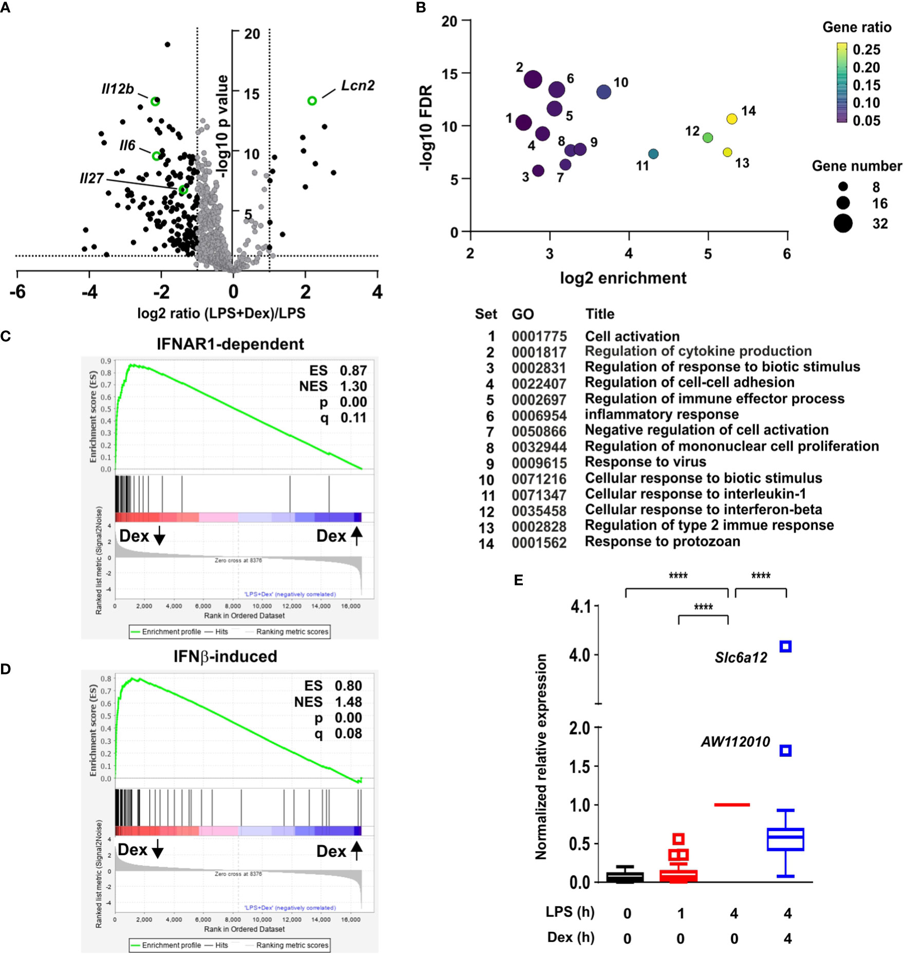
Figure 1 Dexamethasone broadly inhibits IFNβ-dependent gene expression in LPS-treated macrophages. (A) Mouse BMDMs (three independent isolates per condition) were treated with vehicle, 10 ng/ml LPS for 1 h or 4 h, or 10 ng/ml LPS + 100 nM Dex for 4 h. mRNA was harvested, quantified by microarray and analyzed using Partek. Transcripts robustly induced by LPS at 4 h (fold increase > 5, pAdj < 0.05) were identified and their responses to addition of Dex illustrated by volcano plot. Black dots represent LPS-induced transcripts whose expression was significantly altered by addition of Dex (fold change > 2, pAdj < 0.05). The transcripts highlighted in green are discussed in the text. (B) LPS-induced and Dex-suppressed transcripts (black dots in left quadrant of (A) were subjected to GO analysis using default parameters in Panther. Significantly enriched GO terms were filtered manually and using REVIGO to eliminate redundant terms. The top 14 remaining terms were plotted according to enrichment, FDR, gene number and gene ratio. (C) Gene set enrichment analysis was performed using the comparison of LPS vs. LPS + Dex as a quantitative data set, and the top 70 IFNAR1-dependent transcripts from (26) as a categorical gene list. Dex-inhibited transcripts are clustered at the left, and Dex-enhanced transcripts at the right. ES, enrichment score; NES, normalized enrichment score; q, false discovery rate. (D) Gene set enrichment analysis was performed as in C, except using the 93 most robustly IFNβ-inducible genes from (27). (E) For 96 genes that were robustly induced by LPS in our data set and interferon-regulated according to (26) or (27) gene expression was normalized to the 4 h value, and mean expression was plotted in Tukey box-and-whisker format. Outliers exceed the median by more than 1.5x interquartile range. ****, p < 0.001.
Gene Ontology (GO) analysis of GC down-regulated genes revealed highly significant enrichment of terms related to inflammation, cytokine production and responses to biotic stimuli (Figure 1B; Supplemental Table 1). Enrichment of such terms was predictable and uninformative, since the original set of 599 transcripts was selected on the basis of strong induction by LPS. We were interested by the gene sets “Response to virus” (GO:0009615; set 9 in Figure 1B), “Cellular response to interferon beta” (GO:0035458; set 12 in Figure 1B), and “Response to protozoan” (GO:0001562; set 14 in Figure 1B), which displayed 10.4-fold, 31.8-fold and 39.3-fold enrichment, respectively. Enrichment of these terms suggested that Dex inhibited expression of genes involved in antimicrobial responses (which are commonly regulated by IFNβ). However, there was no significant enrichment of GO terms specifically related to responses to intracellular bacteria.
Closer inspection of the microarray data revealed many interferon-stimulated genes (ISGs) amongst the LPS-induced genes that were significantly down-regulated by Dex. To explore this in more detail we made use of two published data sets related to interferon-mediated gene regulation in primary murine macrophages (generated in a similar manner). First, a systems-based analysis of the macrophage transcriptome identified a large number of genes whose delayed induction by lipidA (a highly TLR4-specific LPS moiety) was dependent on autocrine signaling via the interferon receptor IFNAR1 (29). The top 70 IFNAR1-dependent genes from that study were used as a gene set for GSEA (gene set enrichment analysis) (Figure 1C). This confirmed extremely strong enrichment of IFNAR1-dependent genes amongst those negatively regulated by Dex in our own data set, with an enrichment score of 0.866 and p value 0.0. From a study of macrophage regulatory networks (26) we then identified genes robustly induced by treatment of BMDMs with IFNβ alone (fold change > 100 and p < 0.05), and used this list of 93 genes in GSEA (Figure 1D). Once again there was very strong enrichment of IFNβ-inducible genes amongst those suppressed by Dex in our microarray set, with enrichment score 0.800 and p = 0.0. Removal of genes common to the two lists reduced the enrichment score only marginally (ES = 0.775) and did not change the nominal p value of zero.
We generated a list of 96 genes that were robustly induced by LPS in our own data set and interferon-regulated according to one or both of the above-mentioned data sets (26, 29). Mean responses of these genes to LPS and/or Dex were then plotted (Figure 1E). The great majority were expressed in a delayed fashion, with little or no increase of expression 1 h after addition of LPS (Figure 1E). At the 4 h time point, expression of 86 out of 92 genes was significantly inhibited by Dex. Importantly, the inhibitory effect of Dex was not quite universal. The IFNβ-responsive gene Acod1 (aconitate decarboxylase 1), previously known as Irg1 (immune-responsive gene 1), mediates production of the anti-inflammatory metabolite itaconic acid (27, 30). The Acod1 transcript was induced more than 1000-fold by LPS, and this response was spared from inhibition by Dex. Two IFNβ-responsive genes were significantly cooperatively regulated by LPS and Dex (the outliers AW112010 and Slc6a12 in Figure 1E: p = 1.73x10⁻⁴ and p = 1.00 x 10⁻⁷, respectively).
These data suggested that LPS treatment of BMDMs induced an antimicrobial state, likely involving autocrine signaling by IFNβ, and that this process was opposed by Dex. The nature of the antimicrobial state was difficult to define because of lack of knowledge of factors that restrict some pathogens, and because many antimicrobial factors are in fact not specific for pathogen class. We therefore focused on antiviral mediators, because many of these have been systematically identified and annotated on the basis of high throughput functional screens [for example (31)]. First we conducted a systematic screen of our microarray data, identifying genes that were induced by LPS (log2 fold change > 1, p < 0.05); expressed in a delayed manner (at least two-fold higher at 4 h than at 1 h); inhibited by Dex (p < 0.05); labelled as type I interferon-inducible in mouse according to the Interferome database (32); and had plausible roles in the regulation of viral life cycles (at least two supporting publications found in Pubmed using the systematic gene name and “antiviral” as search terms). This generated a list of 107 genes (Supplemental Table 2), which is likely to be under-inclusive because of the use of a single, relatively early time point for analysis of gene expression; shortcomings of the text-mining approach; or shortcomings in functional annotation of genes. For example, the poorly annotated gene AA467197, mouse ortholog of the human C15orf48 gene, has only recently been identified as an interferon regulated gene and a putative antiviral factor (33, 34). It is also important to note that viruses may co-opt cellular antiviral machinery to gain a competitive advantage, therefore not all of the genes in the final list are unequivocally “antiviral” under all circumstances. However, many of these are known to contribute to cell-intrinsic defense against viral pathogens (23, 31). These genes belong to several classes including: GTPases or GTP-binding proteins, which contribute to antimicrobial defense via a number of mechanisms (23, 35) (Gbp2, Gbp4, Gbp5, Gbp9, Gnb4, Iigp1, Irgm2, Mx1, Mx2, Tgtp1, Tgtp2); PRRs involved in the recognition of intracellular pathogens and the initiation of antiviral responses (Aim2, Ddx58 [RIG-I], Dhx58, Eif2ak2 [PKR], Ifi203, Ifi204, Ifih1 [MNDA-5], Mndal, Nlrc5, Pyhin); several members of the poly-ADP ribosyltransferase (PARP) and Schlafen (SLFN) families that contribute to antiviral defenses (36, 37); and diverse restriction factors that act at distinct points of infectious cycles to prevent entry, replication or exit of viruses (Bst2 [tetherin], Ch25h, Cmpk2, Herc6, Ifi35, Ifi44, Ifi47, Ifit1, Ifit2, Ifit3, Isg15, Isg20, Oasl1, Parp14, Rsad2 [viperin], Trim21, Usp18) (23, 24). Patterns of expression of the top 50 most Dex sensitive genes are illustrated in heatmap form in Figure 2A, whilst the full list is included in Supplemental Table 2.
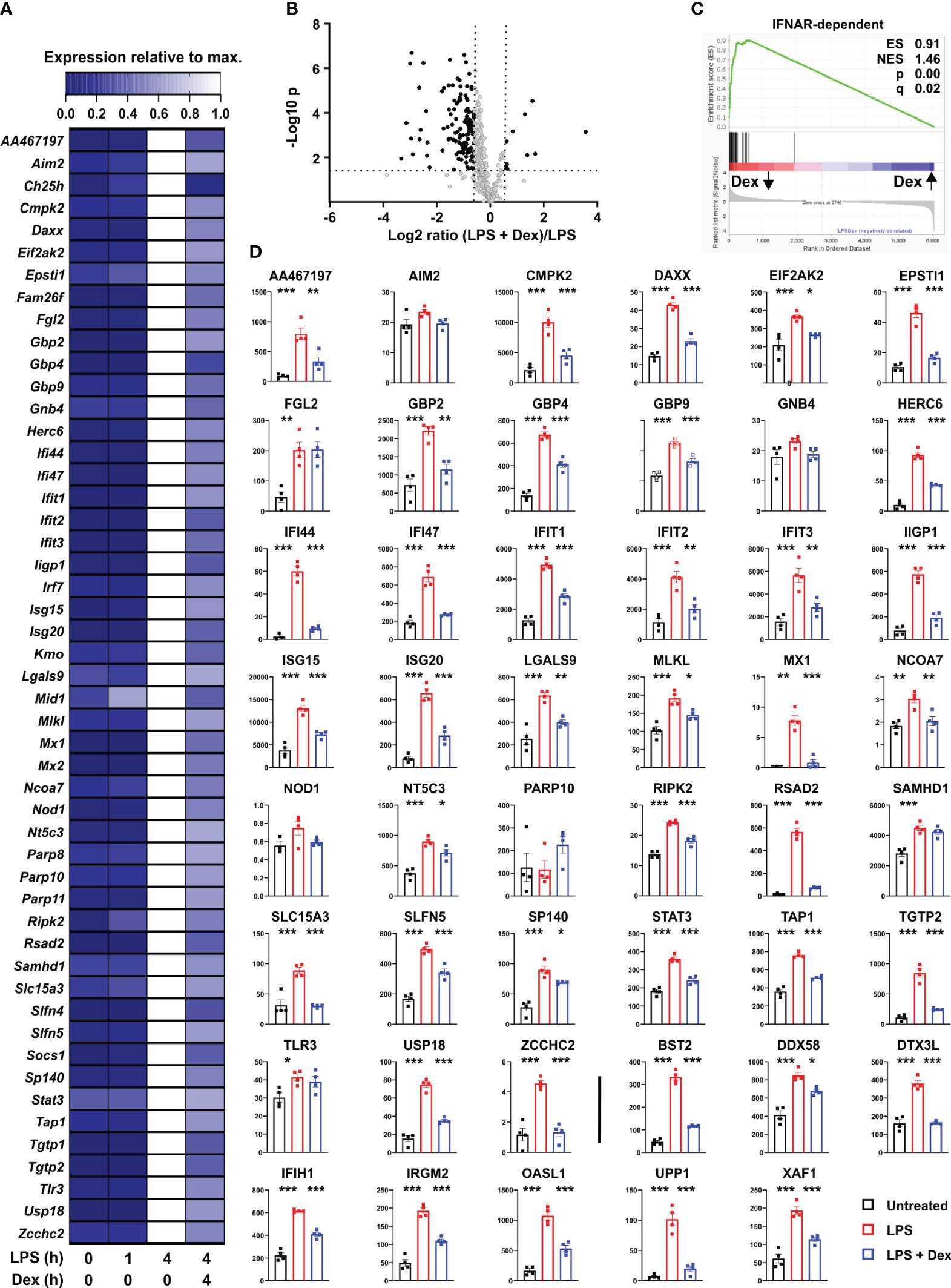
Figure 2 Dexamethasone inhibits expression of multiple antiviral mediators at both mRNA and protein levels. (A) To identify putative interferon-regulated antiviral mediators that are suppressed by Dex we filtered our data set for genes that were 1) well-annotated; 2) robustly induced by LPS (Log2 FC > 1, padj < 0.05); 3) expressed in delayed manner (mean expression at least 2-fold higher at 4h than at 1 h); 4) significantly down-regulated by Dex (padj < 0.05); 5) annotated as type I interferon regulated genes according to the Interferome database (32); 6) with plausible evidence of a role in the regulation of viral life cycles (at least two supporting publications found by PubMed search with appropriate gene symbol and “antiviral”). For these 107 genes (See Supplemental Table 2), mean expression was normalized to expression after 4 h stimulation with LPS. The 50 most Dex-sensitive genes are illustrated in heat map format, ordered alphabetically. (B) Mouse BMDMs (four independent isolates per condition) were treated with vehicle, 10 ng/ml LPS or 10 ng/ml LPS + 100 nM Dex for 24 h. Protein lysates were generated and subjected to data independent acquisition-based quantitative proteomic analysis. The 410 proteins whose levels were increased by LPS treatment (> 1.5-fold change, padj < 0.05) are illustrated in volcano plot according to their response to addition of Dex. Black dots indicate proteins whose levels were significantly affected by Dex (> 1.5-fold increase or decrease, padj <0.05). (C) Gene set enrichment analysis was performed essentially as in 1C. Protein expression data were clustered according to response to Dex, with Dex-suppressed proteins on the left; protein accession numbers were substituted by corresponding gene symbols; and IFNAR-dependent genes from (26) formed the categorical list. (D) Where data were available from the proteomic study, levels of proteins corresponding to the genes listed in A were plotted. Numerical values are protein concentrations in nM. The last eight plots (after the vertical bar) show proteins of interest, whose corresponding transcripts were not in (A) *, p < 0.05; **, p < 0.01; ***, p < 0.005. Absence of symbol indicates lack of statistical significance.
An unbiased proteomic screen was then used to assess protein levels in BMDMs treated with vehicle, LPS or LPS + Dex for 24 hours. Depth of proteome coverage was good, with more than 6,000 proteins detected, and normalized protein concentrations were similar across conditions, indicating an absence of systemic bias (not shown). We identified proteins whose abundance was increased by LPS (> 1.5-fold increase, Padj < 0.05). The lower threshold for fold change was chosen because LPS-induced changes of abundance were generally lower at the protein than at the mRNA level. This generated a list of 410 LPS-regulated proteins, whose responses to Dex were then examined. According to volcano plot, the effect of Dex was again dominated by suppression (Figure 2B). 125 LPS-induced proteins were significantly negatively regulated by Dex, and 96 of these were identified as type I interferon-regulated genes in the Interferome database (32). Amongst these Dex sensitive targets, gene ontology analysis revealed very strong enrichment of terms related to type I interferons (response to interferon-beta, GO:0035456, fold enrichment 41.2, p = 3.2 x 10⁻¹⁹; cellular response to interferon-beta, GO:0035458, fold enrichment 40.9, p = 9.3 x 10⁻¹⁷). Gene set enrichment analysis also revealed exceptional enrichment of IFNAR-dependent genes that were negatively regulated by Dex at the protein level (Figure 2C). Where data were available in the proteomic data set, we examined the expression of individual protein products of the top 50 Dex-sensitive genes (Figure 2D). With a few exceptions, patterns of expression were similar at mRNA and protein levels, with significant increase in response to LPS and significant decrease in response to addition of Dex. The extent of agreement is striking given that mRNAs were sampled at 4 and proteins at 24 hours after stimulation. The selection of the 24 h time point for assessment of protein expression may have led us to miss or underestimate effects of both LPS and Dex in this experiment. Nevertheless, the emerging picture is one of strong and sustained GC-mediated suppression of antiviral programs. Note that some mediators of antiviral responses (e.g., STING1 and MAVS) were constitutively expressed at both mRNA and protein levels and neither up-regulated by LPS nor down-regulated by Dex (data not shown).
Several transcripts encoding secreted or cell-surface immunomodulatory factors were also expressed in delayed fashion, negatively regulated by Dex, and identified as ISGs according to the Interferome database (32) (Figure 3A). Il12b, Il6 and Il27 mRNAs were highlighted in Figure 1A. Levels of the corresponding proteins were measured in supernatants of BMDMs treated under the same conditions (Figure 3B). All of these cytokines displayed no increase of expression 1 h after an LPS stimulus, strong increase at 4h, and strong inhibition by Dex. Some of the immunomodulatory proteins were measurable in the unbiased proteomic screen, up-regulated by LPS and suppressed by Dex (Figure 3C). Here we also confirmed the strong induction of LCN2 protein by the combination of LPS and Dex (Figure 3C).
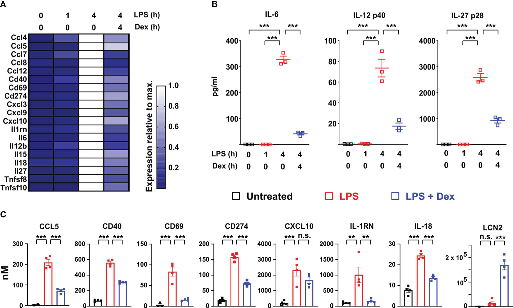
Figure 3 Dexamethasone inhibits expression of multiple cell surface and secreted immuno-modulators at both mRNA and protein levels. (A) Based on the same microarray data as analyzed in Figure 1, expression of selected interferon-regulated immuno-modulatory proteins is illustrated in heat map form as in Figure 2A. (B) In a separate experiment mouse BMDMs (three independent isolates) were treated as indicated, and secreted IL-6, IL-12 p40 and IL-27 p28 were measured in supernatants. ***, p < 0.005. (C). Where data were available from the proteomic study, the expression of immunomodulatory proteins was plotted as in Figure 2D. **, p < 0.01; ***, p < 0.005; n.s., not statistically significant.
ISGF3 plays a central role in the regulation of ISGs (18). Ligand-activated GR was reported to inhibit expression of ISGs by competing with ISGF3 for an essential transcriptional cofactor (38). To investigate the site of action of Dex we therefore used a mouse macrophage cell line stably transfected with an ISGF3-dependent reporter construct. This construct was strongly activated by either LPS or recombinant IFNβ, and in each case the response was ablated by the broad-spectrum JAK inhibitor Ruxolitinib, consistent with dependence on the phosphorylation of STAT1 and STAT2 (Figure 4A). LPS-induced activation of the reporter was dose-dependently inhibited by Dex, whereas the IFNβ-induced response was insensitive to even the highest dose of Dex used (1 μM), suggesting that Dex does not directly interfere with ISGF3 function.
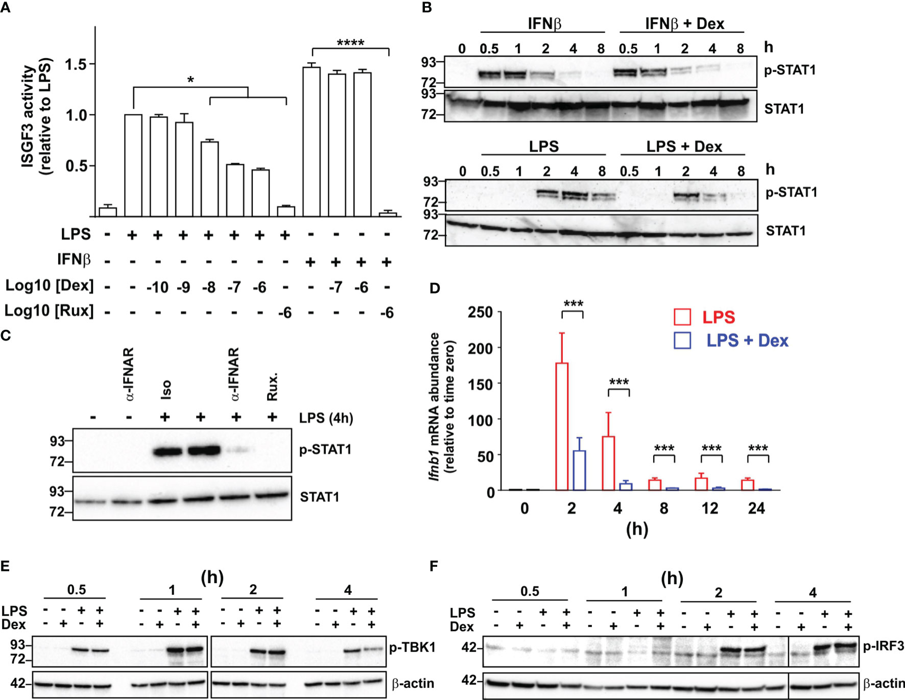
Figure 4 Dexamethasone inhibits expression of IFNβ in LPS-activated mouse macrophages via an unknown mechanism. (A) A RAW264.7 cell line stably transfected with an ISGF3-dependent SEAP (secreted alkaline phosphatase) reporter was treated with combinations of LPS (10 ng/ml), IFNβ (10 ng/ml), Ruxolitinib (Rux) and Dex as indicated for 24 h and reporter activity was assayed. The graph shows mean results from 3-6 independent experiments ± SEM, with normalization against reporter activity in the presence of LPS alone. *, p < 0.05 (Wilcoxon signed rank test); ****, p < 0.001 (ANOVA). (B) Mouse BMDMs were treated with combinations of LPS (10 ng/ml), IFNβ (10 ng/ml) and Dex (100 nM) as indicated. Cell lysates were generated and western blotted using antibodies against phosphorylated (Tyr 701) or total STAT1. Representative of three independent experiments. (C) Mouse BMDMs were stimulated with LPS for 4 h in the absence or presence of an IFNAR neutralizing antibody or isotype control (both 10 μg/ml). Cell lysates were prepared and western blotted as in (B) Representative of three independent experiments. (D) Mouse BMDMs were treated with LPS (10 ng/ml) or LPS + Dex (100nM) for the indicated times, and Ifnb1 mRNA was measured by qPCR. Mean of three independent experiments ± SEM. ***, p < 0.005. (E) Mouse BMDMs were treated with combinations of LPS (10 ng/ml) and Dex (100 nM) for 0.5 – 4 h, cell lysates were prepared and blotted for phosphorylated (activated) TBK1 or β-actin. Representative of two independent experiments. (F) Mouse BMDMs were treated as in E and lysates were blotted for phosphorylated (activated) IRF3 or β-actin.
IFNβ caused a rapid increase in the phosphorylation of STAT1, which was evident by 30 minutes and insensitive to Dex (Figure 4B). In contrast, LPS caused delayed STAT1 phosphorylation, which was not evident until 2 h, and was impaired by Dex, particularly at later time points. The delayed activation of STAT1 in response to LPS was dependent on IFNAR1-mediated signaling, and could be prevented by either an IFNAR1-neutralising antibody or treatment of cells with Ruxolitinib (Figure 4C). These observations suggest that Dex acts upstream of IFNβ biosynthesis to disrupt the IFNβ-mediated autocrine loop that controls macrophage functions. In fact, the Ifnb1 gene appeared in several of the GO terms discussed above (Supplemental Table 1), and its expression was strongly inhibited by Dex in the microarray experiment (see below). Quantitative PCR confirmed rapid, strong and sustained induction of the Ifnb1 gene following LPS treatment of BMDMs (Figure 4D). Throughout a 24 h time course, this response to LPS was inhibited at least 70% by 100 nM Dex.
It was previously reported that pre-treatment of the myeloid cell line U937 with Dex impaired the LPS-induced phosphorylation and activation of TBK1 (39). We therefore considered the hypothesis that inhibition of TBK1 function explains the inhibitory effect of Dex on IFNβ-mediated feedback in primary macrophages. However, simultaneous treatment of primary BMDMs with LPS and Dex caused no impairment of early LPS-induced TBK1 phosphorylation under conditions that resulted in strong inhibition of both Ifnb1 and ISGs (Figure 4E). Dex weakly but consistently reduced TBK1 phosphorylation at the four-hour time point. However, this late effect was unlikely to contribute to changes in expression of Ifnb1, which were strongly declining by four hours. Dex also had no effect on the phosphorylation of IRF3, which mediates the activation of Ifnb1 transcription downstream of TBK1 (Figure 4F).
Glucocorticoids selectively inhibit the expression of certain pro-inflammatory genes by increasing and prolonging the expression of DUSP1 and inhibiting MAPK p38 (7–10). DUSP1 is a negative regulator of Ifnb1 gene expression (40). Both serum IFNβ and hepatic IFNβ-dependent genes (including Ifit2, Rsad2, Iigp1 and many more of the ISGs discussed here) were over-expressed in Dusp1-/- mice following infection with E. coli (41). These observations suggested the hypothesis that Dex impairs expression of IFNβ and IFNβ-dependent genes in LPS-activated BMDMs via the induction of DUSP1. This hypothesis was tested using macrophages generated from Dusp1-/- mice. IFNβ biosynthesis was elevated in Dusp1-/- BMDMs as previously reported (40), but similarly inhibited by Dex in Dusp1+/+ and Dusp1-/- BMDMs (Figure 5A: note different scales of y axes). A previous publication reported that Dex did not impair LPS-induced IFNβ expression in BMDMs (42). The reasons for this discrepancy are not clear, but may include different sources and doses of LPS or different duration of Dex treatment.
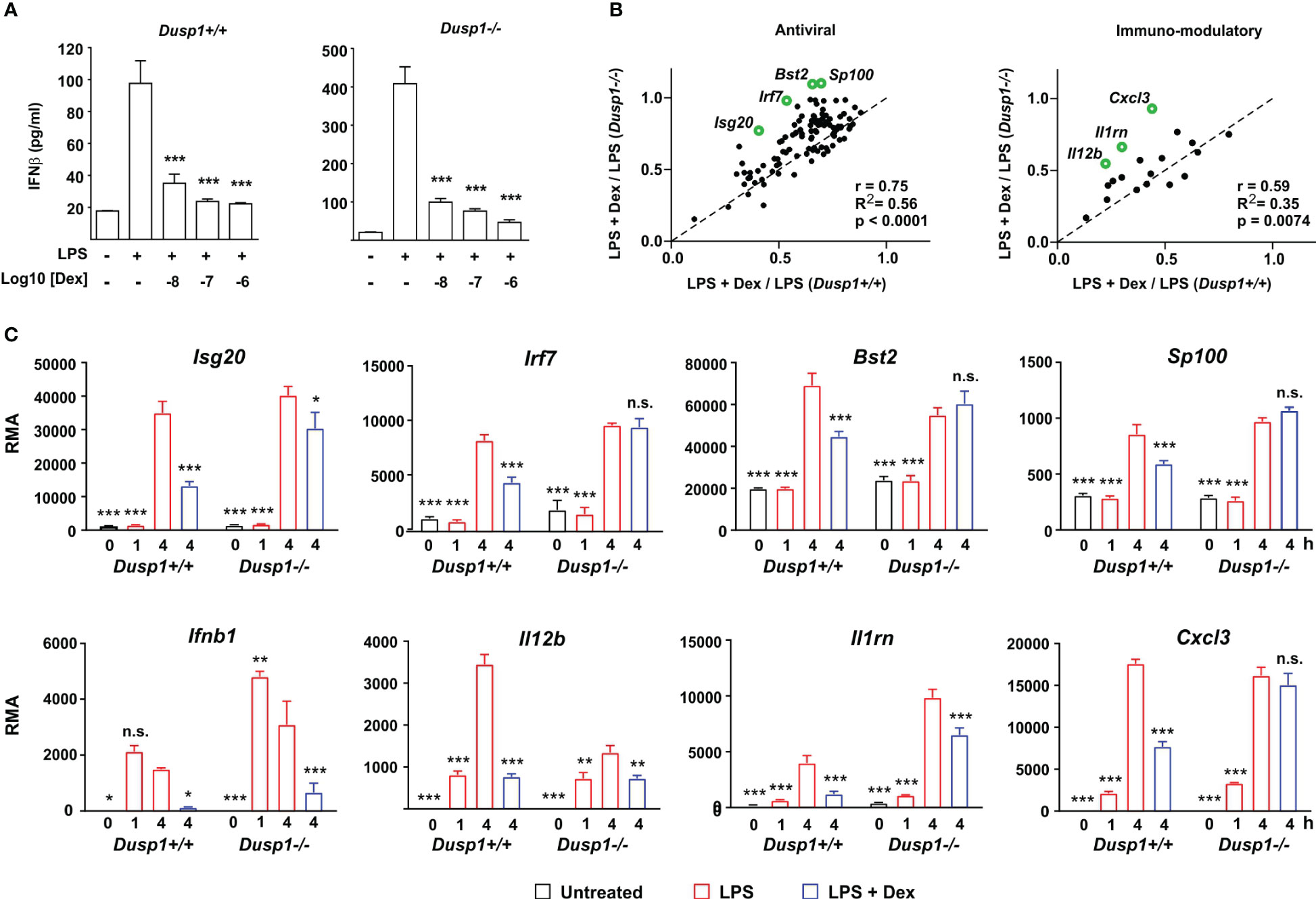
Figure 5 DUSP1 contributes to Dex-mediated suppression of ISGs. (A) Dusp1+/+ or Dusp1-/- BMDMs (three independent isolates of each) were treated with LPS (10 ng/ml) for 4h with or without addition of Dex as indicated. IFNβ protein was measured by Luminex assay. The graph shows mean ± SEM, n = 3. ***, p < 0.005. (B) For each interferon-regulated gene of “antiviral” or “immmuno-modulatory” subsets (see Figures 2, 3), Dex sensitivity was calculated as the ratio of expression in the presence of LPS + Dex vs. expression in the presence of LPS alone. In the plot of Dex sensitivity in Dusp1+/+ and Dusp1-/- BMDMs, the dotted diagonal represents the null hypothesis, that effects of Dex are independent of Dusp1 genotype. Selected outlier genes that do not fit the null hypothesis are highlighted. (C) Microarray-derived expression data for these outlier genes are illustrated. The pattern of expression of Ifnb1 itself is also shown. n.s., not significant; *, p < 0.05; **, p < 0.01; ***, p < 0.005; all in comparison to 4 h LPS treatment.
We then systematically examined the impact of DUSP1 depletion on the Dex sensitivity of IFNβ-regulated antiviral genes (Figure 5B, left) and immunomodulatory genes (Figure 5B, right). For the gene sets discussed above, Dex sensitivity was calculated as average ratio of expression under conditions (LPS + Dex/LPS). In plots of Dex sensitivity in Dusp1+/+ (x axis) and Dusp1-/- (y axis) backgrounds the dotted diagonal line represents the null hypothesis that disruption of the Dusp1 gene has no impact on responsiveness to Dex. For both gene sets, sensitivity to Dex was closely correlated in Dusp1+/+ and Dusp1-/- BMDMs, supporting the null hypothesis and suggesting a minimal impact of DUSP1 depletion. However, there were several outliers whose response to Dex was altered by Dusp1 gene disruption. Some of these are highlighted in Figure 5B, and primary data from the microarray experiment are illustrated in Figure 5C. In the cases of Irf7, Bst2, Sp100 and Cxcl3, significant inhibitory effects of Dex were completely lost in the absence of DUSP1. We conclude that overall DUSP1 plays a minor and highly gene-specific role in the regulation of IFNβ signaling by Dex. Figure 5C also illustrates the behavior of the Ifnb1 gene itself in the microarray experiment, confirming elevated expression in Dusp1-/- BMDMs and strong suppression by Dex in BMDMs of both genotypes.
Collectively, these observations led to a clear and testable hypothesis: that Dex impairs the responses of macrophages to LPS by inhibiting the expression of IFNβ and disrupting the IFNβ-mediated autocrine feedback loop. We predicted that secondary genes, which are induced by LPS in an IFNβ-dependent manner, would be insensitive to Dex if activated directly by IFNβ, by-passing the site of GC action. To test this hypothesis, we first generated a panel of test genes comprising three intracellular antiviral effectors (Ifit2, Rsad2 and Iigp1) and three secreted cytokines or chemokines (Cxcl9, Il6 and Il27). IFNAR1 dependence of all of these genes was confirmed by reference to a published data-set (29); by our own experiments using Ifnar1-/- BMDMs (data not shown); and by use of an IFNAR1-blocking antibody (Figure 6A). We then tested the responses of all six genes to challenge with LPS or IFNβ in the absence or presence of Dex (Figure 6B). All three of the antimicrobial effector genes responded as predicted by the hypothesis: their expression in response to LPS was significantly impaired by Dex, whereas their expression in response to IFNβ was insensitive to Dex. Surprisingly, all three of the cytokine/chemokine genes were suppressed by Dex whether induced by LPS or IFNβ. Dex may therefore act both upstream and downstream of IFNβ to impair LPS responses of BMDMs. The IFNβ-induced expression of Ifit2, Rsad2, Iigp1, Il6 and Cxcl9 was very strongly dependent on ISGF3 (at least 100-fold lower expression in IFNβ-stimulated Irf9-/- BMDMs) (43). Ifit2, Rsad2 and Iigp1 were insensitive to Dex, whilst Il6 and Cxcl9 were suppressed by Dex in IFNβ-stimulated BMDMs. This confirms the conclusion from the reporter gene assay (Figure 4A); that Dex does not regulate IFNβ signaling in BMDMs via broad suppression of ISGF3 function.
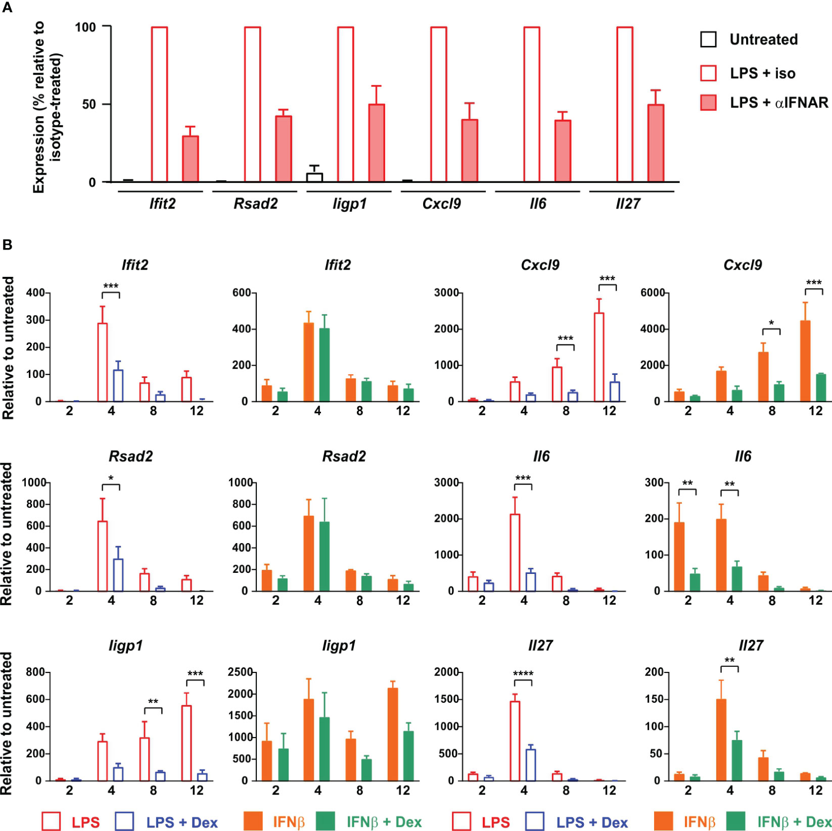
Figure 6 Dex regulates expression of ISGs both upstream and downstream of IFNβ in mouse BMDMs. (A) BMDMs (4-5 independent replicates) were treated for 4 h with vehicle, 10 ng/ml LPS + 10 μg/ml IFNAR neutralizing antibody, or 10 ng/ml LPS + 10 μg/ml isotype control. RNA was isolated and the indicated transcripts were measured by qPCR, with normalization against LPS + isotype control. (B) BMDMs (at least three independent replicates) were treated for 0 - 12 h with LPS (10 ng/ml) ± Dex (100 nM) or IFNβ (10 ng/ml) ± Dex (100 nM). Expression of selected genes was determined by qPCR. *, p < 0.05; **, p < 0.01; ***, p < 0.005; ****, p < 0.001; absence of symbol indicates lack of statistical significance (p > 0.05).
To investigate the same phenomena in human cells we generated monocyte-derived macrophages from peripheral blood of healthy donors and stimulated them with LPS in the absence or presence of 100 nM Dex. Surprisingly there was greater than ten-fold donor-to-donor variation in quantity of IFNβ secreted by LPS-activated MDMs (note logarithmic axis in Figure 7A). However, IFNβ release was consistently inhibited by 100 nM Dex (mean inhibition ± SEM 52% ± 6%, p = 0.001). Basal expression of some ISGs varied between donors by several hundred-fold (not shown), and magnitudes of response to both LPS and IFNβ were also highly variable (Figure 7B). This created a practical problem in generating adequate statistical power to investigate effects of Dex, power calculation suggesting that 100 or more individual donors would be needed for some ISGs. Nevertheless, a preliminary analysis using seven independent isolates of MDMs confirmed that Dex can inhibit expression of the ISGs RSAD2 and CXCL9 whether evoked by either LPS or IFNβ. In human alveolar macrophages the LPS-induced expression of IFNB1 and RSAD2 genes was consistently inhibited by Dex (Figure 7C); however, IFNβ protein was below the limit of detection in supernatants of these cells. Scarcity of samples, low yields of mRNA and lack of statistical power prevented us from carrying out an extensive survey of ISG expression. Yet these preliminary data suggest that Dex may also impair type I interferon signaling in alveolar macrophages.
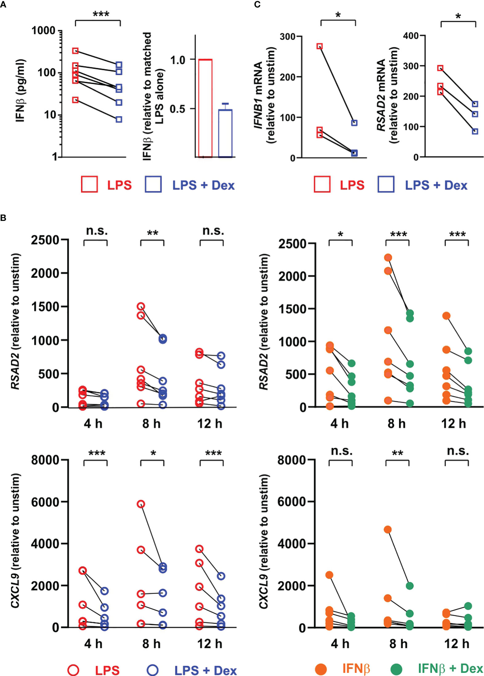
Figure 7 Dex regulates expression of ISGs both upstream and downstream of IFNβ in primary human macrophages. (A) Primary human monocyte-derived macrophages (n=7) were stimulated with 10 ng/ml LPS ± 100 nM Dex for 4 h, and IFNβ protein in supernatants was measured by Luminex assay. Right hand panel illustrates the same data with normalization to LPS alone. (B) Primary human monocyte-derived macrophages (n=7) were stimulated with 10 ng/ml LPS ± 100 nM Dex (left) or with 10 ng/ml IFNβ ± 100 nM Dex (right) for 4, 8 or 12 h. Rsad2 and Cxcl9 mRNAs were measured by qPCR, with normalization against untreated control for each individual macrophage culture. *, p < 0.05; **, p < 0.01; ***, p < 0.005; n.s., p > 0.05. C. Alveolar macrophages were isolated from histologically normal regions of lung resection tissue and treated with 1 μg/ml LPS ± 100 nM Dex for 12 h. IFNB1 and RSAD2 mRNAs were measured by qPCR, with normalization against untreated control for each individual sample. *, p < 0.05.
Glucocorticoids have been reported to impair type I interferon signaling in macrophages, but these studies tended to document expression of relatively few ISGs, at the mRNA level only (38, 39, 42, 44, 45). Here we describe a remarkably broad impact of Dex on the expression of ISGs by LPS-activated BMDMs, occurring at both mRNA and protein levels. Several mechanisms have been suggested to mediate inhibitory effects of GCs on type I interferon signaling, but none of them satisfactorily explain the observations described here. 1) Dex pretreatment impaired the LPS-induced phosphorylation and activation of TBK1 and IRF3 in a myeloid cell line (39). In our hands Dex did not affect LPS-induced TBK1 phosphorylation or downstream IRF3 activation under conditions where the expression of Ifnb1 mRNA and IFNβ protein was strongly inhibited. In any case the impairment of IFNβ production only partially accounts for Dex-mediated impairment of ISG expression in LPS-treated BMDMs, because some ISGs were also sensitive to Dex when directly induced by IFNβ itself. 2) Downstream of the IFNβ receptor, Dex was reported to inhibit ISG expression by reducing the expression of STAT1 (44), or by up-regulating SOCS1 and inhibiting STAT1 phosphorylation (42). In the presence of either LPS or IFNβ, Dex did not influence STAT1 protein expression over 8 hours. Although Dex inhibited the LPS-induced phosphorylation of STAT1 this was almost certainly a consequence of impaired IFNβ secretion since IFNβ-induced STAT1 phosphorylation was insensitive to Dex. Furthermore, Dex reduced rather than increased the LPS-induced expression of Socs1 mRNA (LPS alone, 17,621 ± 912; LPS + Dex, 6,278 ± 914). 3) Another proposed mechanism of action of glucocorticoids is the impairment of ISGF3 function, involving competition between GR and ISGF3 for the essential transcriptional cofactor NCOA2 (nuclear receptor coactivator A2) (38). Several lines of evidence argue against such a mechanism here. First, IFNβ-mediated activation of an ISGF3-dependent reporter construct was insensitive to Dex. In contrast the same reporter was sensitive to Dex when activated by LPS, presumably again due to impairment of IFNβ expression. Amongst our Dex-sensitive ISGs, just over half were reported to have ISGF3 binding sites in their vicinity in a previous study (43) (Supplemental Table 2). However, there was poor correlation between ISGF3-dependence in that study and Dex sensitivity in ours (R2 = 0.09). The discrepancy is illustrated by the LPS-induced gene Oas2 (oligoadenylate synthase 2), which was exceptionally dependent on ISGF3 (43) but unaffected by Dex in our study. Although we cannot rule out that Dex inhibits some ISGs via impairment of ISGF3 function, this mechanism cannot explain the broad impact of Dex. 4) We previously reported that Dex-induced expression of the phosphatase DUSP1 contributes to the suppression of certain LPS-induced genes in macrophages (7, 8, 10). However, the sensitivity of most ISGs to Dex was affected little by disruption of the Dusp1 gene. In only a few cases was there a clear loss of sensitivity to Dex in Dusp1-/- BMDMs. If there is a unifying mechanism by which Dex controls the type I interferon response, it remains to be discovered. Any explanation of this phenomenon must account for its selectivity, and the escape of certain well-known ISGs such as Acod1 and Oas2 (46).
An important question is whether the same suppression of antimicrobial genes occurs in macrophages exposed to a viral mimetic such as poly(I:C), rather than the bacterial component LPS. Our preliminary data indicate that this is the case (data not shown) – as one would predict given that poly(I:C) also induces robust IFNβ expression in macrophages. A second important question is whether GCs similarly affect type I interferon-dependent antimicrobial programs in other cell types. In A549 airway epithelial cells stimulated for 18 h with IL-1β, the expression of 84 genes was significantly impaired by addition of Dex (fold decrease >2, p < 0.05) (47). The majority of these genes are interferon-regulated genes according to the Interferome database (32), and many are orthologs of genes that we found to be suppressed by Dex in LPS-treated BMDMs (e.g., Cmpk2, Herc6, Ifi44, Ifih1, Ifit1, Ifit3, Isg15, Isg20, Usp18). It is likely that Dex also disrupts IFNβ-mediated positive feedback loops in these cells. More than half of the ISGs discussed here were also induced by LPS in primary human alveolar macrophages, suggesting that antimicrobial responses of these cells are also governed by IFNβ-mediated feedback (48) (Supplemental Table 2).
The type I interferon pathway has been described as a two-edged sword in the battle between host and pathogen (49). On one hand interferons are critical for cell-intrinsic defenses against pathogens and initiation of adaptive immune responses to destroy pathogens and infected cells. On the other hand, they can contribute to pathogen-induced inflammatory pathology and consequent tissue damage in infectious diseases such as influenza and tuberculosis (50–52). GC treatment of infectious diseases also has highly variable consequences. In specific contexts or at specific stages of disease GCs may alleviate pathogen-induced inflammation, but otherwise they may exacerbate infection. In the majority of infectious diseases, the use of GCs is controversial, outcomes of clinical trials are often contradictory, and consensus on best practice is difficult to achieve. GC treatment of pandemic (H1N1) influenza was associated with increased mortality, increased length of stay in intensive care, and prolonged virus shedding (53–55). Deleterious outcomes may be related to the complex cross-talk between GCs and type I interferon signaling. For example, GC treatment of bronchial epithelial cells impaired the expression of selected ISGs and promoted infection by influenza A virus or rhinovirus. Treatment with a type I interferon reversed the pro-viral effect of GC treatment both in vitro and in vivo (56, 57), indicating that GCs can favor viral infections by suppressing the type I interferon pathway.
Several lines of evidence reveal the critical role of type I interferon signaling for host defense against the zoonotic coronavirus SARS-CoV-2. 1) Inborn and acquired defects of the type I interferon pathway are associated with increased susceptibility to infection (58). 2) Systematic screens and more targeted approaches have identified several ISGs as restriction factors that limit SARS-CoV-2 infection at different stages of the viral life-cycle (31). 3) Like other viruses (59), SARS-CoV-2 deploys several mechanisms to subvert the type I interferon-mediated defense machinery, both impairing the detection of virus and inhibiting the execution of antiviral programs of gene expression (60). 4) An early and robust type I interferon signature in circulating monocytes was associated with mild disease following SARS-CoV-2 infection (61). Likewise myeloid cell interferon responses correlated with viral clearance in an experimental model of COVID-19 (62). 5) Conversely, low expression of antiviral genes including CMPK2, EPSTI1, HERC6, IFI44, OASL and RSAD2 in peripheral blood was associated with worse clinical outcomes in COVID-19 (63). On the other side of the equation, several components of the damaging SARS-CoV-2-induced cytokine storm are type I interferon-inducible factors that contribute to pathogenesis of COVID-19 (64).
Almost uniquely amongst infectious pathologies, severe COVID-19 is now routinely treated using GCs (65). This makes it an important context in which to discuss the crosstalk between GCs and type I interferon signaling, and the balance between beneficial and harmful effects of GCs. Beneficial effects of GCs in severe COVID-19 are thought to involve suppression of the pro-inflammatory cytokine storm (66), in which the inhibition of type I interferon signaling may play a part (67). For example, elevated levels of the interferon-regulated cytokines IL-27 and IL-6 contribute to a signature that is predictive of poor outcome in COVID-19 (68, 69). GC-mediated suppression of these factors may contribute to improved outcomes. On the negative side, endogenous GC excess is a risk factor for COVID-19 (70). Similarly, prolonged use of synthetic GCs for the treatment of immune-mediated inflammatory diseases both increases the likelihood of contracting COVID-19 and contributes to worse outcomes of the disease (71). Although GCs reduce mortality by approximately a third in patients suffering from severe COVID-19 and requiring respiratory support (72), in mild or early disease there is no evidence of benefit, and some suggestion of harmful effects including delays to viral clearance (72–74). We note that several of the ISGs that we found to be negatively regulated by Dex at mRNA and protein levels are known restriction factors for SARS-CoV-2 (31) (Supplemental Table 2). These include DDX58 (RIG-I) and IFIH1 (MDA5), which are essential for detection of intracellular SARS-CoV-2 (75) and are targeted by the virus as a means of evading interferon responses (76). We therefore hypothesize that GCs are able to promote SARS-CoV-2 infection by opposing type I interferon-mediated antiviral mechanisms. This hypothesis remains to be tested, and is the focus of our ongoing research.
There are clearly many unanswered questions about when and how best to use GCs against SARS-CoV-2 and other viral pathogens. Indeed, it has previously been argued that variable outcomes of GC trials in COVID-19 were related to inconsistent timing of delivery (77). The balance between the harm of suppressing antiviral mechanisms in early disease and the benefit of suppressing the cytokine storm in the later phase is precarious. The tipping point between harm and benefit may also differ between individuals. There is clearly a strong case for application of well-informed patient stratification in COVID-19 (78, 79). We argue that the effects of GCs on type I interferon-mediated host defenses should be part of such a precision medicine approach. Furthermore, there is urgent need for further research on the mechanistic basis of cross-talk between GCs and type I interferon signaling.
All mice were maintained at the Biomedical Services Unit of the University of Birmingham. Animal care and experimental procedures were performed according to Home Office guidelines and approved by the University of Birmingham Local Ethical Review Committee. Bone marrow was isolated from the femurs of humanely culled 6–12-week-old wild type C57BL/6J mice, and BMDMs (bone marrow derived macrophages) generated essentially as described (80) by differentiation in vitro in RPMI 1640 medium with L-glutamine (Gibco Thermo Fisher Scientific 21875034) supplemented with 10% heat-inactivated fetal bovine serum (Sigma F0392) and 50ng/ml recombinant M-CSF (Peprotech 300-25) for 7 days. BMDMs were plated at a density of 1 x 10⁶/ml in the appropriate cell culture plate at least 1 d prior to stimulation. This method generated primary mouse macrophages with purity approaching 99% as measured by flow cytometry using antibodies against F4/80, CD11b, CD14 and CD64 (Figure S1).
The Dusp1-/- strain was a generous gift from Bristol-Myers Squibb, and was back crossed to C57BL/6 for ten generations prior to experiments described here.
Human monocytes from healthy blood donors were isolated from leukapheresis blood cones supplied by the National Blood and Transplant Service (ethical approval ERN_16-0191). Monocytes were enriched by negative selection using StemCell RosetteSep Human Monocyte Enrichment Cocktail (StemCell 15068; 75μl per ml of blood) and Ficoll-Paque (VWR 17144003). Cells were differentiated for 7 days in RPMI 1640 medium with L-glutamine (Gibco Thermo Fisher Scientific 21875034) supplemented with 5% heat-inactivated fetal bovine serum (Biosera FB-1001) and 50ng/ml recombinant M-CSF (Peprotech 300-25). Differentiated macrophages were plated at a density of 1 x 10⁶/ml in the appropriate cell culture plate at least 1 d prior to stimulation. In our hands this method generates primary human macrophages with purity >98% as measured by flow cytometry using antibodies against CD14, CD64 and CD206 (Figure S1).
Ethical approval was obtained to recruit adult patients scheduled for surgery to remove lung tissue as part of their clinical treatment plan (predominantly lobectomy for lung cancer) at the Thoracic Surgery Unit at the Queen Elizabeth Hospital Birmingham (REC 17/WM/0272). Lung tissue samples distant from any tumor, without macroscopically evident pathology, and surplus to requirement for histopathology, were washed through with 500 ml of sterile 0.9% saline (Baxter, UK) using a 14-gauge needle (Vasofix ®). The washed through lavage fluid was collected and centrifuged at 4°C and 560g for 10 minutes. Cell pellets were resuspended in 10 ml of RPMI1640 containing 10% FBS, and overlaid onto 10 ml of Lymphoprep™ (StemCell) prior to centrifugation at 800g for 30 min at 4°C. The interphase layer containing mononuclear cells was aspirated into a sterile 50 ml tube containing PBS with 10% FBS, which was then centrifuged at 300g for 10 minutes at 4°C. AMs were resuspended in RPMI1640 containing 10% FBS, 100U/mL penicillin, 100µg/mL streptomycin and 2mM L-glutamine (Sigma-Aldrich), and plated. This method generates primary human alveolar macrophages with purity >98% as measured by a validated combination of cytospin and flow cytometry using antibodies against CD68 (81).
Stimulations were carried out in appropriate culture plates using the following reagents and concentrations unless otherwise stated: LPS (Enzo ALX-581-010-L002; 10ng/ml); recombinant human IFNβ (Peprotech 300-02BC; 10ng/ml); recombinant mouse IFNβ (BioLegend 581306; 10ng/ml); Ruxolitinib (Selleck S1378; 1μM); Dexamethasone (Sigma D8893; 100nM). Where stated, cells were incubated for 2 hours with an IFNAR1 blocking monoclonal antibody (Fisher Scientific MAR1-5A3; 10 μg/ml), or mouse IgG1 kappa isotype control, (Fisher Scientific P3.6.2.8.1; 10 μg/ml) prior to stimulations.
Upon harvesting of in vitro-differentiated cells for counting and seeding for experiments (mouse BMDMs day 7; human MDMs day 6), a fraction of the cell suspension was used for purity assessment by flow cytometry. Cells were stained with eFluor780 fixable viability dye (eBioscience 65-0865, 1/1000 in PBS) for 20min on ice to allow gating for live cells. Fc receptor blocking was performed for 5min prior to antibody staining using Mouse BD Fc Block (BD 553142) or Human BD Fc Block (BD 564220). Antibody staining was performed for 30min on ice in FACS buffer (PBS+2% FCS+1mM EDTA) using the antibodies and dilutions listed below. Cells were washed and resuspended in FACS buffer and run on a BD LSRFortessa X-20. Analysis was performed using FlowJo v10.
Mouse BMDM staining: PE rat anti-mouse F4/80 Antibody (Biolegend 123110, 1/50); PE rat IgG2a κ Isotype Control Antibody (Biolegend 400507, 1/50); FITC rat anti-mouse/human CD11b Antibody (Biolegend 101205, 1/100); FITC rat IgG2b Isotype Control Antibody (eBioscience 11-4031-82, 1/100); PerCP-eFluor710 rat anti-mouse CD14 Antibody (eBioscience 46-0141-80, 1/40); PerCP-eFluor710 rat IgG2a κ Isotype Control Antibody (eBioscience 46-4321-82, 1/40); APC rat anti-mouse CD64 Antibody (Biolegend 139305, 1/20); APC rat IgG1 κ Isotype Control Antibody (eBioscience 17-4301-81, 1/20).
Human MDM staining: PE mouse anti-human CD14 Antibody (Biolegend 367104, 1/100); PE mouse IgG1 κ Isotype Control Antibody (Biolegend 400113, 1/20); PerCP-eFluor710 mouse anti-human CD64 Antibody (eBioscience 46-0649-41, 1/20); PerCP-eFluor710 mouse IgG1 κ Isotype Control Antibody (eBioscience 46-4714-80, 1/20); APC mouse anti-human CD206 Antibody (Biolegend 321110, 1/20); APC mouse IgG1 κ Isotype Control Antibody (Biolegend 400119, 1/20).
The RAW-Blue ISG macrophage cell line stably transfected with an ISGF3-dependent reporter construct was cultured and maintained according to the manufacturer’s instructions (RAW-Blue ISG cells, Invivogen raw-isg). ISGF3 reporter activity was determined from cell culture supernatants using QUANTI-Blue detection reagents according to the manufacturer’s instructions (Invivogen).
RNA was isolated from mouse BMDMs and human monocyte derived macrophages using Norgen Total RNA Purification Plus kit (Geneflow P4-0016) according to manufacturer’s instructions. cDNA was synthesized using the iScript cDNA Synthesis Kit (Biorad 1708891). mRNA was detected by RT-qPCR using SYBR TB Green Premix Ex Taq (Takara RR820W) and primers supplied by Eurofins Genomics or Sigma Aldrich. UBC (human) or B2M (mouse) were used to normalize mRNA measurements via 2^-ΔΔCt method. Primers are listed in Table 1.
Microarray analysis was performed using SurePrint G3 Mouse GE 8x60K slides (Agilent) and Partek Genomics Suite version 6.6, build 6.13.0315 (Partek) as previously described (82). For generation of volcano plots, transcripts were first filtered for significant upregulation in response to LPS (> 5-fold increase, adjusted p-value < 0.05) and weakly expressed transcripts were removed by application of an arbitrary filter of 200 RMA. Plots (log2 fold difference of expression vs -log10 ANOVA p-value) were constructed using Prism (GraphPad Software), with subset cut-offs at p-value < 0.05 and fold difference of expression > 2. Microarray data discussed in this paper were deposited at Gene Expression Omnibus (http://www.ncbi.nlm.nih.gov/geo/) with the accession number GSE68449.
Gene Ontology analysis was performed using the Panther v16 online toolset (83). Redundant GO terms were filtered using REVIGO (84). Gene Set Enrichment Analysis (GSEA) was performed using GSEA_4.1.0 (85).
Conditioned medium samples from cultured macrophages were subjected to custom multiplex Luminex assay (Bio-Rad Custom Bio-Plex Assays), and Invitrogen ProcartaPlex Mouse or Human IFNβ simplexes, (EXP01A-26044-901 or EXP01A-12088-901, respectively), according to manufacturer’s instructions.
For whole cell lysates, cells were harvested in RIPA buffer and samples passed through a Qiashredder column to disrupt genomic DNA (Qiagen 79656). Protein was quantified by Pierce BCA Assay (Thermo Fisher Scientific 23225). Laemmli or XT sample buffer was added and samples heated to 95°C for 5min. Western blotting was performed using Criterion TGX protein gels (BioRad) and Tris-Glycine SDS buffer (Geneflow B9-0032) or 4–12% Criterion™ XT Bis-Tris Protein Gels (BioRad) and XT MES running buffer (BioRad 1610789). Protein was transferred to BioRad Trans-Blot PVDF membranes (BioRad 1704157) using BioRad Trans-Blot Turbo transfer system. Blots were imaged using Clarity Western ECL Substrate (BioRad 1705061) and BioRad ChemiDoc MP Imaging System. Densitometry for Western blot quantification was performed using ImageJ. Antibodies used in western blotting are listed in Table 2.
Following derivation and stimulation of mouse BMDMs (as described above) cells were washed with PBS and lysed in proteomic lysis buffer (5% SDS, 10mM TCEP, 50mM TEAB). Unbiased proteome analysis was carried out by data-independent acquisition (DIA) mass spectrometry proteomics utilizing S-Trap on-column digestion and purification, following the methods detailed by Baker et al. (86). Protein copy number was determined using Perseus software (86, 87) with normalization for histone protein content. Data were analyzed as described in the above methods for microarray analysis and bioinformatics.
GraphPad Prism software (Version 6) was used for statistical analysis. Mann Whitney U test was used for comparison of two groups. For analysis of multiple groups, ANOVA was used with Bonferroni correction for multiple comparisons. The following marks are used throughout: *,p<0.05; **,p<0.01;***,p<0.005; n.s., not statistically significant. N numbers specified in figure legends indicate biological replicates. In human alveolar macrophages, where expression of ISGs was highly variable between donors, ratio paired t test was used to test for consistent effect of Dex.
The datasets presented in this study can be found in online repositories. The names of the repository/repositories and accession number(s) can be found below: https://www.ncbi.nlm.nih.gov/geo/, GSE68449 https://www.ebi.ac.uk/pride/archive/, PXD041375.
The studies involving humans were approved by West Midlands Research Ethics Committee. The studies were conducted in accordance with the local legislation and institutional requirements. The participants provided their written informed consent to participate in this study. Ethical approval was not required for the study involving animals in accordance with the local legislation and institutional requirements because bone marrow was obtained from mice after Schedule 1 procedure, not requiring ethical approval.
AC secured funding for the study and wrote the manuscript. AC, JO, RH and JA contributed to conception and design of the study. JO, OB, SC, TT, KD, SL-R, JW and CM performed experiments and analyzed data. RM provided essential clinical samples. All authors contributed to the article and approved the submitted version.
This work was supported by a Program Grant (21802) and Research into Inflammatory Arthritis Centre Versus Arthritis (22072), both from Versus Arthritis (UK). OB was supported by an MB-PhD studentship from the Kennedy Trust (UK).
The authors declare that the research was conducted in the absence of any commercial or financial relationships that could be construed as a potential conflict of interest.
All claims expressed in this article are solely those of the authors and do not necessarily represent those of their affiliated organizations, or those of the publisher, the editors and the reviewers. Any product that may be evaluated in this article, or claim that may be made by its manufacturer, is not guaranteed or endorsed by the publisher.
The Supplementary Material for this article can be found online at: https://www.frontiersin.org/articles/10.3389/fimmu.2023.1190261/full#supplementary-material
1. Hillier SG. Diamonds are forever: the cortisone legacy. J Endocrinol (2007) 195(1):1–6. doi: 10.1677/JOE-07-0309
2. Schacke H, Docke WD, Asadullah K. Mechanisms involved in the side effects of glucocorticoids. Pharmacol Ther (2002) 96(1):23–43. doi: 10.1016/S0163-7258(02)00297-8
3. Cain DW, Cidlowski JA. Immune regulation by glucocorticoids. Nat Rev Immunol (2017) 17(4):233–47. doi: 10.1038/nri.2017.1
4. De Bosscher K, Vanden Berghe W, Haegeman G. Mechanisms of anti-inflammatory action and of immunosuppression by glucocorticoids: negative interference of activated glucocorticoid receptor with transcription factors. J Neuroimmunol (2000) 109(1):16–22. doi: 10.1016/S0165-5728(00)00297-6
5. Clark AR, Belvisi MG. Maps and legends: the quest for dissociated ligands of the glucocorticoid receptor. Pharmacol Ther (2012) 134(1):54–67. doi: 10.1016/j.pharmthera.2011.12.004
6. Oh KS, Patel H, Gottschalk RA, Lee WS, Baek S, Fraser IDC, et al. Anti-inflammatory chromatinscape suggests alternative mechanisms of glucocorticoid receptor action. Immunity (2017) 47(2):298–309.e5. doi: 10.1016/j.immuni.2017.07.012
7. Lasa M, Abraham SM, Boucheron C, Saklatvala J, Clark AR. Dexamethasone causes sustained expression of mitogen-activated protein kinase (Mapk) phosphatase 1 and phosphatase-mediated inhibition of mapk P38. Mol Cell Biol (2002) 22(22):7802–11. doi: 10.1128/MCB.22.22.7802-7811.2002
8. Abraham SM, Lawrence T, Kleiman A, Warden P, Medghalchi M, Tuckermann J, et al. Antiinflammatory effects of dexamethasone are partly dependent on induction of dual specificity phosphatase 1. J Exp Med (2006) 203(8):1883–9. doi: 10.1084/jem.20060336
9. Bhattacharyya S, Brown DE, Brewer JA, Vogt SK, Muglia LJ. Macrophage glucocorticoid receptors regulate toll-like receptor 4-mediated inflammatory responses by selective inhibition of P38 map kinase. Blood (2007) 109(10):4313–9. doi: 10.1182/blood-2006-10-048215
10. Joanny E, Ding Q, Gong L, Kong P, Saklatvala J, Clark AR. Anti-inflammatory effects of selective glucocorticoid receptor modulators (Sgrms) are partially dependent on upregulation of dual specificity phosphatase 1 (Dusp1). Br J Pharmacol (2011) 165(4b):1124–36. doi: 10.1111/j.1476-5381.2011.01574.x
11. Wang X, Nelin LD, Kuhlman JR, Meng X, Welty SE, Liu Y. The role of map kinase phosphatase-1 in the protective mechanism of dexamethasone against endotoxemia. Life Sci (2008) 83(19-20):671–80. doi: 10.1016/j.lfs.2008.09.003
12. Udalova IA, Mantovani A, Feldmann M. Macrophage heterogeneity in the context of rheumatoid arthritis. Nat Rev Rheumatol (2016) 12(8):472–85. doi: 10.1038/nrrheum.2016.91
13. Kurowska-Stolarska M, Alivernini S. Synovial tissue macrophages in joint homeostasis, rheumatoid arthritis and disease remission. Nat Rev Rheumatol (2022) 18(7):384–97. doi: 10.1038/s41584-022-00790-8
14. Merad M, Martin JC. Pathological inflammation in patients with covid-19: A key role for monocytes and macrophages. Nat Rev Immunol (2020) 20(6):355–62. doi: 10.1038/s41577-020-0331-4
15. Knoll R, Schultze JL, Schulte-Schrepping J. Monocytes and macrophages in Covid-19. Front Immunol (2021) 12:720109. doi: 10.3389/fimmu.2021.720109
16. Fenton C, Martin C, Jones R, Croft A, Campos J, Naylor AJ, et al. Local steroid activation is a critical mediator of the anti-inflammatory actions of therapeutic glucocorticoids. Ann Rheum Dis (2021) 80(2):250–60. doi: 10.1136/annrheumdis-2020-218493
17. Amit I, Garber M, Chevrier N, Leite AP, Donner Y, Eisenhaure T, et al. Unbiased reconstruction of a mammalian transcriptional network mediating pathogen responses. Science (2009) 326(5950):257–63. doi: 10.1126/science.1179050
18. Platanitis E, Decker T. Regulatory networks involving stats, irfs, and nfκb in inflammation. Front Immunol (2018) 9:2542. doi: 10.3389/fimmu.2018.02542
19. Hirotani T, Yamamoto M, Kumagai Y, Uematsu S, Kawase I, Takeuchi O, et al. Regulation of lipopolysaccharide-inducible genes by myd88 and toll/il-1 domain containing adaptor inducing ifn-beta. Biochem Biophys Res Commun (2005) 328(2):383–92. doi: 10.1016/j.bbrc.2004.12.184
20. Karaghiosoff M, Steinborn R, Kovarik P, Kriegshauser G, Baccarini M, Donabauer B, et al. Central role for type I interferons and tyk2 in lipopolysaccharide-induced endotoxin shock. Nat Immunol (2003) 4(5):471–7. doi: 10.1038/ni910
21. Sheikh F, Dickensheets H, Gamero AM, Vogel SN, Donnelly RP. An essential role for ifn-beta in the induction of ifn-stimulated gene expression by lps in macrophages. J Leukoc Biol (2014) 96(4):591–600. doi: 10.1189/jlb.2A0414-191R
22. Thomas KE, Galligan CL, Newman RD, Fish EN, Vogel SN. Contribution of interferon-beta to the murine macrophage response to the toll-like receptor 4 agonist, lipopolysaccharide. J Biol Chem (2006) 281(41):31119–30. doi: 10.1074/jbc.M604958200
23. MacMicking JD. Interferon-inducible effector mechanisms in cell-autonomous immunity. Nat Rev Immunol (2012) 12(5):367–82. doi: 10.1038/nri3210
24. Fitzgerald KA. The interferon inducible gene: viperin. J Interferon Cytokine Res (2011) 31(1):131–5. doi: 10.1089/jir.2010.0127
25. Weijtens O, Schoemaker RC, Cohen AF, Romijn FP, Lentjes EG, van Rooij J, et al. Dexamethasone concentration in vitreous and serum after oral administration. Am J Ophthalmol (1998) 125(5):673–9. doi: 10.1016/s0002-9394(98)00003-8
26. Raza S, Barnett MW, Barnett-Itzhaki Z, Amit I, Hume DA, Freeman TC. Analysis of the transcriptional networks underpinning the activation of murine macrophages by inflammatory mediators. J Leukoc Biol (2014) 96(2):167–83. doi: 10.1189/jlb.6HI0313-169R
27. Mills EL, Ryan DG, Prag HA, Dikovskaya D, Menon D, Zaslona Z, et al. Itaconate is an anti-inflammatory metabolite that activates nrf2 via alkylation of keap1. Nature (2018) 556(7699):113–7. doi: 10.1038/nature25986
28. Yamazaki S, Akira S, Sumimoto H. Glucocorticoid augments lipopolysaccharide-induced activation of the Iκbζ-dependent genes encoding the anti-microbial glycoproteins lipocalin 2 and pentraxin 3. J Biochem (2015) 157(5):399–410. doi: 10.1093/jb/mvu086
29. Tong AJ, Liu X, Thomas BJ, Lissner MM, Baker MR, Senagolage MD, et al. A stringent systems approach uncovers gene-specific mechanisms regulating inflammation. Cell (2016) 165(1):165–79. doi: 10.1016/j.cell.2016.01.020
30. Ferreira AV, Netea MG, Domínguez-Andrés J. Itaconate as an immune modulator. Aging (Albany NY) (2019) 11(12):3898–9. doi: 10.18632/aging.102057
31. Martin-Sancho L, Lewinski MK, Pache L, Stoneham CA, Yin X, Becker ME, et al. Functional landscape of sars-cov-2 cellular restriction. Mol Cell (2021) 81(12):2656–68.e8. doi: 10.1016/j.molcel.2021.04.008
32. Rusinova I, Forster S, Yu S, Kannan A, Masse M, Cumming H, et al. Interferome V2.0: an updated database of annotated interferon-regulated genes. Nucleic Acids Res (2013) 41(Database issue):D1040–6. doi: 10.1093/nar/gks1215
33. Clayton SA, Daley KK, MacDonald L, Fernandez-Vizarra E, Bottegoni G, O'Neil JD, et al. Inflammation causes remodeling of mitochondrial cytochrome C oxidase mediated by the bifunctional gene C15orf48. Sci Adv (2021) 7(50):eabl5182. doi: 10.1126/sciadv.abl5182
34. Sorouri M, Chang T, Jesudhasan P, Pinkham C, Elde NC, Hancks DC. Signatures of host-pathogen evolutionary conflict reveal mistr-a conserved mitochondrial stress response network. PloS Biol (2020) 18(12):e3001045. doi: 10.1371/journal.pbio.3001045
35. Rafeld HL, Kolanus W, van Driel IR, Hartland EL. Interferon-induced gtpases orchestrate host cell-autonomous defence against bacterial pathogens. Biochem Soc Trans (2021) 49(3):1287–97. doi: 10.1042/bst20200900
36. Kim ET, Weitzman MD. Schlafens can put viruses to sleep. Viruses (2022) 14(2):442. doi: 10.3390/v14020442
37. Malgras M, Garcia M, Jousselin C, Bodet C, Lévêque N. The antiviral activities of poly-adp-ribose polymerases. Viruses (2021) 13(4):582. doi: 10.3390/v13040582
38. Flammer JR, Dobrovolna J, Kennedy MA, Chinenov Y, Glass CK, Ivashkiv LB, et al. The type I interferon signaling pathway is a target for glucocorticoid inhibition. Mol Cell Biol (2010) 30(19):4564–74. doi: 10.1128/mcb.00146-10
39. McCoy CE, Carpenter S, Palsson-McDermott EM, Gearing LJ, O'Neill LA. Glucocorticoids inhibit irf3 phosphorylation in response to toll-like receptor-3 and -4 by targeting tbk1 activation. J Biol Chem (2008) 283(21):14277–85. doi: 10.1074/jbc.M709731200
40. McGuire VA, Rosner D, Ananieva O, Ross EA, Elcombe SE, Naqvi S, et al. Beta interferon production is regulated by P38 mitogen-activated protein kinase in macrophages via both msk1/2- and tristetraprolin-dependent pathways. Mol Cell Biol (2017) 37(1):e00454-16. doi: 10.1128/mcb.00454-16
41. Kirk SG, Murphy PR, Wang X, Cash CJ, Barley TJ, Bowman BA, et al. Knockout of mapk phosphatase-1 exaggerates type I ifn response during systemic escherichia coli infection. J Immunol (2021) 206(12):2966–79. doi: 10.4049/jimmunol.2001468
42. Bhattacharyya S, Zhao Y, Kay TW, Muglia LJ. Glucocorticoids target suppressor of cytokine signaling 1 (Socs1) and type 1 interferons to regulate toll-like receptor-induced stat1 activation. Proc Natl Acad Sci USA (2011) 108(23):9554–9. doi: 10.1073/pnas.1017296108
43. Platanitis E, Demiroz D, Schneller A, Fischer K, Capelle C, Hartl M, et al. A molecular switch from stat2-irf9 to isgf3 underlies interferon-induced gene transcription. Nat Commun (2019) 10(1):2921. doi: 10.1038/s41467-019-10970-y
44. Ballegeer M, Van Looveren K, Timmermans S, Eggermont M, Vandevyver S, Thery F, et al. Glucocorticoid receptor dimers control intestinal stat1 and tnf-induced inflammation in mice. J Clin Invest (2018) 128(8):3265–79. doi: 10.1172/jci96636
45. Reily MM, Pantoja C, Hu X, Chinenov Y, Rogatsky I. The grip1:Irf3 interaction as a target for glucocorticoid receptor-mediated immunosuppression. EMBO J (2006) 25(1):108–17. doi: 10.1038/sj.emboj.7600919
46. Li Y, Gong W, Li W, Liu P, Liu J, Jiang H, et al. The irg1-itaconate axis: A regulatory hub for immunity and metabolism in macrophages. Int Rev Immunol (2023) 42(5):364–78. doi: 10.1080/08830185.2022.2067153
47. King EM, Chivers JE, Rider CF, Minnich A, Giembycz MA, Newton R. Glucocorticoid repression of inflammatory gene expression shows differential responsiveness by transactivation- and transrepression-dependent mechanisms. PloS One (2013) 8(1):e53936. doi: 10.1371/journal.pone.0053936
48. Pinilla-Vera M, Xiong Z, Zhao Y, Zhao J, Donahoe MP, Barge S, et al. Full spectrum of lps activation in alveolar macrophages of healthy volunteers by whole transcriptomic profiling. PloS One (2016) 11(7):e0159329. doi: 10.1371/journal.pone.0159329
49. Trinchieri G. Type I interferon: friend or foe? J Exp Med (2010) 207(10):2053–63. doi: 10.1084/jem.20101664
50. Wu W, Metcalf JP. The role of type I ifns in influenza: antiviral superheroes or immunopathogenic villains? J Innate Immun (2020) 12(6):437–47. doi: 10.1159/000508379
51. McNab F, Mayer-Barber K, Sher A, Wack A, O'Garra A. Type I interferons in infectious disease. Nat Rev Immunol (2015) 15(2):87–103. doi: 10.1038/nri3787
52. Moreira-Teixeira L, Mayer-Barber K, Sher A, O'Garra A. Type I interferons in tuberculosis: foe and occasionally friend. J Exp Med (2018) 215(5):1273–85. doi: 10.1084/jem.20180325
53. Gottlieb M, Seagraves T, Gore SR. Do corticosteroids benefit patients with influenza pneumonia? Ann Emerg Med (2020) 75(1):100–1. doi: 10.1016/j.annemergmed.2019.06.021
54. Lansbury L, Rodrigo C, Leonardi-Bee J, Nguyen-Van-Tam J, Lim WS. Corticosteroids as adjunctive therapy in the treatment of influenza. Cochrane Database Syst Rev (2019) 2(2):CD010406. doi: 10.1002/14651858.CD010406.pub3
55. Giannella M, Alonso M, Garcia de Viedma D, Lopez Roa P, Catalán P, Padilla B, et al. Prolonged viral shedding in pandemic influenza a(H1n1): clinical significance and viral load analysis in hospitalized patients. Clin Microbiol Infect (2011) 17(8):1160–5. doi: 10.1111/j.1469-0691.2010.03399.x
56. Marcellini A, Swieboda D, Guedán A, Farrow SN, Casolari P, Contoli M, et al. Glucocorticoids impair type I ifn signalling and enhance rhinovirus replication. Eur J Pharmacol (2021) 893:173839. doi: 10.1016/j.ejphar.2020.173839
57. Thomas BJ, Porritt RA, Hertzog PJ, Bardin PG, Tate MD. Glucocorticosteroids enhance replication of respiratory viruses: effect of adjuvant interferon. Sci Rep (2014) 4:7176. doi: 10.1038/srep07176
58. Zhang Q, Bastard P, Cobat A, Casanova JL. Human genetic and immunological determinants of critical Covid-19 pneumonia. Nature (2022) 603(7902):587–98. doi: 10.1038/s41586-022-04447-0
59. Zhu J, Chiang C, Gack MU. Viral evasion of the interferon response at a glance. J Cell Sci (2023) 136(12):jcs260682. doi: 10.1242/jcs.260682
60. Minkoff JM, tenOever B. Innate immune evasion strategies of Sars-Cov-2. Nat Rev Microbiol (2023) 21(3):178–94. doi: 10.1038/s41579-022-00839-1
61. Schulte-Schrepping J, Reusch N, Paclik D, Baßler K, Schlickeiser S, Zhang B, et al. Severe Covid-19 is marked by a dysregulated myeloid cell compartment. Cell (2020) 182(6):1419–40.e23. doi: 10.1016/j.cell.2020.08.001
62. Singh DK, Aladyeva E, Das S, Singh B, Esaulova E, Swain A, et al. Myeloid cell interferon responses correlate with clearance of Sars-Cov-2. Nat Commun (2022) 13(1):679. doi: 10.1038/s41467-022-28315-7
63. Dong Z, Yan Q, Cao W, Liu Z, Wang X. Identification of key molecules in Covid-19 patients significantly correlated with clinical outcomes by analyzing transcriptomic data. Front Immunol (2022) 13:930866. doi: 10.3389/fimmu.2022.930866
64. Ramasamy S, Subbian S. Critical determinants of cytokine storm and type I interferon response in Covid-19 pathogenesis. Clin Microbiol Rev (2021) 34(3):e00299-20. doi: 10.1128/cmr.00299-20
65. Amati F, Tonutti A, Huston J, Dela Cruz CS. Glucocorticoid therapy in Covid-19. Semin Respir Crit Care Med (2023) 44(1):100–17. doi: 10.1055/s-0042-1759778
66. Kino T, Burd I, Segars JH. Dexamethasone for severe Covid-19: how does it work at cellular and molecular levels? Int J Mol Sci (2021) 22(13):6764. doi: 10.3390/ijms22136764
67. Kalinina O, Golovkin A, Zaikova E, Aquino A, Bezrukikh V, Melnik O, et al. Cytokine storm signature in patients with moderate and severe Covid-19. Int J Mol Sci (2022) 23(16):8879. doi: 10.3390/ijms23168879
68. Angioni R, Sánchez-Rodríguez R, Munari F, Bertoldi N, Arcidiacono D, Cavinato S, et al. Age-severity matched cytokine profiling reveals specific signatures in Covid-19 patients. Cell Death Dis (2020) 11(11):957. doi: 10.1038/s41419-020-03151-z
69. Ozger HS, Karakus R, Kuscu EN, Bagriacik UE, Oruklu N, Yaman M, et al. Serial measurement of cytokines strongly predict Covid-19 outcome. PloS One (2021) 16(12):e0260623. doi: 10.1371/journal.pone.0260623
70. Vogel F, Reincke M. Endocrine risk factors for covid-19: endogenous and exogenous glucocorticoid excess. Rev Endocr Metab Disord (2022) 23(2):233–50. doi: 10.1007/s11154-021-09670-0
71. Hasseli R, Mueller-Ladner U, Hoyer BF, Krause A, Lorenz HM, Pfeil A, et al. Older age, comorbidity, glucocorticoid use and disease activity are risk factors for Covid-19 hospitalisation in patients with inflammatory rheumatic and musculoskeletal diseases. RMD Open (2021) 7(1):e001464. doi: 10.1136/rmdopen-2020-001464
72. Horby P, Lim WS, Emberson JR, Mafham M, Bell JL, Linsell L, et al. Dexamethasone in hospitalized patients with Covid-19. N Engl J Med (2021) 384(8):693–704. doi: 10.1056/NEJMoa2021436
73. Qiao W, Meng L, Zhang Y, Li D, Chen J, Wang J, et al. Safety and efficacy of glucocorticoids in the treatment of Covid-19: A meta-analysis of randomized control trials. Expert Rev Respir Med (2023) 17(1):81–96. doi: 10.1080/17476348.2023.2177155
74. Tang X, Feng YM, Ni JX, Zhang JY, Liu LM, Hu K, et al. Early use of corticosteroid may prolong Sars-Cov-2 shedding in non-intensive care unit patients with Covid-19 pneumonia: A multicenter, single-blind, randomized control trial. Respiration (2021) 100(2):116–26. doi: 10.1159/000512063
75. Yang DM, Geng TT, Harrison AG, Wang PH. Differential roles of rig-I like receptors in Sars-Cov-2 infection. Mil Med Res (2021) 8(1):49. doi: 10.1186/s40779-021-00340-5
76. Oh SJ, Shin OS. Sars-Cov-2-mediated evasion strategies for antiviral interferon pathways. J Microbiol (2022) 60(3):290–9. doi: 10.1007/s12275-022-1525-1
77. Cain DW, Cidlowski JA. After 62 years of regulating immunity, dexamethasone meets Covid-19. Nat Rev Immunol (2020) 20(10):587–8. doi: 10.1038/s41577-020-00421-x
78. Amati F, Dela Cruz CS. One size does not fit all: moving towards a personalized approach for steroids in Covid-19. Chest (2021) 159(5):1693–5. doi: 10.1016/j.chest.2021.01.043
79. Sinha P, Furfaro D, Cummings MJ, Abrams D, Delucchi K, Maddali MV, et al. Latent class analysis reveals Covid-19-related acute respiratory distress syndrome subgroups with differential responses to corticosteroids. Am J Respir Crit Care Med (2021) 204(11):1274–85. doi: 10.1164/rccm.202105-1302OC
80. Davis BK. Derivation of macrophages from mouse bone marrow. Methods Mol Biol (2019) 1960:41–55. doi: 10.1007/978-1-4939-9167-9_3
81. Mahida RY, Lax S, Bassford CR, Scott A, Parekh D, Hardy RS, et al. Impaired alveolar macrophage 11β-hydroxysteroid dehydrogenase type 1 reductase activity contributes to increased pulmonary inflammation and mortality in sepsis-related ARDS. Front Immunol (2023) 14:1159831. doi: 10.3389/fimmu.2023.1159831
82. Smallie T, Ross EA, Ammit AJ, Cunliffe HE, Tang T, Rosner DR, et al. Dual-specificity phosphatase 1 and tristetraprolin cooperate to regulate macrophage responses to lipopolysaccharide. J Immunol (2015) 195(1):277–88. doi: 10.4049/jimmunol.1402830
83. Mi H, Ebert D, Muruganujan A, Mills C, Albou LP, Mushayamaha T, et al. Panther version 16: A revised family classification, tree-based classification tool, enhancer regions and extensive api. Nucleic Acids Res (2021) 49(D1):D394–403. doi: 10.1093/nar/gkaa1106
84. Supek F, Bošnjak M, Škunca N, Šmuc T. Revigo summarizes and visualizes long lists of gene ontology terms. PloS One (2011) 6(7):e21800. doi: 10.1371/journal.pone.0021800
85. Subramanian A, Tamayo P, Mootha VK, Mukherjee S, Ebert BL, Gillette MA, et al. Gene set enrichment analysis: A knowledge-based approach for interpreting genome-wide expression profiles. Proc Natl Acad Sci USA (2005) 102(43):15545–50. doi: 10.1073/pnas.0506580102
86. Baker CP, Phair IR, Brenes AJ, Atrih A, Ryan DG, Bruderer R, et al. Dia label-free proteomic analysis of murine bone-marrow-derived macrophages. STAR Protoc (2022) 3(4):101725. doi: 10.1016/j.xpro.2022.101725
Keywords: glucocorticoid, dexamethasone, macrophage, type I interferon, interferon β, innate immunity, virus, SARS-CoV-2
Citation: O’Neil JD, Bolimowska OO, Clayton SA, Tang T, Daley KK, Lara-Reyna S, Warner J, Martin CS, Mahida RY, Hardy RS, Arthur JSC and Clark AR (2023) Dexamethasone impairs the expression of antimicrobial mediators in lipopolysaccharide-activated primary macrophages by inhibiting both expression and function of interferon β. Front. Immunol. 14:1190261. doi: 10.3389/fimmu.2023.1190261
Received: 20 March 2023; Accepted: 11 October 2023;
Published: 24 October 2023.
Edited by:
Oliver Planz, University of Tübingen, GermanyReviewed by:
Tünde Fekete, University of Debrecen, HungaryCopyright © 2023 O’Neil, Bolimowska, Clayton, Tang, Daley, Lara-Reyna, Warner, Martin, Mahida, Hardy, Arthur and Clark. This is an open-access article distributed under the terms of the Creative Commons Attribution License (CC BY). The use, distribution or reproduction in other forums is permitted, provided the original author(s) and the copyright owner(s) are credited and that the original publication in this journal is cited, in accordance with accepted academic practice. No use, distribution or reproduction is permitted which does not comply with these terms.
*Correspondence: Andrew R. Clark, YS5yLmNsYXJrQGJoYW0uYWMudWs=
†These authors contributed equally to this work and share first authorship
Disclaimer: All claims expressed in this article are solely those of the authors and do not necessarily represent those of their affiliated organizations, or those of the publisher, the editors and the reviewers. Any product that may be evaluated in this article or claim that may be made by its manufacturer is not guaranteed or endorsed by the publisher.
Research integrity at Frontiers

Learn more about the work of our research integrity team to safeguard the quality of each article we publish.