95% of researchers rate our articles as excellent or good
Learn more about the work of our research integrity team to safeguard the quality of each article we publish.
Find out more
CORRECTION article
Front. Immunol. , 31 March 2023
Sec. Cancer Immunity and Immunotherapy
Volume 14 - 2023 | https://doi.org/10.3389/fimmu.2023.1175360
This article is part of the Research Topic Fibrosis, inflammation, and cancers: dangerous liaisons to be depicted and targeted View all 10 articles
This article is a correction to:
Single-Cell RNA-seq Analysis Reveals Cellular Functional Heterogeneity in Dermis Between Fibrotic and Regenerative Wound Healing Fates
 Cao-Jie Chen1
 Hiroki Kajita1
 Kento Takaya1
 Noriko Aramaki-Hattori1
 Shigeki Sakai1
 Toru Asou2*
 Kazuo Kishi1*
A corrigendum on
Single-cell RNA-seq analysis reveals cellular functional heterogeneity in dermis between fibrotic and regenerative wound healing fates
by Chen C-J, Kajita H, Takaya K, Aramaki-Hattori N, Sakai S, Asou T and Kishi K (2022) . 13:875407. doi: 10.3389/fimmu.2022.875407
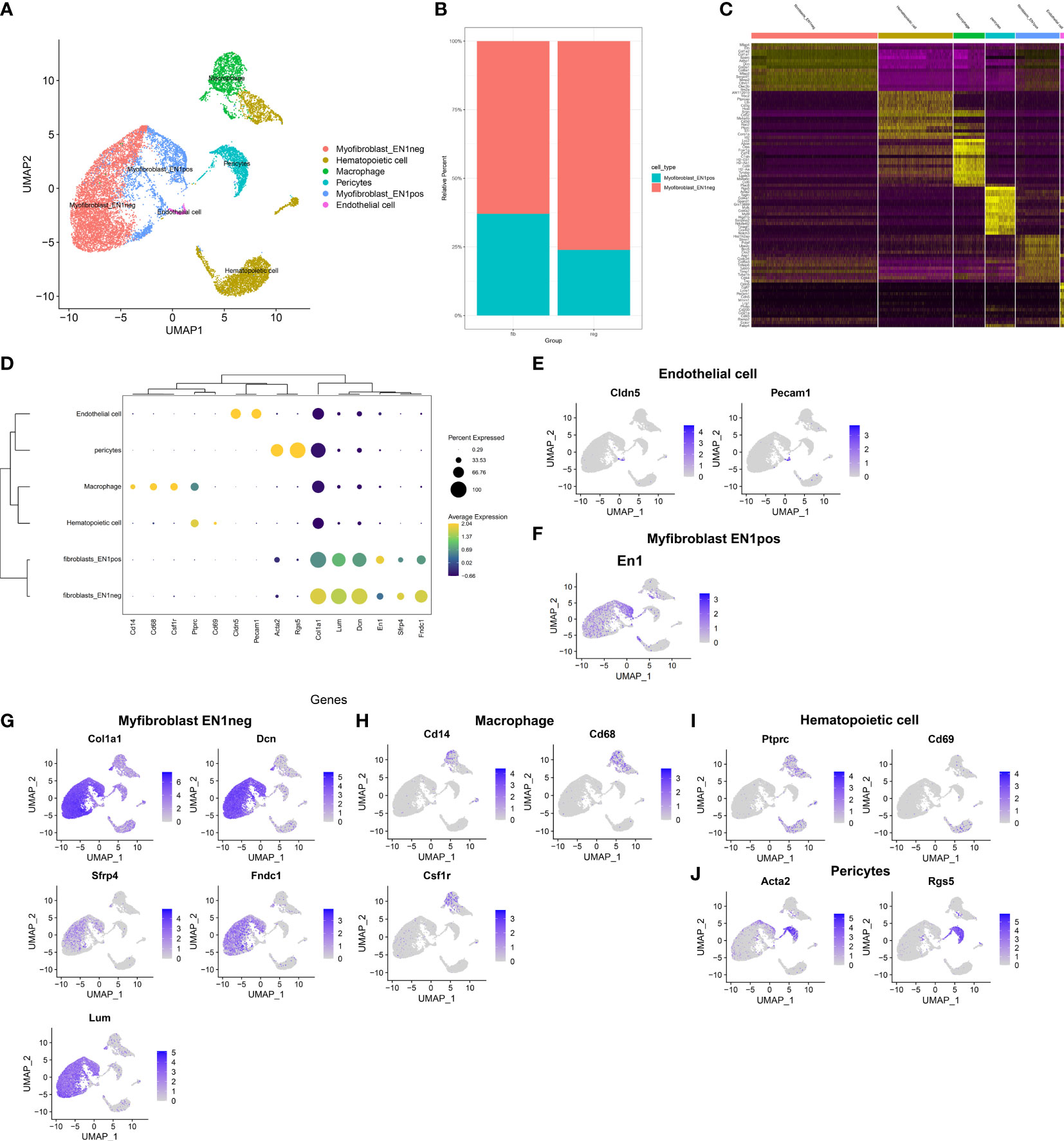
In the published article, there was an error in Figures 2C, D and F as published. The dot size for EN1 gene in different cell types in Figure 2D was wrong because we mislabeled the gene name during the production of the picture. Due to the same reason, the Figure 2F was also wrongly placed. In addition, we want to replace Figure 2C to add more feature genes (top 15, previously was top 10) in the heatmap to better characterize cell-type-specific gene expression patterns. The corrected Figures 2C, D and F appear below.

Figure 2 Identification of cell types and their marker genes across fibrotic and regenerative wound dermal cells. (A) UMAP plots showing cell types identified by marker genes. Each cell type was colored by a unique color. (B) The cell ratio of EN1-negative and -positive myofibroblasts among fibrotic and regenerative wound dermal cells. (C) Heatmap visualizing cell-type-specific gene expression patterns. Each column represented the average expression after cells were grouped. (D) Integrated analysis showing marker genes across cell types. The size of each circle reflected the percentage of cells in each cell type where the gene was detected, and the color shadow reflected the average expression level within each cell type. (E–J) UMAP plots of expression of the marker genes for endothelial cells, EN1-negative and -positive myofibroblasts, macrophages, hematopoietic cells, and pericytes.
The authors apologize for this error and state that this does not change the scientific conclusions of the article in any way. The original article has been updated.
All claims expressed in this article are solely those of the authors and do not necessarily represent those of their affiliated organizations, or those of the publisher, the editors and the reviewers. Any product that may be evaluated in this article, or claim that may be made by its manufacturer, is not guaranteed or endorsed by the publisher.
Keywords: skin wound healing, fibrosis, regeneration, myofibroblast, macrophage, single-cell RNA sequencing
Citation: Chen C-J, Kajita H, Takaya K, Aramaki-Hattori N, Sakai S, Asou T and Kishi K (2023) Corrigendum: Single-cell RNA-seq analysis reveals cellular functional heterogeneity in dermis between fibrotic and regenerative wound healing fates. Front. Immunol. 14:1175360. doi: 10.3389/fimmu.2023.1175360
Received: 27 February 2023; Accepted: 20 March 2023;
Published: 31 March 2023.
Edited and Reviewed by:
Tian Li, Independent Researcher, Xi’an, ChinaCopyright © 2023 Chen, Kajita, Takaya, Aramaki-Hattori, Sakai, Asou and Kishi. This is an open-access article distributed under the terms of the Creative Commons Attribution License (CC BY). The use, distribution or reproduction in other forums is permitted, provided the original author(s) and the copyright owner(s) are credited and that the original publication in this journal is cited, in accordance with accepted academic practice. No use, distribution or reproduction is permitted which does not comply with these terms.
*Correspondence: Kazuo Kishi, a2tpc2hpQGE3LmtlaW8uanA=; Toru Asou, bW9yaUBpZGVhamFwYW4uY29t
Disclaimer: All claims expressed in this article are solely those of the authors and do not necessarily represent those of their affiliated organizations, or those of the publisher, the editors and the reviewers. Any product that may be evaluated in this article or claim that may be made by its manufacturer is not guaranteed or endorsed by the publisher.
Research integrity at Frontiers

Learn more about the work of our research integrity team to safeguard the quality of each article we publish.