- Division of Molecular Oncology and Immunology, Oncode Institute, Netherlands Cancer Institute, Amsterdam, Netherlands
Functional genetic screens to uncover tumor-intrinsic nodes of immune resistance have uncovered numerous mechanisms by which tumors evade our immune system. However, due to technical limitations, tumor heterogeneity is imperfectly captured with many of these analyses. Here, we provide an overview of the nature and sources of heterogeneity that are relevant for tumor-immune interactions. We argue that this heterogeneity may actually contribute to the discovery of novel mechanisms of immune evasion, given a sufficiently large and heterogeneous set of input data. Taking advantage of tumor cell heterogeneity, we provide proof-of-concept analyses of mechanisms of TNF resistance. Thus, consideration of tumor heterogeneity is imperative to increase our understanding of immune resistance mechanisms.
Introduction
The utility of functional, CRISPR-Cas9 genetic screens in understanding immune resistance mechanisms and, by extension, their value in identifying novel therapeutic targets has become increasingly clear in recent years. Multiple research groups have used such screens to elucidate immunologically active pathways in tumor cells and presented strategies to (therapeutically) exploit them to combat cancer, both in vitro (1–11) and in vivo (5, 12–15). In vitro, such screens have almost invariably been performed with genome-scale libraries in one (or few) tumor cell line(s), whereas in vivo screens have been performed using smaller, focused libraries in single tumor cell lines. The reason that screens have largely been limited to single cell lines in publications is a technical one: to ensure maintenance of library complexity (i.e., sufficient replication of each genetic perturbation), and thus fidelity and confidence of the hits identified, a(n extremely) large number of cells need to be used in such screens, making the inclusion of multiple cell lines labor-intensive. Despite this limitation, their success and fidelity were demonstrated by virtue of their identification of common pathways by several groups. By and large they comprise the TNF, IFNγ, antigen presentation and autophagy pathways [reviewed by us (16) and others (17, 18)].
However, these genetic screens do occasionally differ in terms of the exact nodes that they discover within the identified pathways, offering glimpses at potential context-dependent vulnerabilities. This is seen most prominently in one of the few publications in which multiple cell lines were employed (5). Because the screens were performed in the same lab, technical and methodological variation is limited. In those parallel screens, the loss of TRAF2 was able to sensitize all but one tumor cell line to T cell attack. For this gene in particular, we validated that different tumor cell lines may indeed not all be equally dependent on TRAF2 for their immune resistance, with some cell lines relying (more) on BIRC2, whereas others require inactivation of both genes in order to be sensitized to T cell challenge (1). These observations thus underscore the need to scale up these screens to add to their fidelity and offer insight into the context of identified hits. Because of their limited scale, heterogeneity between tumor cell lines in terms of intrinsic immune resistance mechanisms is currently largely ignored in the design of CRISPR-Cas9 screens, limiting our understanding of immune-resistance mechanisms and preventing us from predicting which cell lines and, by extension, which tumors will respond to specific forms of immunotherapy. In this perspective we will outline sources of tumor heterogeneity, how this may negatively influence CRISPR-Cas9 screens and how to take advantage of those mechanisms in the design of these screens.
Heterogeneity: nature and causes
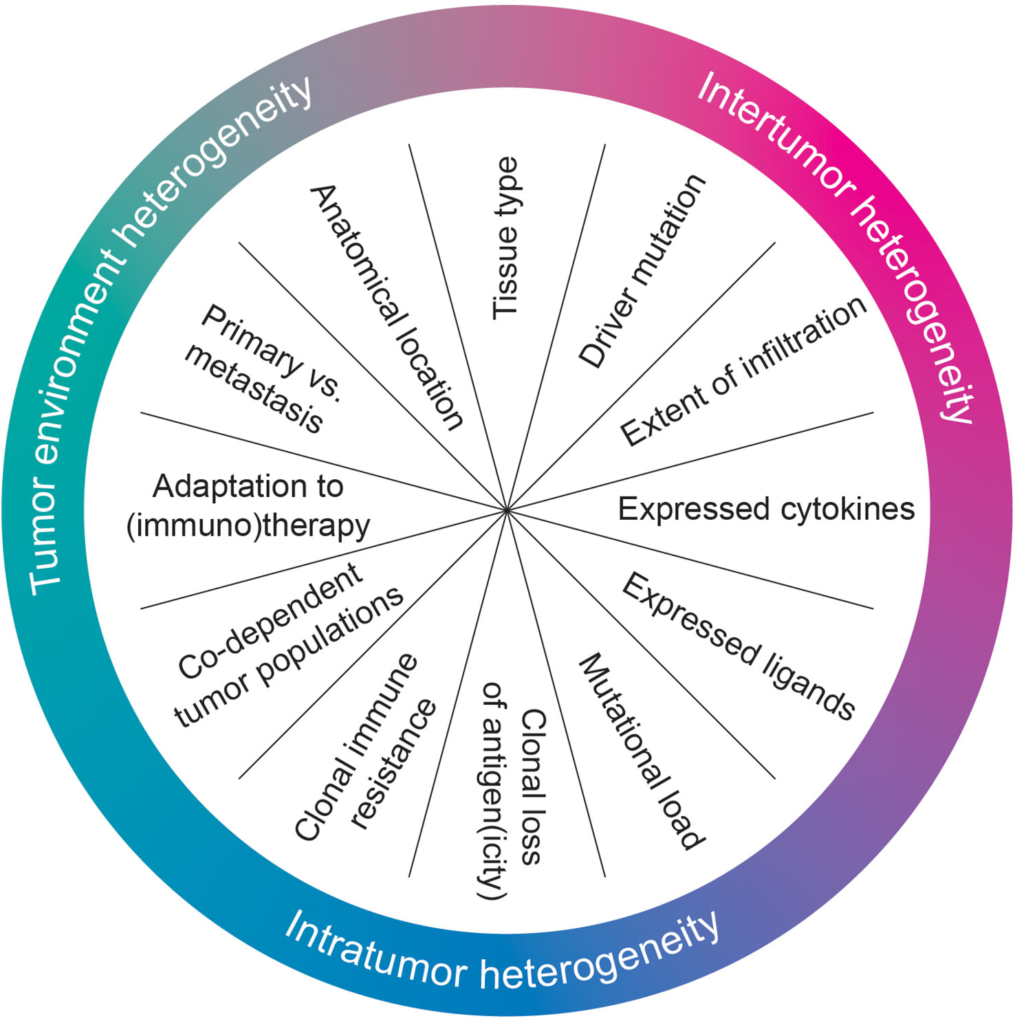
Tumor heterogeneity exists in different forms and is caused by multiple processes. Intertumor heterogeneity (i.e., the differences between different tumors), intratumor heterogeneity (i.e., the difference between different tumor cells/clones/populations/regions of the same tumor) and heterogeneity in the tumor micro-environment (i.e., the difference in the anatomical location and non-tumor cell infiltration between [different] tumors and/or metastases) all contribute to the smorgasbord we term cancer. These mechanisms of heterogeneity not only co-exist, but frequently also actively influence one another. For example, different metastases of the same tumor in distinct anatomical locations may experience different growth signals and thus display preferential outgrowth of different subpopulations (19–21). In addition, the genetic heterogeneity within tumors can surpass even that between tumors of different individuals (22, 23). Even different single cells within the same tumor can have remarkably different characteristics [reviewed in (24)].
This heterogeneity is manifested through a variety of different mechanisms. They can be summarized in four, central concepts: germline differences, genomic instability, selection by exogenous means and obligate co-dependency of tumor subpopulations. For each of these, clinical evidence illustrates how they can result in tumor heterogeneity. Germline differences are perhaps best characterized within hereditary cancers. For example, hereditary breast cancer cases generally have poorer prognoses than sporadic cases (25). Genomic instability also, a core hallmark of cancer-causing mutations and other genomic aberrations such as genetic duplications or deletions, can lead to inter- and intratumor heterogeneity. This can be driven by, for example, enhanced APOBEC3 activity in late-stage cancers which promotes the stochastic mutation of the tumor genome (26). Furthermore, non-tumor driven selection, for example through therapy, can result in heterogeneity as tumor subclones with therapy-resistant traits are selected for (27, 28). Lastly, tumors can also evolve to be heterogeneous through the common, co-dependent evolution of different tumor cell subpopulations. In such a symbiotic relationship, one population within the tumor provides growth stimuli to another and, in some cases, this may even be reciprocated (29–31).
Heterogeneity affects immune sensitivity of tumors
Heterogeneity can also affect the sensitivity of tumors challenged by multiple different inflammatory cytokines and/or cells of the immune system. This may occur in a general sense, but could also impact specific immune effector pathways. The same general concepts of tumor heterogeneity are involved in these processes (Figure 1).
Intertumor heterogeneity is perhaps most evident for the tissue from which a tumor arises. The identity of this tissue in and of itself can already determine immune sensitivity. For example, cancers arising from intrinsically (more) hypoxic tissues, such as melanoma, have heightened expression of cIAP1. These tumors therefore display enhanced resistance against TNF (32). Extending these observations, a recent meta-analysis of tumor-intrinsic determinants of ICB sensitivity identified multiple strong predictors of response for individual tumor types, but those factors fail to predict well in a tumor type-agnostic fashion (33), implying tumor-type specific mechanisms to be at play. Intertumor heterogeneity also manifests through heterogeneity in driver mutations, which can differentially affect the antitumor immune response. An example of this is the generation of an immunosuppressive TME driven by the loss of PTEN (34). KRASG12C and several p53 mutations, too, alter immune sensitivity (35, 36).
Intertumor heterogeneity can also more broadly influence immune status, being associated with both mutational load (37) and immune infiltrate (38). Each of these may influence which type of immune pressure, and of what strength, a tumor encounters. Another determinant concerns the expression of activating and inhibitory immune ligands, which also differ between tumors and/or tumor types. This heterogeneity in receptor expression may occur upon induction by signals from the TME, such as the differential strength of induction of PD-L1 in different tumors (and tumor cell lines) (39, 40). This phenomenon is particularly of interest as PD-L1, being the main ligand for the inhibitory T cell checkpoint PD-1, is a key target for immune-checkpoint blockade (41–43). Diversity in receptor expression may also be more deeply ingrained, such as the genetically-encoded, patient-specific repertoire of inhibitory receptors for NK cells (immune effector cells that rely on a combined input of activating and inhibitory ligands for their activation) (44–47). This is not only true for cell-surface bound ligands, but equally for tumor cell-derived cytokines or other soluble factors secreted (only) by specific tumors. For example, tumor cell-derived CCL2 indirectly dampens CD8+ T cell responses (48) while, additionally, induction of the Wnt/β-catenin signaling pathway leads to T cell exclusion (49).
Intratumor heterogeneity can equally influence immune sensitivity. While some of the above mechanisms may also be evident within a tumor, such as local expression of cytokines and/or immune ligands, other phenomena are also at play. For example, regions within the tumor can lose components of the antigen-presentation machinery, specific T cell antigens or HLA alleles, limiting T cell recognition (50–52). At the same time, such tumor adaptations may (locally) attract otherwise absent immune cells, as was recently shown for Vδ1 and Vδ3 T cells in B2M MUT colorectal cancer (53). Additionally, tumor subclones can contain mutations in key immune signaling nodes, even before onset of therapy. They include mutations in JAK1, responsible for transmitting IFNγ signals, and in CASP8, responsible for the final, decisive step in the apoptotic cascade initiated by TNF (54, 55). Furthermore, different, interdependent subpopulations may contribute to intratumor heterogeneity. In a particularly elegant study, it was demonstrated that IFNγ pathway-mutant tumors are more sensitive to CD8+ T cell-mediated eradication due to the loss of protection by IFNγ-induced PD-L1, but become more resistant when intermixed with PD-L1-producing wildtype tumor cells (56). This intratumor heterogeneity is enhanced once (immuno)therapy is administered to the patient tumor, with ample opportunity for selection of escape mutants (52, 57–64).
Lastly, the anatomical location of the tumor may affect immune sensitivity. First, there is a purely technical consideration: the way in which immune sensitivity mechanisms are studied influences how the biology of the pathway manifests. For example, IFNγ has seemingly opposing effects on tumor cell viability in vitro and in vivo: the cytostatic effects of IFNγ largely inhibit tumor cell growth in vitro, whereas in vivo, the induction of PD-L1 by IFNγ provides a strong, cytoprotective effect that overcomes those inhibitory effects (16, 65, 66). Additionally, and perhaps obviously, some immune pathways cannot be studied at all in vitro because of the use of simplified model systems: either cell types, ligands or cytokines can be missing. The influence of tumor location on heterogeneity has also been demonstrated clinically: different distant metastases may have entirely different TMEs, (neo)antigen burden and immune resistance mechanisms (19, 63, 67, 68). Along these lines, a recent meta-analysis of >2,000 patients showed that genetic alterations in IFNγ signaling components that are present prior to treatment do not necessarily diminish ICB response (69).
Approach to counteract heterogeneity in CRISPR-Cas9 immune screens
Heterogeneity thus has near limitless influence on the sensitivity of tumors to eradication by the immune system. How can we meaningfully combat, and perhaps even exploit, this heterogeneity in CRISPR-Cas9 screens for tumor-intrinsic, immune sensitivity modifiers? By integrating large amounts of functional screening and omics data from many different settings and contexts, one can more precisely annotate tumor cell nodes of immune sensitivity. Specifically, this integration will yield either biomarkers, which mark cell lines in which a particular immune sensitivity node is active, or will generate mechanistic hypotheses that explain why a given node is seemingly inactive in a given cell line. Based on the mechanistic sources of heterogeneity described above, ideally one would derive omics and screening data from as many sources as possible. These would include (epi)genomic, transcriptomic and proteomic omics data. At the same time, the screening data should be derived from both in vitro and in vivo screens from as many genetic backgrounds as possible [reviewed in (16)]. Such an undertaking however, would require immense investments of both time and funding.
Proof-of-concept analyses exploiting cell line-to-cell line heterogeneity
While a comprehensive catalogue of screening data is currently lacking, other domains of research have already embraced the concept of heterogeneity more comprehensively. In fact, in order to find an Achilles’ heel for specific cancers, many cell lines have already been deeply characterized. A multi-decade, multi-national effort, collected within the DepMap database, has screened >1800 cell lines using genome-scale perturbation libraries to identify cancer (type)-specific dependencies. Aside from these functional genetic screens, the cell lines used in these studies have also been extensively characterized, including the collection of RNA, DNA, epigenetic, metabolic and drug-sensitivity metrics (70–72). The use of these databases has allowed investigators to identify novel therapeutic targets in a variety of cancer indications (73–75). An important element lacking from this database then, is an annotation of which genes can be considered immune sensitivity modifiers.
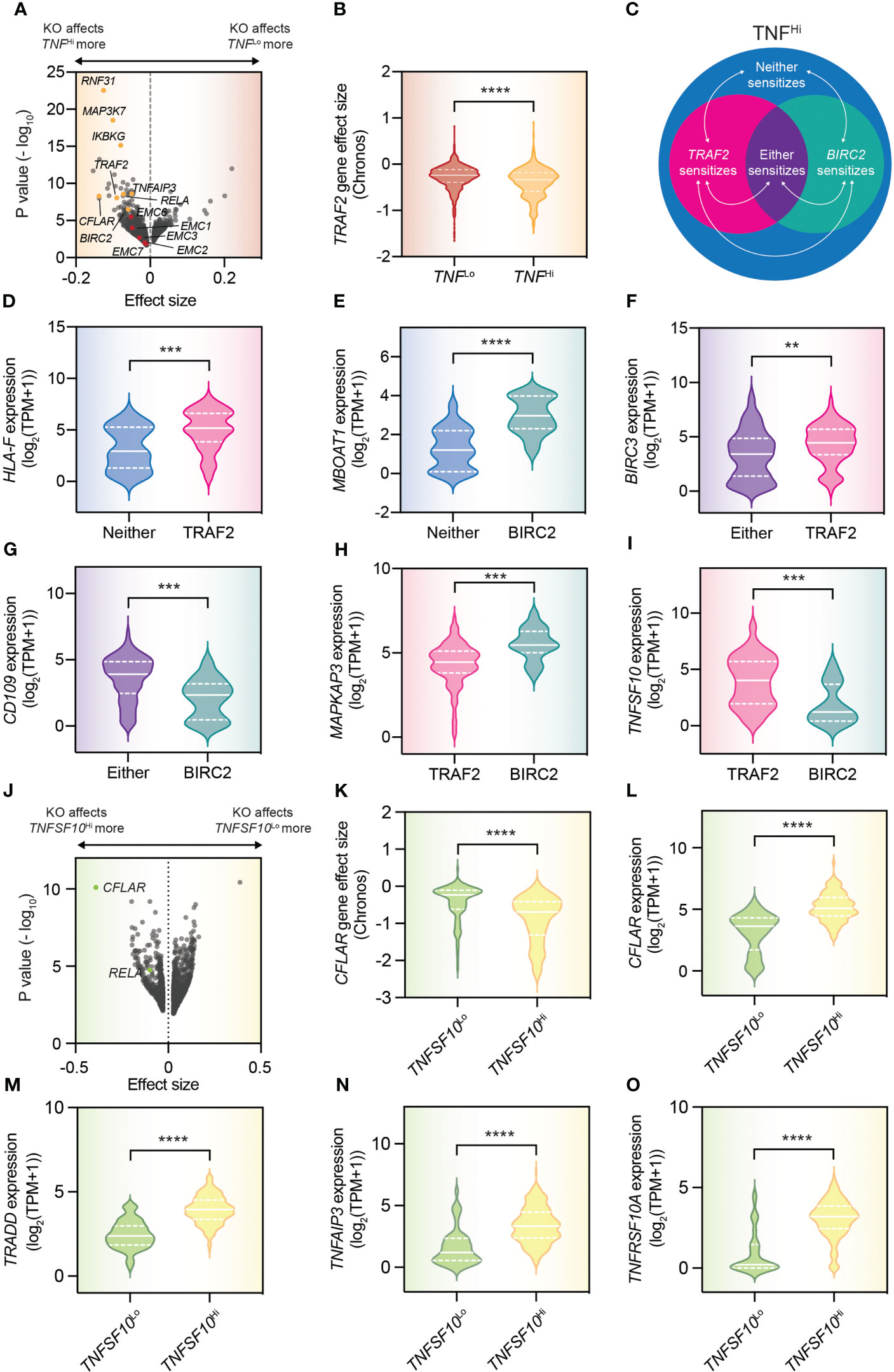
Interestingly, because of the extent of this database, both in terms of cell line number and cell line characterization, we can perform a proof-of-concept analysis for immune sensitivity modifiers that exploit heterogeneity. Specifically, we can look at modifiers of TNF sensitivity. As more than 300 cell lines in the DepMap produce TNF, we can compare the effects of gene knockouts in these cell lines compared to those that do not produce TNF, to identify factors sensitizing tumor cells to TNF (which, using the excellent portal is trivial to accomplish). By performing this analysis, we could find factors whose ablation reduces viability of TNFHi cell lines specifically (Figure 2A). Indeed, many of those we had already identified and validated ourselves, including TRAF2, BIRC2 (encoding cIAP1) and RNF31 (Figures 2A, B) (1, 2). However, with such an approach we could identify also novel, potential TNF sensitivity modifiers, such as the EMC family of genes which, though currently not yet validated, we also identified in our meta-analysis of immune sensitivity screens (Figure 2A) (16).

Figure 2 DepMap dependency analyses allow understanding of heterogeneity in immune resistance mechanisms. (A) Volcano plot that compares the gene perturbation effects in TNFHi [i.e., >0.5 log2(TPM+1)] and TNFLo (i.e., 0 read counts for TNF) cell lines. (B) Comparison of the effect of TRAF2 knockout in TNFHi and TNFLo cell lines. (C) Schematic diagram indicating the populations analyzed in the panels that follow. Only TNFHi cell lines were used in the analyses. (D–I) Violin plots of the expression of indicated genes for the indicated populations (cell lines were deemed sensitive when their CERES score was < -0.3 and insensitive when their CERES score was > -0.1). Statistics were performed by Student t test. The solid white line indicates the population median, with the bottom and top dashed white lines indicating the first and third quartiles, respectively. (J) Volcano plot comparing the gene perturbation effects in TNFSF10Hi [i.e., >5 log2(TPM+1)] and TNFSF10Lo (i.e., 0 read counts for TNFSF10) cell lines. (K) Comparison of the effect of CFLAR knockout in TNFSF10Hi and TNFSF10Lo cell lines. (L–O) Violin plots of the expression of indicated genes for the indicated populations. Statistics were performed by Student t test. The solid white line indicates the population median, with the bottom and top dashed white lines indicating the first and third quartiles respectively. **p < 0.01, ***p < 0.001, ****p < 0.0001.
Having established the fidelity of this approach, we could continue by also taking advantage of the size and heterogeneity of the particular database used. In our previous work, we have identified a differential reliance on TRAF2 and BIRC2 to establish resistance to TNF in different tumor cell lines. While it had been difficult to fully comprehend this differential sensitivity before, given that TRAF2 and cIAP1 are thought to signal in a linear fashion, we could now make transcriptomic comparisons between TNFHi cell lines in which both TRAF2 and BIRC2 sensitize, those in which solely BIRC2 knockout sensitizes, those in which solely TRAF2 knockout sensitizes, or those cell lines in which neither the loss of TRAF2 nor the loss of BIRC2 reduces the viability of the affected cell line (Figure 2C). These analyses can yield biomarkers of specific populations (Figures 2D–J). For example, high HLA-F expression marks populations that will respond solely to TRAF2 inhibition (Figure 2D). These analyses can also provide mechanistic insight. For example, the observation that BIRC3 expression is higher in cell lines that respond solely to the loss of TRAF2 compared to those that respond to either TRAF2 or BIRC2 loss, implies that this protein compensates for the loss of its paralog BIRC2 (Figure 2F).
As a second proof-of-concept for discovery of immune sensitivity modifiers that exploit heterogeneity using the DepMap, we performed a similar analysis for cells producing TNF-related apoptosis-inducing ligand (TRAIL, encoded by the gene TNFSF10; Figure 2J). Here, we identified the loss of both CFLAR and RELA to specifically sensitize those cells capable of producing TRAIL, in line with published literature (Figures 2J, K) (76, 77). Using the transcriptomic data of those same cell lines, we may even begin to speculate as to how these cells are capable of surviving in the presence of TRAIL. These cells seemingly induce transcription of genes that protect against TRAIL-induced cell death, including the aforementioned CFLAR, but also TRADD and TNFAIP3 (Figures 2L–N) (76–78). In doing so, they may gain a previously described proliferative advantage of TRAIL signaling (78), which may explain their higher level of expression of the TRAIL receptor, TNFRSF10A (Figure 2O), but this prediction awaits functional validation.
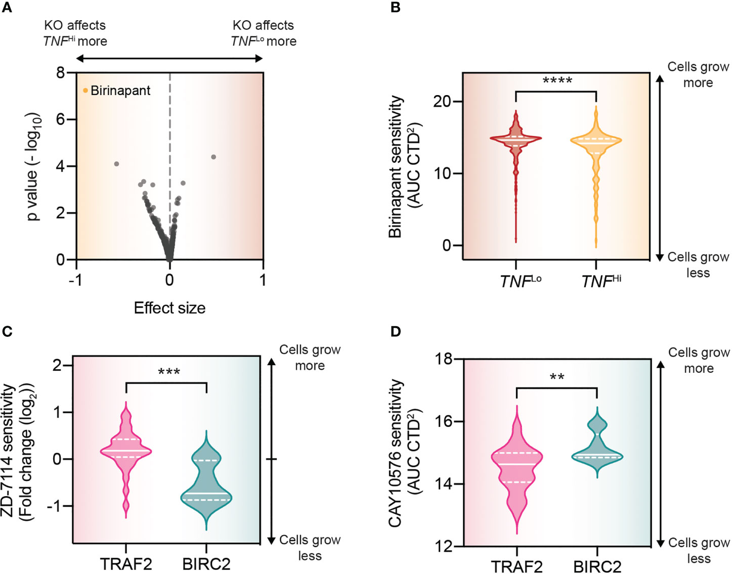
Beyond these transcriptomic comparisons, we can exploit the DepMap to find ways of targeting these specific tumor cell subpopulations. Again, to probe the fidelity of such an approach, we compared the drug sensitivity between the TNFLo and TNFHi cell lines in the DepMap. With this analysis, we could, at least in part, recapitulate the genetic analysis, identifying birinapant, an inhibitor of cIAP1 to sensitize TNFHi cell lines more than TNFLo cell lines (Figures 3A, B). We validated this therapeutic approach previously in conditions of high concentrations of TNF (i.e., T cell attack) (1). Using the drug sensitivity database, we could also identify specific inhibitors for the cell lines differentially dependent on TRAF2 and BIRC2 for their resistance against TNF. For those cell lines that particularly depend on BIRC2 we found that ZD-7114, a β3-adrenoceptor agonist, is a potential pharmaceutical strategy (Figure 3C). In cell lines that depend on TRAF2, we could find a specific sensitivity to CAY10576, an IKKϵ inhibitor (Figure 3D). IKKϵ is known interact with TRAF2, and its identification may thus have a clear mechanistic basis (79).

Figure 3 DepMap drug analyses allow for the potential exploitation of heterogeneity in immune resistance mechanisms. (A) Volcano plot that compares the drug treatment effects in TNFHi (i.e., >0.5 log2(TPM+1)) and TNFLo (i.e., 0 read counts for TNF) cell lines. (B) Comparison of the effect of birinapant in TNFHi and TNFLo cell lines. (C) Violin plots of the drug effects of ZD-7114 for the indicated populations. Statistics were performed by Student t test. The solid white line indicates the population median, with the bottom and top dashed white lines indicating the first and third quartiles respectively. (D) As (C), but for CAY10576. **p < 0.01, ***p < 0.001, **** p < 0.0001..
Considerations for the future
While the above analyses show the promise of integrating heterogeneity in target discovery, they are preliminary and marred by assumptions (e.g., can we realistically assume that TNF-producing cells are a good model for cell experiencing T cell-derived TNF? Can we assume that protein levels of TNF scale linearly with TNF mRNA expression)? Therefore, and as mentioned above, the true complexity of tumor-immune interactions, and forms and mechanisms of heterogeneity at play require more data to be integrated in these models. Firstly, and perhaps most easy to accomplish, the field should invest in performing more tumor : T cell screens, to complement those that have already been reported in key publications in the recent past (1, 3–7, 12–16, 18, 75, 80). These screens, combined with deep characterization as performed for the DepMap, should result in a more granular understanding of genotype – phenotype interactions, as demonstrated here with our proof-of-concept analyses (Figure 2). An analogous approach was already taken for NK sensitivity (75). Obviously, such screens only scratch the surface of the different types of heterogeneity outlined above. One could imagine that with time, and significant investment, the screens can be performed in parallel in a large number of settings. For example, they can be performed with different (e.g., NK cells, as was done in (75), or ‘exhausted’ vs. polyfunctional T cells), or more complex co-culture systems (e.g., tumor : T cell : NK cell combinations), more environmental perturbations (e.g., nutrient starvation, hypoxia, highly acidic conditions), in in vivo mouse models (as in (5, 12, 13), in isogenic tumor cell lines with specific alterations [as was done in (5)] or even in combination with specific therapeutics (e.g. anti-CTLA-4 or anti-PD-1). Ultimately, such genetic screens will improve our understanding of important immune resistance mechanisms, aiming to have as many patients as possible benefit from (personalized) immunotherapy.
Author contributions
DV performed the analyses. DV and DP wrote the manuscript. All authors contributed to the article and approved the submitted version.
Funding
This study was supported by Oncode Institute and the Dutch Cancer Society (KWF).
Conflict of interest
DP is a co-founder, shareholder and advisor of Immagene B.V. DV is currently employed at Genmab B.V., which is unrelated to this study.
Publisher’s note
All claims expressed in this article are solely those of the authors and do not necessarily represent those of their affiliated organizations, or those of the publisher, the editors and the reviewers. Any product that may be evaluated in this article, or claim that may be made by its manufacturer, is not guaranteed or endorsed by the publisher.
References
1. Vredevoogd DW, Kuilman T, Ligtenberg MA, Boshuizen J, Stecker KE, de Bruijn B, et al. Augmenting immunotherapy impact by lowering tumor TNF cytotoxicity threshold. Cell (2019) 178:585–599.e15. doi: 10.1016/j.cell.2019.06.014
2. Zhang Z, Kong X, Ligtenberg MA, van Hal-van Veen SE, Visser NL, de Bruijn B, et al. RNF31 inhibition sensitizes tumors to bystander killing by innate and adaptive immune cells. Cell Rep Med (2022) 3:100655. doi: 10.1016/j.xcrm.2022.100655
3. Pan D, Kobayashi A, Jiang P, Ferrari de Andrade L, Tay RE, Luoma AM, et al. A major chromatin regulator determines resistance of tumor cells to T cell-mediated killing. Sci (1979) (2018) 359:770–5. doi: 10.1126/science.aao1710
4. Kearney CJ, Vervoort SJ, Hogg SJ, Ramsbottom KM, Freeman AJ, Lalaoui N, et al. Tumor immune evasion arises through loss of TNF sensitivity. Sci Immunol (2018) 3. doi: 10.1126/sciimmunol.aar3451
5. Lawson KA, Sousa CM, Zhang X, Kim E, Akthar R, Caumanns JJ, et al. Functional genomic landscape of cancer-intrinsic evasion of killing by T cells. Nature (2020) 586:120–6. doi: 10.1038/s41586-020-2746-2
6. Patel SJ, Sanjana NE, Kishton RJ, Eidizadeh A, Vodnala SK, Cam M, et al. Identification of essential genes for cancer immunotherapy. Nature (2017) 548:537–42. doi: 10.1038/nature23477
7. Hou J, Wang Y, Shi L, Chen Y, Xu C, Saeedi A, et al. Integrating genome-wide CRISPR immune screen with multi-omic clinical data reveals distinct classes of tumor intrinsic immune regulators. J Immunother Cancer (2021) 9:e001819. doi: 10.1136/jitc-2020-001819
8. Dufva O, Koski J, Maliniemi P, Ianevski A, Klievink J, Leitner J, et al. Integrated drug profiling and CRISPR screening identify essential pathways for CAR T-cell cytotoxicity. Blood (2020) 135:597–609. doi: 10.1182/blood.2019002121
9. Joung J, Kirchgatterer PC, Singh A, Cho JH, Nety SP, Larson RC, et al. CRISPR activation screen identifies BCL-2 proteins and B3GNT2 as drivers of cancer resistance to T cell-mediated cytotoxicity. Nat Commun (2022) 13:1–14. doi: 10.1038/s41467-022-29205-8
10. Zhuang X, Veltri DP, Long EO. Genome-wide CRISPR screen reveals cancer cell resistance to NK cells induced by NK-derived IFN-γ. Front Immunol (2019) 10:2879. doi: 10.3389/fimmu.2019.02879
11. Singh N, Lee YG, Shestova O, Ravikumar P, Hayer KE, Hong SJ, et al. Impaired death receptor signaling in leukemia causes antigen-independent resistance by inducing CAR T-cell dysfunction. Cancer Discov (2020) 10:552–67. doi: 10.1158/2159-8290.CD-19-0813
12. Manguso RT, Pope HW, Zimmer MD, Brown FD, Yates KB, Miller BC, et al. In vivo CRISPR screening identifies Ptpn2 as a cancer immunotherapy target. Nature (2017) 547:413–8. doi: 10.1038/nature23270
13. Dubrot J, Lane-Reticker SK, Kessler EA, Ayer A, Mishra G, Wolfe CH, et al. In vivo screens using a selective CRISPR antigen removal lentiviral vector system reveal immune dependencies in renal cell carcinoma. Immunity (2021) 54:571–585.e6. doi: 10.1016/j.immuni.2021.01.001
14. Ishizuka JJ, Manguso RT, Cheruiyot CK, Bi K, Panda A, Iracheta-Vellve A, et al. Loss of ADAR1 in tumours overcomes resistance to immune checkpoint blockade. Nature (2019) 565:43–8. doi: 10.1038/s41586-018-0768-9
15. Li F, Huang Q, Luster TA, Hu H, Zhang H, Ng WL, et al. In vivo epigenetic crispr screen identifies asf1a as an immunotherapeutic target in kras-mutant lung adenocarcinoma. Cancer Discov (2020) 10:270–87. doi: 10.1158/2159-8290.CD-19-0780
16. Vredevoogd DW, Apriamashvili G, Peeper DS. The (re)discovery of tumor-intrinsic determinants of immune sensitivity by functional genetic screens. Immuno-Oncology Technol (2021) 11:100043. doi: 10.1016/j.iotech.2021.100043
17. Liu D, Zhao X, Tang A, Xu X, Liu S, Zha L, et al. CRISPR screen in mechanism and target discovery for cancer immunotherapy. Biochim Biophys Acta Rev Cancer (2020) 1874:188378. doi: 10.1016/j.bbcan.2020.188378
18. Freeman AJ, Kearney CJ, Silke J, Oliaro J. Unleashing TNF cytotoxicity to enhance cancer immunotherapy. Trends Immunol (2021) 42:1128–42. doi: 10.1016/j.it.2021.10.003
19. Dang HX, Krasnick BA, White BS, Grossman JG, Strand MS, Zhang J, et al. The clonal evolution of metastatic colorectal cancer. Sci Adv (2020) 6:9691–701. doi: 10.1126/sciadv.aay9691
20. Frankell AM, Dietzen M, Al Bakir M, Lim EL, Karasaki T, Ward S, et al. The evolution of lung cancer and impact of subclonal selection in TRACERx. Nature (2023) 616:525–33. doi: 10.1038/s41586-023-05783-5
21. Martínez-Ruiz C, Black JRM, Puttick C, Hill MS, Demeulemeester J, Larose Cadieux E, et al. Genomic-transcriptomic evolution in lung cancer and metastasis. Nature (2023) 616:543–52. doi: 10.1038/s41586-023-05706-4
22. Martinez P, Birkbak NJ, Gerlinger M, McGranahan N, Burrell RA, Rowan AJ, et al. Parallel evolution of tumour subclones mimics diversity between tumours. J Pathol (2013) 230:356–64. doi: 10.1002/path.4214
23. Al Bakir M, Huebner A, Martínez-Ruiz C, Grigoriadis K, Watkins TBK, Pich O, et al. The evolution of non-small cell lung cancer metastases in TRACERx. Nature (2023) 616:534–42. doi: 10.1038/s41586-023-05729-x
24. Burrell RA, McGranahan N, Bartek J, Swanton C. The causes and consequences of genetic heterogeneity in cancer evolution. Nature (2013) 501:338–45. doi: 10.1038/nature12625
25. Van Der Groep P, Bouter A, van der Zanden R, Siccama I, Menko FH, Gille JJP, et al. Distinction between hereditary and sporadic breast cancer on the basis of clinicopathological data. J Clin Pathol (2006) 59:611–7. doi: 10.1136/jcp.2005.032151
26. Swanton C, McGranahan N, Starrett GJ, Harris RS. APOBEC enzymes: mutagenic fuel for cancer evolution and heterogeneity. Cancer Discov (2015) 5:704–12. doi: 10.1158/2159-8290.CD-15-0344
27. Kemper K, Krijgsman O, Cornelissen-Steijger P, Shahrabi A, Weeber F, Song J-Y, et al. Intra- and inter-tumor heterogeneity in a vemurafenib-resistant melanoma patient and derived xenografts. EMBO Mol Med (2015) 7:1104–18. doi: 10.15252/emmm.201404914
28. Boshuizen J, Vredevoogd DW, Krijgsman O, Ligtenberg MA, Blankenstein S, de Bruijn B, et al. Reversal of pre-existing NGFR-driven tumor and immune therapy resistance. Nat Commun (2020) 11:1–13. doi: 10.1038/s41467-020-17739-8
29. Li X, Thirumalai D. Cooperation among tumor cell subpopulations leads to intratumor heterogeneity. Biophys Rev Lett (2020) 15:99–119. doi: 10.1142/s1793048020300042
30. Marusyk A, Tabassum DP, Altrock PM, Almendro V, Michor F, Polyak K. Non-cell-autonomous driving of tumour growth supports sub-clonal heterogeneity. Nature (2014) 514:54–8. doi: 10.1038/nature13556
31. Cleary AS, Leonard TL, Gestl SA, Gunther EJ. Tumour cell heterogeneity maintained by cooperating subclones in wnt-driven mammary cancers. Nature (2014) 508:113–7. doi: 10.1038/nature13187
32. Samanta D, Huang TYT, Shah R, Yang Y, Pan F, Semenza GL. BIRC2 expression impairs anti-cancer immunity and immunotherapy efficacy. Cell Rep (2020) 32:108073. doi: 10.1016/j.celrep.2020.108073
33. Litchfield K, Reading JL, Puttick C, Thakkar K, Abbosh C, Bentham R, et al. Meta-analysis of tumor- and T cell-intrinsic mechanisms of sensitization to checkpoint inhibition. Cell (2021) 184:596–614.e14. doi: 10.1016/j.cell.2021.01.002
34. Peng W, Chen JQ, Liu C, Malu S, Creasy C, Tetzlaff MT, et al. Loss of PTEN promotes resistance to T cell–mediated immunotherapy. Cancer Discov (2016) 6:202–16. doi: 10.1158/2159-8290.CD-15-0283
35. Wellenstein MD, Coffelt SB, Duits DEM, van Miltenburg MH, Slagter M, de Rink I, et al. Loss of p53 triggers WNT-dependent systemic inflammation to drive breast cancer metastasis. Nature (2019) 572:538–42. doi: 10.1038/s41586-019-1450-6
36. Mugarza E, van Maldegem F, Boumelha J, Moore C, Rana S, Llorian Sopena M, et al. Therapeutic KRAS G12C inhibition drives effective interferon-mediated antitumor immunity in immunogenic lung cancers. Sci Adv (2022) 8:2021. doi: 10.1126/sciadv.abm8780
37. Yarchoan M, Hopkins A, Jaffee EM. Tumor mutational burden and response rate to PD-1 inhibition. New Engl J Med (2017) 377:2500–1. doi: 10.1056/nejmc1713444
38. Zuo S, Wei M, Wang S, Dong J, Wei J. Pan-cancer analysis of immune cell infiltration identifies a prognostic immune-cell characteristic score (ICCS) in lung adenocarcinoma. Front Immunol (2020) 11:1218. doi: 10.3389/fimmu.2020.01218
39. Apriamashvili G, Vredevoogd DW, Krijgsman O, Bleijerveld OB, Ligtenberg MA, de Bruijn B, et al. Ubiquitin ligase STUB1 destabilizes IFNγ-receptor complex to suppress tumor IFNγ signaling. Nat Commun (2022) 13:1–16. doi: 10.1038/s41467-022-29442-x
40. Hänze J, Wegner M, Noessner E, Hofmann R, Hegele A. Co-Regulation of immune checkpoint PD-L1 with interferon-gamma signaling is associated with a survival benefit in renal cell cancer. Target Oncol (2020) 15:377–90. doi: 10.1007/s11523-020-00728-8
41. Topalian SL, Hodi FS, Brahmer JR, Gettinger SN, Smith DC, McDermott DF, et al. Safety, activity, and immune correlates of anti–PD-1 antibody in cancer. New Engl J Med (2012) 366:2443–54. doi: 10.1056/nejmoa1200690
42. Sharma P, Hu-Lieskovan S, Wargo JA, Ribas A. Primary, adaptive, and acquired resistance to cancer immunotherapy. Cell (2017) 168:707–23. doi: 10.1016/j.cell.2017.01.017
43. Freeman GJ, Long AJ, Iwai Y, Bourque K, Chernova T, Nishimura H, et al. Engagement of the PD-1 immunoinhibitory receptor by a novel B7 family member leads to negative regulation of lymphocyte activation. J Exp Med (2000) 192:1027–34. doi: 10.1084/jem.192.7.1027
44. Subedi N, Verhagen LP, Bosman EM, van Roessel I, Tel J. Understanding natural killer cell biology from a single cell perspective. Cell Immunol (2022) 373:104497. doi: 10.1016/j.cellimm.2022.104497
45. Ruggeri L, Capanni M, Urbani E, Perruccio K, Shlomchik WD, Tosti A, et al. Effectiveness of donor natural killer cell aloreactivity in mismatched hematopoietic transplants. Sci (1979) (2002) 295:2097–100. doi: 10.1126/science.1068440
46. Ruggeri L, Vago L, Eikema DJ, de Wreede LC, Ciceri F, Diaz MA, et al. Natural killer cell alloreactivity in HLA-haploidentical hematopoietic transplantation: a study on behalf of the CTIWP of the EBMT. Bone Marrow Transplant (2021) 56:1900–7. doi: 10.1038/s41409-021-01259-0
47. Valiante NM, Uhrberg M, Shilling HG, Lienert-Weidenbach K, Arnett KL, D’Andrea A, et al. Functionally and structurally distinct NK cell receptor repertoires in the peripheral blood of two human donors. Immunity (1997) 7:739–51. doi: 10.1016/S1074-7613(00)80393-3
48. Kersten K, Coffelt SB, Hoogstraat M, Verstegen NJM, Vrijland K, Ciampricotti M, et al. Mammary tumor-derived CCL2 enhances pro-metastatic systemic inflammation through upregulation of IL1β in tumor-associated macrophages. Oncoimmunology (2017) 6. doi: 10.1080/2162402X.2017.1334744
49. Spranger S, Bao R, Gajewski TF. Melanoma-intrinsic β-catenin signalling prevents anti-tumour immunity. Nature (2015) 523:231–5. doi: 10.1038/nature14404
50. Rosenthal R, Cadieux EL, Salgado R, Al-Bakir M, Moore DA, Hiley CT, et al. Neoantigen-directed immune escape in lung cancer evolution. Nature (2019) 567:479–85. doi: 10.1038/s41586-019-1032-7
51. McGranahan N, Rosenthal R, Hiley CT, Rowan AJ, Watkins TBK, Wilson GA, et al. Allele-specific HLA loss and immune escape in lung cancer evolution. Cell (2017) 171:1259–1271.e11. doi: 10.1016/j.cell.2017.10.001
52. Sade-Feldman M, Jiao YJ, Chen JH, Rooney MS, Barzily-Rokni M, Eliane JP, et al. Resistance to checkpoint blockade therapy through inactivation of antigen presentation. Nat Commun (2017) 8:1–11. doi: 10.1038/s41467-017-01062-w
53. de Vries NL, van de Haar J, Veninga V, Chalabi M, Ijsselsteijn ME, van der Ploeg M, et al. γδ T cells are effectors of immunotherapy in cancers with HLA class I defects. Nature (2023) 613:743–50. doi: 10.1038/s41586-022-05593-1
54. Rooney MS, Shukla SA, Wu CJ, Getz G, Hacohen N. Molecular and genetic properties of tumors associated with local immune cytolytic activity. Cell (2015) 160:48–61. doi: 10.1016/j.cell.2014.12.033
55. Shin DS, Zaretsky JM, Escuin-Ordinas H, Garcia-Diaz A, Hu-Lieskovan S, Kalbasi A, et al. Primary resistance to PD-1 blockade mediated by JAK1/2 mutations. Cancer Discov (2017) 7:188–201. doi: 10.1158/2159-8290.CD-16-1223
56. Williams JB, Li S, Higgs EF, Cabanov A, Wang X, Huang H, et al. Tumor heterogeneity and clonal cooperation influence the immune selection of IFN-γ-signaling mutant cancer cells. Nat Commun (2020) 11:1–14. doi: 10.1038/s41467-020-14290-4
57. Riaz N, Havel JJ, Makarov V, Desrichard A, Urba WJ, Sims JS, et al. Tumor and microenvironment evolution during immunotherapy with nivolumab. Cell (2017) 171:934–949.e15. doi: 10.1016/j.cell.2017.09.028
58. Luksza M, Riaz N, Makarov V, Balachandran VP, Hellmann MD, Solovyov A, et al. A neoantigen fitness model predicts tumour response to checkpoint blockade immunotherapy. Nature (2017) 551:517–20. doi: 10.1038/nature24473
59. Snyder A, Makarov V, Merghoub T, Yuan J, Zaretsky JM, Desrichard A, et al. Genetic basis for clinical response to CTLA-4 blockade in melanoma. New Engl J Med (2014) 371:2189–99. doi: 10.1056/nejmc1508163
60. Hugo W, Zaretsky JM, Sun L, Song C, Moreno BH, Hu-Lieskovan S, et al. Genomic and transcriptomic features of response to anti-PD-1 therapy in metastatic melanoma. Cell (2016) 165:35–44. doi: 10.1016/j.cell.2016.02.065
61. Zaretsky JM, Garcia-Diaz A, Shin DS, Escuin-Ordinas H, Hugo W, Hu-Lieskovan S, et al. Mutations associated with acquired resistance to PD-1 blockade in melanoma. N Engl J Med (2016) 375:819–29. doi: 10.1056/NEJMoa1604958
62. Gao J, Shi LZ, Zhao H, Chen J, Xiong L, He Q, et al. Loss of IFN-γ pathway genes in tumor cells as a mechanism of resistance to anti-CTLA-4 therapy. Cell (2016) 167:397–404.e9. doi: 10.1016/j.cell.2016.08.069
63. Liu D, Lin JR, Robitschek EJ, Kasumova GG, Heyde A, Shi A, et al. Evolution of delayed resistance to immunotherapy in a melanoma responder. Nat Med (2021) 27:985–92. doi: 10.1038/s41591-021-01331-8
64. Jerby-Arnon L, Shah P, Cuoco MS, Rodman C, Su MJ, Melms JC, et al. A cancer cell program promotes T cell exclusion and resistance to checkpoint blockade. Cell (2018) 175:984–997.e24. doi: 10.1016/j.cell.2018.09.006
65. Benci JL, Johnson LR, Choa R, Xu Y, Qiu J, Zhou Z, et al. Opposing functions of interferon coordinate adaptive and innate immune responses to cancer immune checkpoint blockade. Cell (2019) 178:933–948.e14. doi: 10.1016/j.cell.2019.07.019
66. Benci JL, Xu B, Qiu Y, Wu TJ, Dada H, Twyman-Saint Victor C, et al. Tumor interferon signaling regulates a multigenic resistance program to immune checkpoint blockade. Cell (2016) 167:1540–1554.e12. doi: 10.1016/j.cell.2016.11.022
67. Zhao Y, Fu X, Lopez JI, Rowan A, Au L, Fendler A, et al. Selection of metastasis competent subclones in the tumour interior. Nat Ecol Evol (2021) 5:1033–45. doi: 10.1038/s41559-021-01456-6
68. Jiménez-Sánchez A, Memon D, Pourpe S, Veeraraghavan H, Li Y, Vargas HA, et al. Heterogeneous tumor-immune microenvironments among differentially growing metastases in an ovarian cancer patient. Cell (2017) 170:927–938.e20. doi: 10.1016/j.cell.2017.07.025
69. Song E, Chow RD. Mutations in IFN-γ signaling genes sensitize tumors to immune checkpoint blockade. Cancer Cell (2023) 41:651–2. doi: 10.1016/j.ccell.2023.02.013
70. Garnett MJ, Edelman EJ, Heidorn SJ, Greenman CD, Dastur A, Lau KW, et al. Systematic identification of genomic markers of drug sensitivity in cancer cells. Nature (2012) 483:570–5. doi: 10.1038/nature11005
71. Barretina J, Caponigro G, Stransky N, Venkatesan K, Margolin AA, Kim S, et al. The cancer cell line encyclopedia enables predictive modelling of anticancer drug sensitivity. Nature (2012) 483:603–7. doi: 10.1038/nature11003
72. Dempster JM, Pacini C, Pantel S, Behan FM, Green T, Krill-Burger J, et al. Agreement between two large pan-cancer CRISPR-Cas9 gene dependency data sets. Nat Commun (2019) 10:1–14. doi: 10.1038/s41467-019-13805-y
73. Bondeson DP, Paolella BR, Asfaw A, Rothberg MV, Skipper TA, Langan C, et al. Phosphate dysregulation via the XPR1–KIDINS220 protein complex is a therapeutic vulnerability in ovarian cancer. Nat Cancer (2022) 3:681–95. doi: 10.1038/s43018-022-00360-7
74. Adane B, Alexe G, Seong BKA, Lu D, Hwang EE, Hnisz D, et al. STAG2 loss rewires oncogenic and developmental programs to promote metastasis in Ewing sarcoma. Cancer Cell (2021) 39:827–844.e10. doi: 10.1016/j.ccell.2021.05.007
75. Sheffer M, Lowry E, Beelen N, Borah M, Amara SNA, Mader CC, et al. Genome-scale screens identify factors regulating tumor cell responses to natural killer cells. Nat Genet (2021) 53:1196–206. doi: 10.1038/s41588-021-00889-w
76. Safa AR, Pollok KE. Targeting the anti-apoptotic protein c-FLIP for cancer therapy. Cancers (Basel) (2011) 3:1639–71. doi: 10.3390/cancers3021639
77. Geismann C, Hauser C, Grohmann F, Schneeweis C, Bölter N, Gundlach JP, et al. NF-κB/RelA controlled A20 limits TRAIL-induced apoptosis in pancreatic cancer. Cell Death Dis (2023) 14:3. doi: 10.1038/s41419-022-05535-9
78. Cao X, Pobezinskaya YL, Morgan MJ, Liu Z. The role of TRADD in TRAIL-induced apoptosis and signaling. FASEB J (2011) 25:1353–8. doi: 10.1096/fj.10-170480
79. Zhou AY, Shen RR, Kim E, Lock YJ, Xu M, Chen ZJ, et al. IKKϵ-mediated tumorigenesis requires K63-linked polyubiquitination by a cIAP1/cIAP2/TRAF2 E3 ubiquitin ligase complex. Cell Rep (2013) 3:724–33. doi: 10.1016/j.celrep.2013.01.031
Keywords: heterogeneity, CRISPR-Cas9, genetic screens, therapy resistance, TNF
Citation: Vredevoogd DW and Peeper DS (2023) Heterogeneity in functional genetic screens: friend or foe? Front. Immunol. 14:1162706. doi: 10.3389/fimmu.2023.1162706
Received: 09 February 2023; Accepted: 30 May 2023;
Published: 16 June 2023.
Edited by:
Harald Wajant, University Hospital Würzburg, GermanyReviewed by:
Kevin Litchfield, University College London, United KingdomSanju Sinha, National Institutes of Health (NIH), United States
Copyright © 2023 Vredevoogd and Peeper. This is an open-access article distributed under the terms of the Creative Commons Attribution License (CC BY). The use, distribution or reproduction in other forums is permitted, provided the original author(s) and the copyright owner(s) are credited and that the original publication in this journal is cited, in accordance with accepted academic practice. No use, distribution or reproduction is permitted which does not comply with these terms.
*Correspondence: Daniel S. Peeper, ZC5wZWVwZXJAbmtpLm5s
 David W. Vredevoogd
 Daniel S. Peeper