 Zhiwei Li
 Frieder Hadlich
 Klaus Wimmers
 Eduard Murani
95% of researchers rate our articles as excellent or good
Learn more about the work of our research integrity team to safeguard the quality of each article we publish.
Find out more
ORIGINAL RESEARCH article
Front. Immunol. , 24 November 2022
Sec. Comparative Immunology
Volume 13 - 2022 | https://doi.org/10.3389/fimmu.2022.976454
This article is part of the Research Topic Pig Translational Model in Immunological Research View all 8 articles
The consequences of glucocorticoid receptor (GR) hypersensitivity during infection have so far received little attention. We previously discovered that a natural gain-of-function Ala610Val substitution in the porcine GR aggravates response of pigs to lipopolysaccharide (LPS)-induced endotoxemia, which can be alleviated by dexamethasone (DEX) pretreatment. In this work, we investigated the relevant molecular basis of these phenotypes by transcriptomic profiling of porcine peripheral blood mononuclear cells (PBMCs) carrying different GR genotypes, in unstimulated conditions or in response to DEX and/or LPS in vitro. The Val allele differentially regulated abunda+nt genes in an additive-genetic manner. A subset of more than 200 genes was consistently affected by the substitution across treatments. This was associated with upregulation of genes related i.a. to endo-lysosomal system, lipid and protein catabolism, and immune terms including platelet activation, and antigen presentation, while downregulated genes were mainly involved in cell cycle regulation. Most importantly, the set of genes constitutively upregulated by Val includes members of the TLR4/LPS signaling pathway, such as LY96. Consequently, when exposing PBMCs to LPS treatment, the Val variant upregulated a panel of additional genes related to TLR4 and several other pattern recognition receptors, as well as cell death and lymphocyte signaling, ultimately amplifying the inflammatory responses. In contrast, when stimulated by DEX treatment, the Val allele orchestrated several genes involved in anti-inflammatory responses during infection. This study provides novel insights into the impact of GR hypersensitivity on the fate and function of immune cells, which may be useful for endotoxemia therapy.
Glucocorticoids (GCs) are a class of steroid hormones produced by the hypothalamic–pituitary–adrenal (HPA) axis (1). As end products of the HPA axis, these molecules participate in a range of biological processes that help organisms cope with stress conditions and restore homeostasis (2). Proper GC action is essential for efficient control of many immune reactions, and to prevent damage from maladapted responses (3). Clinically, GCs are long-standing anti-inflammatory agents that are among the most commonly prescribed drugs in human and veterinary medicine, and are widely used for the treatment of inflammatory and autoimmune diseases (4). Accordingly, dysregulation of GC signaling is observed in several inflammatory diseases, such as sepsis - one of the leading causes of mortality worldwide, and complicates their therapy (5, 6). Endotoxin (Lipopolysaccharide – LPS) is a component of the outer membrane of gram-negative bacteria, and is the most potent pathogen-associated molecular pattern (PAMP) inducing a host of physiological responses including a strong inflammatory reaction as well as a homeostatic activation of the HPA axis (7). Endotoxemia is frequently observed in patients with sepsis (8), and experimentally-induced endotoxemia via LPS administration is commonly used to study the pathobiology of sepsis, even though there are some limitations (9).
GCs exert their effects mainly via the glucocorticoid receptor (GR), a ligand-inducible transcription factor (TF) belonging to the nuclear receptor superfamily (10). Thus, relevant mechanisms of GR signaling and their regulation are under intense investigation using a variety of approaches (11). These studies have demonstrated the necessity of retaining GR dimerization, which is important for receptor activation, and established the importance of enhanced GR expression and TF activity for adequate anti-inflammatory effects. For example, GR dimerization deficiency in GRdim mice resulted in more severe inflammation and sepsis (12, 13), whereas enhancement of GR expression and transactivation activity attenuated LPS-induced endotoxic shock and lethal inflammation (14, 15). These examples emphasize the positive functional role of the GR in anti-inflammatory processes.
In contrast, our previous findings revealed that this association is not always favorable. We observed that the gain-of-function of GR caused by a natural Ala610Val substitution (GRAla610Val) increased the susceptibility of pigs to LPS-induced endotoxemia (16, 17). This GR variant showed higher ligand-binding affinity and transactivation activity in vitro (17). However, it caused aggravated cytokine production, thrombocytopenia, sickness behaviors, anorexia, and metabolic alterations in pigs upon endotoxin challenge (16). The underlying mechanisms behind this negative influence of GR hypersensitivity remain unclear. Their exploration may shed new light on glucocorticoid receptor dysregulation in inflammatory diseases. Importantly, under baseline conditions hypersensitivity of the mutated GR manifests largely only by decreased cortisol level (18), suggesting that GR hypersensitivity may be a complication in inflammatory diseases that has remained largely unrecognized. GRAla610Val provides an exceptional model not only because it is a unique natural gain-of-function GR variant, but the pig model appears particularly relevant because, unlike rodents, pigs feature similar sensitivity to endotoxemia compared to humans (19).
This study set out to investigate the influence of the gain-of-function GRAla610Val genetic variant on the transcriptomic programs of porcine peripheral blood mononuclear cells (PBMCs), at baseline and in response to stimulation by dexamethasone (DEX) and LPS, separately and jointly. To achieve this, porcine PBMCs carrying each of three GR genotypes (Ala/Ala, Ala/Val, Val/Val) were divided into four groups and treated with either vehicle (CON), DEX, LPS, or LPS+DEX for two hours. Next, mRNA sequencing was carried out to elucidate Val allele-associated transcriptomic responses, and the relevant signaling pathways and biological events were subsequently explored using different computational tools. The results are anticipated to improve our understanding of the impact of GR hypersensitivity in the contexts of GC-mediated therapy, immune stimulation, and crosstalk of immune and GR signaling. The knowledge gained may benefit the health and welfare of both humans and farm animals.
The mRNA sequencing data presented and analyzed in the current report were first published and all the sampling and treatment procedures were done in our previous study (20). Briefly, PBMCs were obtained from 24 purebred German Landrace pigs (≈170 days old) with equal numbers of males and females and balanced for the three GR genotypes (Ala/Ala, Ala/Val, and Val/Val). The animals were reared at the FBN experimental pig farm (Dummerstorf, Germany) according to the German Law of Animal Protection. PBMCs from each animal were distributed into four treatment groups and treated for 2 h at 37°C in a 5% CO2 atmosphere with: vehicle (CON group); DEX (5 nM); LPS (10 μg/mL); and LPS + DEX, respectively. The mRNA sequencing was performed using the Illumina HiSeq 2500 platform at the FBN sequencing facility as described previously (20). The resultant sequencing data were first submitted to the ArrayExpress repository (https://www.ebi.ac.uk/arrayexpress) with accession number E-MTAB-9808, as outlined in our earlier study (20).
Prior to analysis, genes with normalized counts ≥5 in fewer than eight of the samples were removed. Differential expression analysis was then performed using the R package DESeq2 version 1.32.0 (21). Principal component analysis (PCA) identified four outlier animals, which were then omitted from downstream analysis. After preprocessing, the final count table retained 14,809 gene entries across 80 samples (20 animals × 4 treatment groups). To ascertain the effect of GRAla610Val under different treatment conditions, differentially expressed genes (DEGs) in response to the Ala610Val substitution were analyzed separately within each group (CON, DEX, LPS, and LPS+DEX). Two variables—sex (male or female), and GR genotypes (Ala/Ala, Ala/Val, or Val/Val)—were included in the design formula, with the latter as the variable of interest. To calculate the log2 fold change (LFC) of genes induced by dosage of the Val allele, the GR genotypes of Ala/Ala, Ala/Val, and Val/Val were expressed as continuous variables with values of zero, one, and two respectively. Genes with a p-value <0.05 were considered differentially expressed. Volcano plots were made using the R package EnhancedVolcano version 1.10.0 (22).
To further investigate the specific effects of the Val allele under different conditions, the Val-regulated DEGs for the four treatment groups were analyzed using the Venn diagram module in the TBtools toolkit (23). Five gene modules were identified: one module of common Val-regulated DEGs across all four treatment groups (shared genes), and four modules of treatment-specific Val-regulated genes. Furthermore, Val-regulated DEGs unique to the LPS group, and common to the LPS and LPS+DEX groups, were referred to as LPS-specific genes. Similarly, Val-regulated DEGs unique to the DEX group, and common to the DEX and LPS+DEX groups, were referred to as DEX-specific genes. The heatmap was generated using the R package pheatmap version 1.0.12 (24), based on the LFC values of shared genes induced by dosage of the Val allele.
Genes exhibiting additive changes in expression associated with the Val allele were identified using the degPatterns function implemented in the R package DEGreport version 1.28.0 (25). To this end, Val-regulated DEGs within each treatment group were subjected to hierarchical clustering. This allowed for identification of genes upregulated (Val-up) or downregulated (Val-down) by the Val allele in an additive manner (i.e., dosage of the Val allele). Within each treatment group, GR targets were identified using the GR targets dataset from Ingenuity Pathway Analysis (IPA; Qiagen, Hilden, Germany) and Harmonizome (26). PCA was conducted based on Val-up and Val-down genes. Separate protein–protein interaction (PPI) networks were constructed for Val-up and Val-down genes, respectively, using the STRING database (27), and visualized by Cytoscape version 3.9.0 (28). Hub genes showing a high degree of connectivity within each PPI network were extracted by the cytoHubba plugin (29).
Functional enrichment analysis was conducted using Metascape (30) and IPA. For Metascape analysis, the ontology sources used were GO Biological Processes, Reactome Gene Sets, KEGG Pathway, and WikiPathways. Metascape terms with a BH-adjusted p-value <0.05 were regarded as significantly enriched. When using IPA, z-scores were calculated using the LFC of genes induced by each copy of Val allele within the respective treatment groups. Three types of IPA terms were included in the analysis: canonical pathways, diseases and biological functions, and upstream regulators. IPA terms having a Fisher exact test p-value <0.05 and an absolute z-score ≥2 were regarded as having statistical significance and a definite regulation direction. The results of functional enrichment analysis were visualized using GraphPad Prism 8.2.1 (GraphPad Software, Inc., San Diego, CA) and the R package ggplot2 version 3.3.5 (31).
To study the effect of the GRAla610Val variant on PBMCs in different states of immune activation (CON, DEX, LPS, and LPS+DEX), DEGs associated with per copy of the Val allele were determined within each group. This analysis identified 1018, 1630, 1473, and 1081 DEGs, respectively, in the four treatment groups (Figures 1A–D). The top DEGs (according to p-values and LFC) are labeled on the volcano plots, and indicate that the Val allele potentially influences immune response (CAMK2A, CCL17, SIAE, TREML1, and TRIM7); lipid metabolism (CERT1, CYP7A1, ELOVL2, and KDSR); protein synthesis and processing (BBS10, CALU, DOLK, GALNT16, and WDR83OS); the endo-lysosomal system (ATP6V1E1, LAPTM4A, and TRIP10); and cell division and survival (EYA1, FGF1, FGF13, MCUR1, REC114, SETD8, SH3GLB1, and ZNF331). It is also of note that the top DEG profiles differed between treatment groups, as indicated by the observation of sets of unique DEGs in the CON, DEX, LPS, and LPS+DEX groups (310, 664, 629, and 303, respectively). These data are presented in the Venn diagram (Figure 1E; Table S1). Nevertheless, there is also a group of shared, constitutive DEGs (248) regulated by the Val allele in the same direction across all four groups (Figures 1E, F). These findings demonstrate that the different states of immune activation modulate and manifest the distinct effects of the Val allele on PBMCs, as further emphasized by functional annotation of the DEGs presented below.
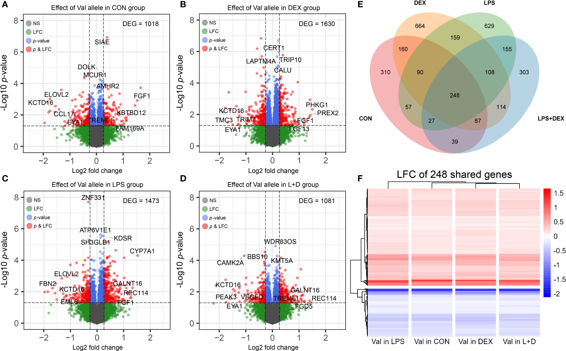
Figure 1 Differential expression analysis. (A-D) Volcano plots of differentially expressed genes (DEGs) induced by dosage of the Val allele in (A) CON, (B) DEX, (C) LPS, and (D) LPS+DEX groups. The top four most significant (p-value) and most up and down regulated DEGs (LFC) were displayed. (E) Venn diagram illustrating shared and unique DEGs induced by dosage of the Val allele among four treatment groups. (F) Heatmap constructed based on LFC of Val DEGs shared by four treatment groups. Val, Valine; CON, control; DEX: dexamethasone; LPS, lipopolysaccharide; LFC, log2 fold change.
Hierarchical clustering was then applied to the DEGs to identify genes up- or downregulated by the Val allele in an additive manner. The CON, DEX, LPS, and LPS+DEX groups contained 551, 983, 873, and 613 upregulated genes (Val-up), respectively, while 433, 615, 561, and 443 downregulated (Val-down) genes, respectively, were identified (Figures 2A-D; Table S1). Within each group, PCA revealed that these Val-up and Val-down genes drive the separation in transcriptional programs of the samples bearing different GR genotypes (Figure 2E).
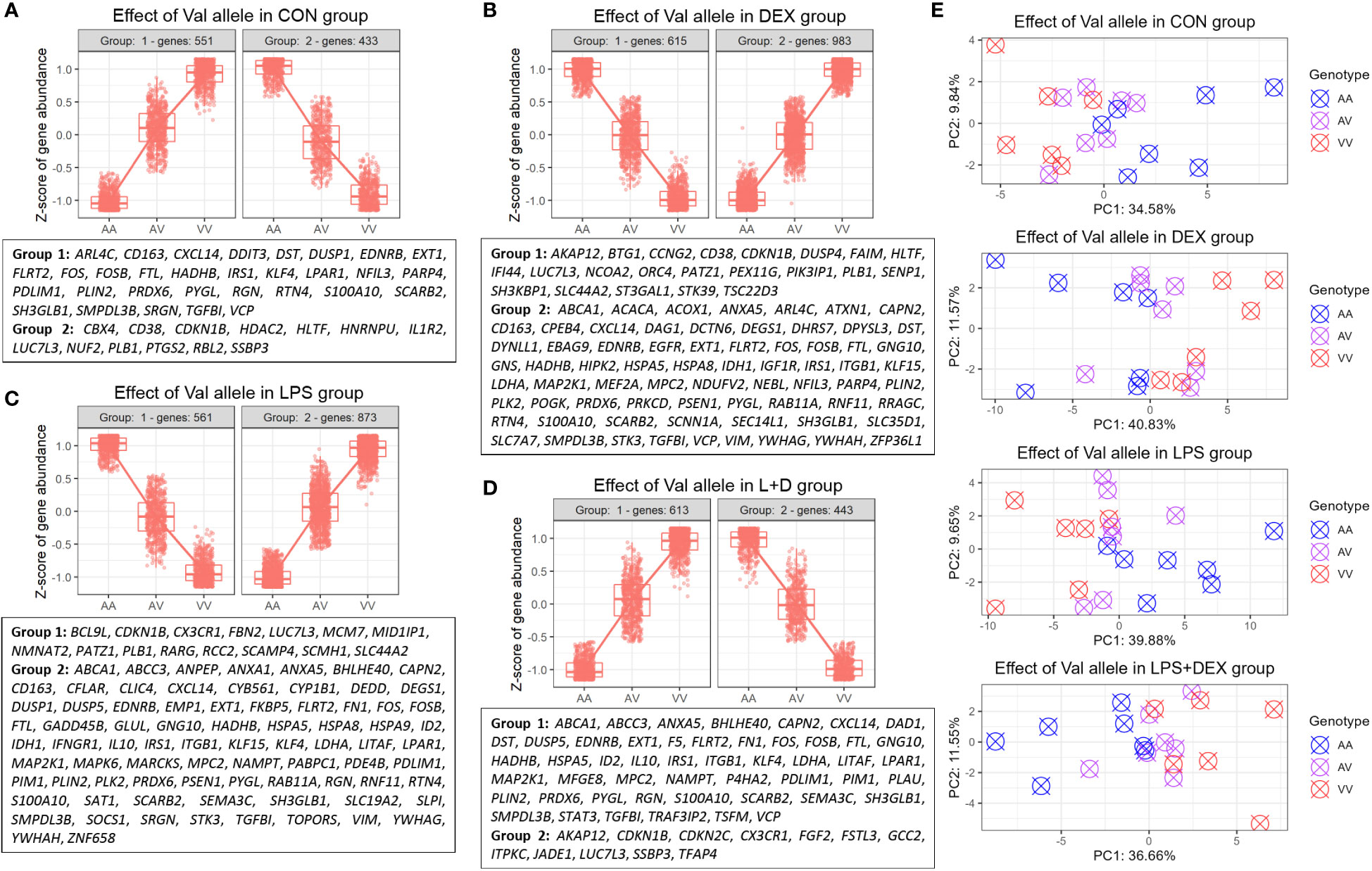
Figure 2 Identification of DEGs regulated by the Val allele additively (Val-up and Val-down genes). (A-D) Hierarchical clustering of expression profiles of Val-regulated DEGs in (A) CON, (B) DEX, (C) LPS, and (D) LPS+DEX groups. Previously reported glucocorticoid receptor (GR) targets identified through the database of Harmonizome (26) and Ingenuity Pathway Analysis (IPA) were displayed for each cluster. (E) Principal component analysis (PCA) of expression profiles of Val-up and Val-down genes in each treatment group. AA, GR genotype AlaAla; AV, GR genotype AlaVal; VV, GR genotype ValVal.
Furthermore, many known GR targets were found in these Val-up and Val-down gene clusters (Figures 2A–D; Table S2). At baseline, without stimulation, Val-up genes included several GR targets involved in inflammatory responses (CD163, CXCL14, DUSP1, FOS, LPAR1, NFIL3, S100A10, SCARB2, SMPDL3B, SRGN, and VCP), suggesting an intrinsic role of the Val allele in immunomodulation (Figure 2A). Additionally, DEX treatment introduced Val-up GR targets related to anti-inflammatory processes (ANXA5, KLF15, and RNF11); lipid metabolism (ABCA1, ACACA, ACOX1, DEGS1, DHRS7, and GNS); mitochondrial function (IDH1, LDHA, MPC2, NDUFV2, PRKCD, and VIM); and protein folding (CPEB4, HSPA5, and HSPA8) (Figure 2B). Several distinct Val-up GR targets associated with inflammatory responses (ANPEP, ANXA1, DUSP5, FKBP5, IFNGR1, IL10, LITAF, NAMPT, SLPI, and SOCS1) were identified upon LPS stimulation, together with GR targets involved in cell death (CAPN2, CFLAR, DEDD, EMP1, GADD45B, GLUL, ID2, MAP2K1, PIM1, SAT1, STK3, and TOPORS; Figure 2C). Considering the role of damage-associated molecular patterns (DAMPs) in coupling cell death to inflammatory responses (32), the identification of these two DEG classes suggested that the Val allele enhances inflammatory signaling associated with the release of DAMPs. Finally, eight Val-up GR targets emerged only when the PBMCs were co-treated with LPS and DEX, namely: DAD1, F5, MFGE8, P4HA2, PLAU, STAT3, TRAF3IP2, and TSFM (Figure 2D).
Notably, many of the Val-down GR targets were related to cell cycle and division, such as CBX4, CDKN1B, HDAC2, HLTF, HNRNPU, NUF2, and RBL2 (CON group); BTG1, CCNG2, DUSP4, and ORC4 (DEX group); MCM7 and RCC2 (LPS group); and CDKN2C, FGF2, and JADE1 (LPS+DEX group; Figures 2A–D; Table S2). Besides this, DEX stimulation induced additional Val-down GR targets important for function of the GR (e.g. NCOA2) and B cells (e.g. FAIM, SH3KBP1, and PIK3IP1; Figure 2B).
Because of their distinct implications, enrichment analysis was carried out independently for Val-up and Val-down genes. The Val-up genes exhibited a variety of functions with many of them common to non-stimulated and stimulated conditions, and thus taken to represent constitutive roles of the Val allele. For example, in common DEGs these functional terms included lysosome, protein and lipid catabolic processes, transport of molecules, and ion homeostasis. They also encompassed a series of signaling pathways involved in immune responses: neutrophil degranulation, MHC mediated antigen processing and presentation, platelet activation, sphingolipid metabolism, and Salmonella infection (Figures 3A–D; Table S3). Accordingly, when analyzing treatment-specific genes, Val DEGs common to all treatment groups supported the above constitutive roles of the Val allele (Figure 4A; Table S4). Consistent with this, the IPA disease and function database predicted that lysosomal storage disease and accumulation of sphingolipid were inhibited, whereas inflammatory response and endocytosis were activated for Val-up genes in all four groups (Table S5).
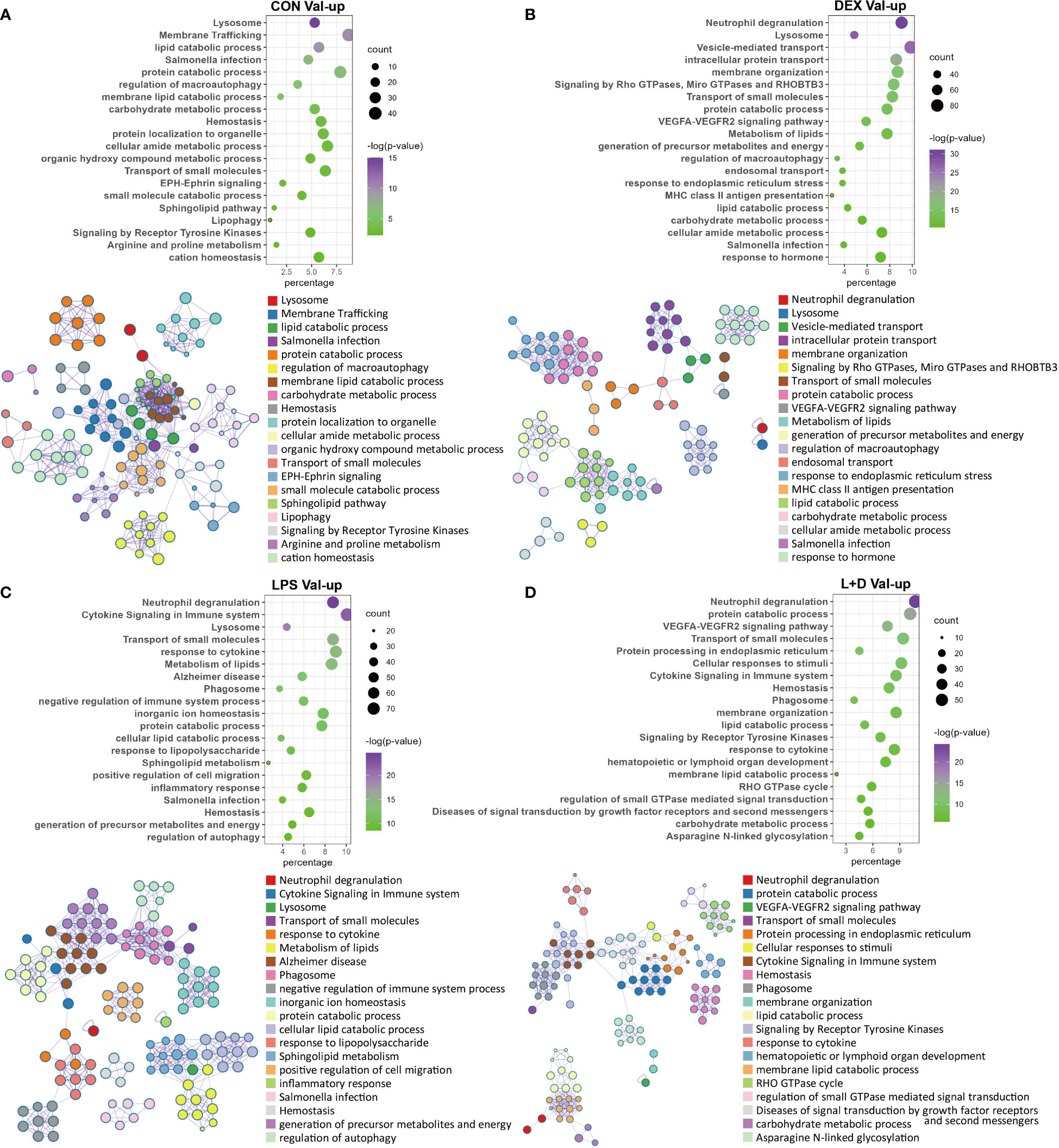
Figure 3 Enrichment analysis of Val-up genes in each treatment group. (A) Functional terms enriched for Val-up genes in CON group. (B) Functional terms enriched for Val-up genes in DEX group. (C) Functional terms enriched for Val-up genes in LPS group. (D) Functional terms enriched for Val-up genes in LPS+DEX group. Enrichment analysis was performed using the Metascape. Bubble plots illustrate significance and gene counts of top enriched terms, where purple indicates higher significance and green indicates lower significance and bubble size is positively correlated with gene counts. Networks illustrate the relationship between terms, where each node represents one functional term and terms with Kappa similarities above 0.3 are connected by edges. Each term cluster is labeled and represented by its most significant member and all members in the same term cluster are indicated by the same color.
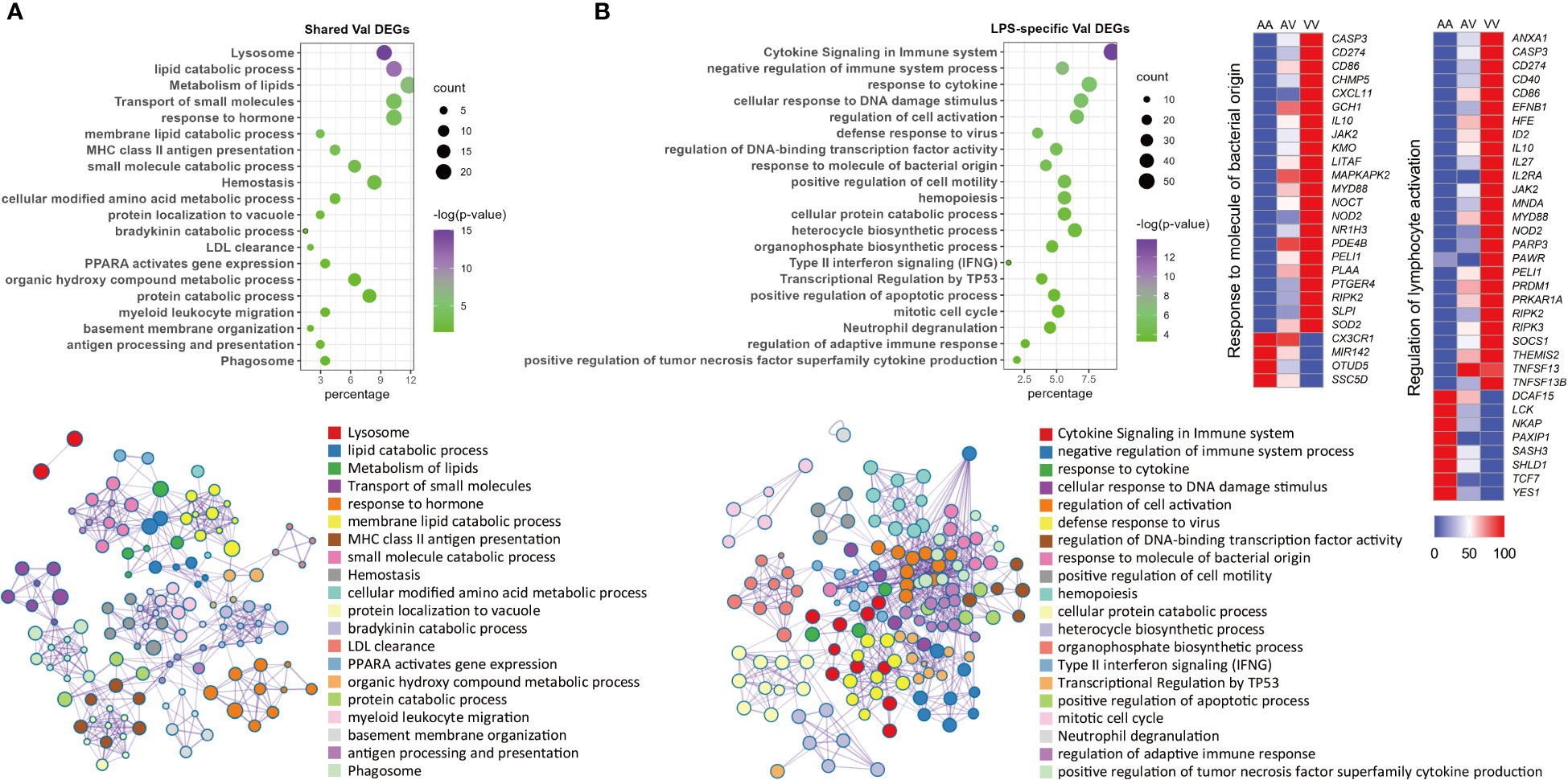
Figure 4 Enrichment analysis of shared and LPS-specific Val-regulated DEGs. (A) Functional terms enriched for Val-regulated DEGs common in all the four groups. (B) Functional terms enriched for LPS-specific Val-regulated DEGs with relative gene expression profiles for two representative LPS-specific terms. Enrichment analysis was performed using the Metascape. Bubble plots illustrate significance and gene counts of top enriched terms, where purple indicates higher significance and green indicates lower significance and bubble size is positively correlated with gene counts. Networks illustrate the relationship between terms, where each node represents one functional term and terms with Kappa similarities above 0.3 are connected by edges. Each term cluster is labeled and represented by its most significant member and all members in the same term cluster are indicated by the same color.
Two additional types of pathway emerged for Val-up genes upon treatment with DEX. Specifically, these were related to protein processing in the endoplasmic reticulum (ER) and mitochondrial energy generation (Figure 3B; Table S3). Additional support was again provided by IPA predictions: activation of the unfolded protein response, TCA cycle, mitochondrial dysfunction, and production of reactive oxygen species (ROS; Tables S5, S6). These results indicate a potential way of the Val allele in regulating cellular activities, since mitochondria have been shown to contact lysosomes and the ER, and these interactions are essential for many cellular functions including cell fate decisions (33).
Compared with DEX, the Val allele exhibited genuine, albeit weaker, influence on mitochondrial dysfunction and unfolded protein response when stimulated with LPS (Table S6). Notably, LPS also triggered a set of immune pathways found among the Val-up genes, including cytokine signaling, response to LPS, T cell receptor (TCR) signaling, B cell receptor (BCR) signaling, and ROS and RNS production in phagocytes (Figure 3C; Table S3). Accordingly, aside from the activated terms already identified for the CON and DEX groups—such as antigen presentation, endocytosis, phagocytosis, and leukocyte migration—the Val-up genes of the LPS group included an additional series of predicted terms involving activation of lymphocytes and T cells (Table S5). Furthermore, IPA predictions identified multiple molecules crucial for inflammatory signaling as activated upstream regulators for Val-up genes in the presence of LPS (such as CD40, EIF2AK2, IL27, JAK2, MYD88, NOD2, RIPK2, STAT2, TLR4, and TNFSF13B), with their predicted activation also corresponding to increased expression under control of the Val allele (Table S1; Table S7). The above results suggest that the Val allele might amplify or skew cellular responses to pro-inflammatory factors like LPS, resulting in a more severe inflammatory phenotype associated with enhanced PRR-mediated inflammatory signaling and signal transduction from antigen-presenting cells (APCs) to T cells. This was apparently confirmed by the identification of a suite of immune mediators, along with enrichment of various immune signaling pathways, among the LPS-specific Val-mediated DEGs. These included response to molecules of bacterial origin, and regulation of lymphocyte activation (Figure 4B; Table S4).
Another key finding for Val-up genes in the LPS group was enrichment of programmed cell death (Table S3), which was further categorized by the IPA database into different subtypes, revealing that ferroptosis, autophagy, and necroptosis were activated by the Val allele, whereas apoptosis was inhibited (Table S5; Table S6). This might be associated with the enriched terms of iron homeostasis, ER stress, mitochondrial dysfunction, and fatty acid oxidation (34) (Table S6). In fact, some of these cell death terms were already identified in the CON and DEX groups (Table S6). However, cell death signaling induced by LPS might have more severe consequences due to increased expression of PRRs such as DDX58 and NOD2 (Table S1), because DAMPs released by damaged cells are tightly associated with inflammation via their recognition by PRRs (32).
Several enriched terms in the LPS+DEX group rationalized the improved response to DEX therapy seen in Val carriers during endotoxemia (16). For example, activation of necroptosis was identified among the Val-up genes in both the LPS and DEX groups, but was not enriched in the LPS+DEX group (Table S6). In contrast, necrosis was predicted by IPA to be inhibited exclusively in this latter group (Table S5). The VEGFA–VEGFR2 signaling pathway plays a key role in the activation of endothelial nitric oxide synthase (eNOS) and eNOS-mediated nitric oxide (NO) production (35), which has been found to be significantly reduced in sepsis (36). This pathway was enriched only in the presence of DEX (DEX and LPS+DEX groups; Figures 3B, D). Several molecules with pivotal roles in anti-inflammatory responses—such as IL6R (37), IRF2 (38), KLF4 (39), and NCOA3 (40)—were predicted as activated upstream regulators only when co-treated with LPS and DEX (Table S7).
In contrast to the myriad functions exhibited by Val-up genes, these were less diverse for the Val-down genes. The most obvious feature was their consistent association with inhibition of cell cycle progression and cell division, regardless of the treatment group (Figures 5A–D). This classification harbored a variety of pathways, involving cell cycle regulation and phase transition, chromatin and cytoskeleton organization, DNA replication and repair, and organelle fission (Figures 5A–D). These effects on cell cycle and cell division were further confirmed by IPA, which predicted inhibition of cell cycle control of chromosomal replication, organization of cytoskeleton, repair of DNA, transcription, and segregation of chromosomes (Table S5; Table S6). Besides this, several positive regulators of cell cycle progression, such as CKAP2L (41), E2F1 (42), and EP400 (43), were predicted as inhibited upstream regulators (Table S7), whereas activation was predicted for the retinoblastoma (Rb) protein, which plays a pivotal role in negative regulation of the cell cycle (44).
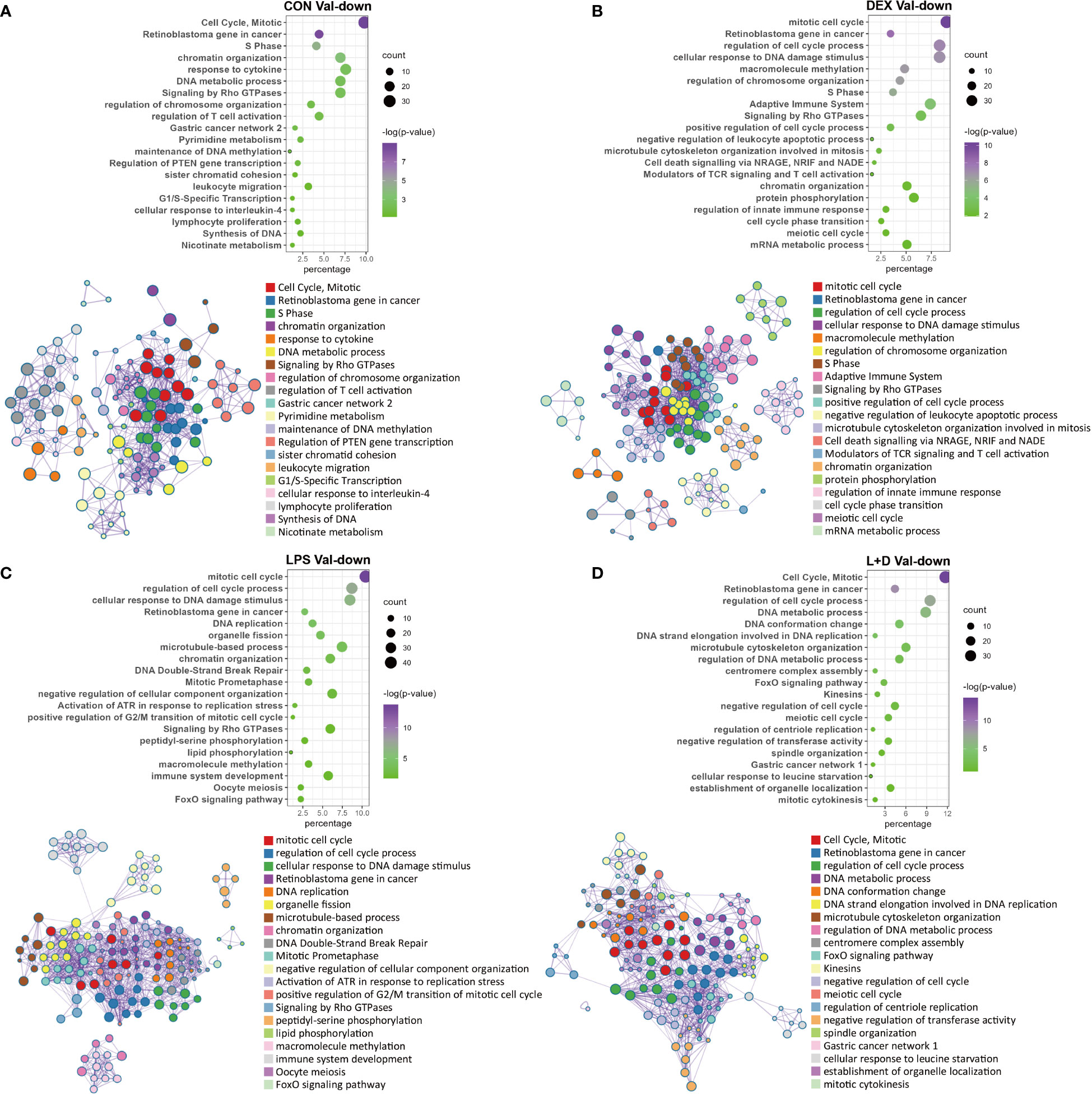
Figure 5 Enrichment analysis of Val-down genes in each treatment group. (A) Functional terms enriched for Val-down genes in CON group. (B) Functional terms enriched for Val-down genes in DEX group. (C) Functional terms enriched for Val-down genes in LPS group. (D) Functional terms enriched for Val-down genes in LPS+DEX group. Enrichment analysis was performed using the Metascape. Bubble plots illustrate significance and gene counts of top enriched terms, where purple indicates higher significance and green indicates lower significance and bubble size is positively correlated with gene counts. Networks illustrate the relationship between terms, where each node represents one functional term and terms with Kappa similarities above 0.3 are connected by edges. Each term cluster is labeled and represented by its most significant member and all members in the same term cluster are indicated by the same color.
Another key theme that emerged among Val-down genes was negative regulation of the immune system. In the absence of LPS (CON and DEX groups), Val-down genes were found to be related to both innate and adaptive immune signaling, with the former mainly involving response to cytokines and the latter associated with T cells (Figures 5A, B). These results were supported by IPA enrichment, which showed inhibition of a series of terms implicated in the quantity, homeostasis, maturation, and development of T cells (Table S5). In addition, inflammatory mediators such as LPS and IFNG were predicted by IPA as inhibited upstream regulators in the CON and DEX groups, while Irgm1—which has been found to prevent endotoxemia in mice (45)—was predicted to be activated under these conditions (Table S7). There is currently no report describing Irgm1 in the pig, however, its predicted activation indicates that Val-down genes altogether induce anti-inflammatory signaling.
In LPS treatment, none of the above inflammatory mediators (LPS, IFNG, and Irgm1) were enriched for the Val-down genes (Table S7), indicating that LPS abolished Val allele-mediated anti-inflammatory responses. However, activation of the anti-inflammatory factor Irgm1 was predicted under co-treatment with LPS and DEX (Table S7).
Hub genes, meaning genes showing a high degree of connectivity in PPI networks of Val-regulated DEGs, also exhibited different profiles among treatment groups and supported the key functions of respective clusters (Figure 6; Table S2). Without stimulation, the identified Val-up hub genes were associated with lysosomal function (CD68, CTSA, CTSB, M6PR, MCOLN1, NPC2, and SCARB2); vesicle-mediated transport (ACTR1A, AGFG1, AP2M1, ARF1, CD63, EXOC7, RAB10, RAB7A, and RAC1); and sphingolipid metabolism (ASAH1, GALC, GBA, GLA, and SGPL1; Figure 6A). In comparison, many Val-up hub genes in the DEX group were functionally distinct, and were related to protein folding, processing, and transport (CALR, CANX, CLTC, DNM2, HSP90B1, HSPA5, HSPA8, LAMP2, PDIA3, RAB11A, RAB7B, and VPS35) and mitochondrial respiration (CS, GSK3B, LDHA, MDH1, MDH2, and VDAC1; Figure 6C). Stimulation by LPS induced hub genes related to pattern recognition receptor (PRR) and inflammatory signaling (ANXA2, ANXA5, CD40, CD44, CD86, DDX58, FN1, HSP90AA1, ICAM1, IL10, JAK2, MYD88, and TLR4; Figure 6E). Given the cell death-related GR targets and signaling pathways found in this cluster, and the role of PRRs in sensing DAMPs as well as PAMPs (46), the hub genes found here apparently confirm Val allele-induced amplification of inflammatory signaling resulting from DAMP and PAMP recognition by PRRs. In the LPS+DEX group, two anti-inflammatory hub genes were identified—STAT3 (47) and PPARA (48) (Figure 6G)—which might partially explain the superior response to DEX therapy in Val carriers among endotoxin-challenged pigs (16).
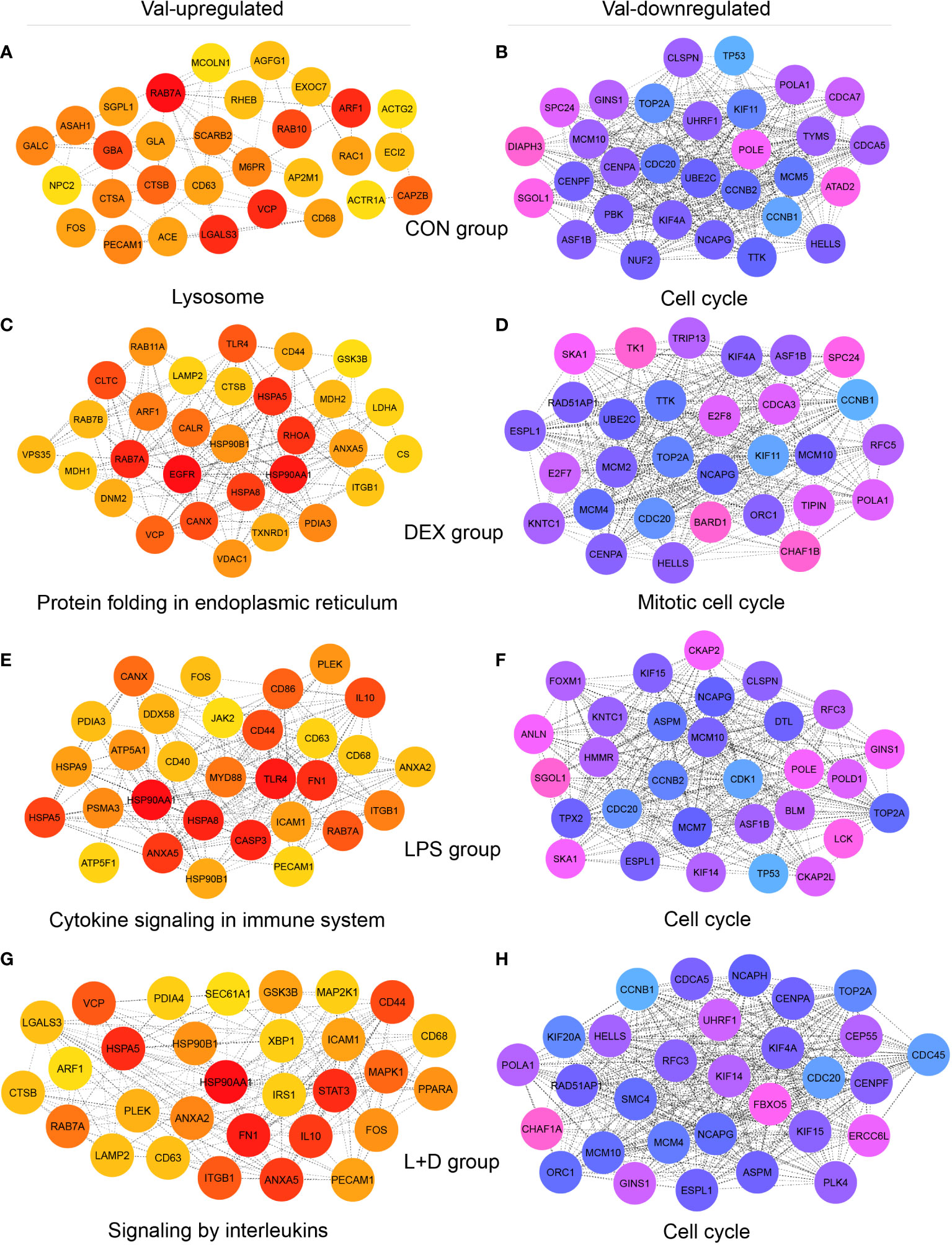
Figure 6 Identification of hub genes of Val-up and Val-down clusters in each treatment group. (A-H) Protein-protein interaction (PPI) networks showing top 30 hub genes with high connectivity within (A) Val-up cluster in CON group, (B) Val-down cluster in CON group, (C) Val-up cluster in DEX group, (D) Val-down cluster in DEX group, (E) Val-up cluster in LPS group, (F) Val-down cluster in LPS group, (G) Val-up cluster in LPS+DEX group, and (H) Val-down cluster in LPS+DEX group. PPI networks were constructed by the STRING database (27) and top 30 hub genes of each network were identified by the Cytoscape plugin cytoHubba (28, 29). Connectivity was correlated with color of circles where red and blue indicate higher degree whereas yellow and pink indicate lower degree. The most significant functional terms of hub genes of each cluster were identified using the Metascape (30) and displayed.
Most of the 30 Val-down hub genes identified in the four treatment groups centered unanimously around cell cycle, mitosis, and DNA replication, suggesting substantial inhibition of these processes (Figures 6B, D, F, H). Among them, CDC20, MCM10, NCAPG, and TOP2A were shared by all groups. Each group also contained unique hub genes, including ATAD2, CDCA7, DIAPH3, MCM5, NUF2, PBK, and TYMS (CON group); BARD1, CDCA3, CHAF1B, E2F7, E2F8, MCM2, RFC5, TIPIN, TK1, and TRIP13 (DEX group); ANLN, BLM, CDK1, CKAP2, CKAP2L, DTL, FOXM1, HMMR, MCM7, POLD1, and TPX2 (LPS group); and CDC45, CEP55, CHAF1A, ERCC6L, FBXO5, KIF20A, NCAPH, PLK4, and SMC4 (LPS+DEX group; Figures 6B, D, F, H).
This study has focused on determining the impact of a previously discovered natural Ala610Val substitution in the porcine glucocorticoid receptor (GRAla610Val) (49) on the transcriptional profiles of porcine PBMCs under non-stimulated and stimulated conditions. By utilizing PBMCs carrying all three GR genotypes (Ala/Ala, Ala/Val, and Val/Val), we identified a subset of genes whose transcription is altered by the Val variant in an additive manner, thus showing a genuine genetic effect. The resultant findings provide a clear evidence for a distinct transcriptome signature of the Val allele, which alters the response to immune stimuli, as seen through upregulated inflammatory responses and downregulation of cell cycle progression and cell division. These results offer a possible explanation and mutual support for the aggravated consequences of LPS-induced endotoxemia associated with the GRAla610Val variant (16), as in more detail elaborated below.
Among the various functions being represented by the Val-up genes, enhanced inflammatory response stands out as the most prominent theme. This is evidenced by the enrichment of signaling pathways involving endocytosis, antigen presentation, and particularly, PRR-mediated recognition and signal transduction of DAMPs and PAMPs among Val-up DEGs.
The first critical impact of the Val allele on immune signaling is upregulation of genes involved in endocytosis and antigen presentation. The three major types of endocytosis are pinocytosis, receptor-mediated endocytosis, and phagocytosis, of which the latter serves as a front-line defense against microbes, and is a crucial function of professional phagocytes including macrophages, monocytes, and neutrophils (50). In this process, extracellular microbial materials are internalized into phagosomes and ultimately subjected to degradation in the lysosome. Endocytosis and lysosomal degradation are both required for effective pathogen elimination (51). The Rab GTPases, such as Rab5 and Rab7, play key regulatory roles in endocytosis and exhibit substantial interactions with immune signals (50). Rab5 regulates fusion of phagosomes with early endosomes, whereas Rab7 facilitates the fusion of late endosomes and phagosomes with lysosomes; and both proteins can be upregulated in response to cytokine stimulation (50). In this study, we found that RAB5C, RAB7A, and RAB7B were all upregulated by the Val allele, supporting increased constitutive microbe internalization induced by the GR variant. Along with endocytosis, lysosomal functioning was also significantly enriched among Val-up genes. This is consistent with the Val-associated upregulation of ATP6V1E1, which encodes a subunit of the vacuolar ATPase (V-ATPase), an ATP hydrolysis-driven proton pump crucial for acidification of lysosomes (52).
Aside from endocytosis, the lysosome is also associated with antigen presentation. Professional APCs such as dendritic cells (DCs), B cells, and macrophages all constitutively express major histocompatibility complex (MHC) class II molecules (53). They internalize and process antigens, then present antigenic peptides to T cells (54). The antigen processing step relies on proteolysis in endosomal–lysosomal antigen-processing compartments, which is tightly regulated in APCs to generate proper peptides (53). In the current study, we found that MHC-mediated antigen presentation was enriched for Val-up genes regardless of treatment, and corresponded with enrichment of lysosomal terms. Antigen presentation is usually considered important for initiating T cell-dependent adaptive responses, but many studies have also observed interplay with TLR-mediated innate immune signaling (55). For example, LPS treatment increased MHC class II expression in DCs, in a manner dependent on an AP-1 enhancer (56), whereas deficiency in MHC class II molecules alleviated TLR-induced cytokine production and endotoxic shock (57). Further investigation revealed that intracellular MHC class II molecules interact with the tyrosine kinase Btk and promote its activation, which facilitates TLR signaling via interaction with MyD88 and TRIF (57). Furthermore, Btk activation requires a costimulatory molecule, CD40 (57), which was identified as a Val-up gene in our study, but only in the presence of LPS.
The major finding of this study is the upregulation of LPS signaling pathway, including TLR4 and its related molecules, by the Val variant. Toll-like receptors (TLRs) are one of the most widely studied molecules in the innate immune system. In this study, we found that the Val allele upregulated TLR4, TLR8, and CD180 of the TLR family. TLR4 is the main receptor for LPS (58). TLR4 signaling in response to LPS relies on cooperation of myeloid differentiation factor 2 (MD2) and CD14, subsequently leading to stimulation of MyD88-dependent and TRIF-dependent signaling, in turn causing activation of NF-κB, AP-1, MAPK, and IRF3 cascades (59). Along with TLR4, other key molecules in TLR4-LPS signaling such as MYD88 and LY96 (encoding MD2) were identified as Val-up genes. Among these, LY96 was constitutively upregulated by the Val allele across treatments, perfectly in line with the predisposition of Val carriers to enhanced LPS response. Unlike LY96, MYD88 was affected by Val only in the presence of LPS. CD180 mediates B cell activation by LPS (60). CD180 enhances TLR4 signaling in B cells but inhibits it in macrophages and DCs; the opposite effect appears associated with TLR4 abundance (61). Thus, CD180 likely contributes to Val enhancement of LPS signaling in porcine B cells. LPS signaling molecules upregulated by the Val allele include TIRAP, FOS, HSP90AA1, LITAF, PELI1, and PLAA. HSP90AA1 was identified as a Val-up GR target gene in our study. Among other functions, HSP 90-alpha was implicated in LPS signal transduction by serving as a CD14-independent LPS receptor (62). PELI1 encoding an E3 ubiquitin ligase participates in LPS-mediated induction of CD86 and MHCII in B cells and Its deficiency renders mice resistant to LPS-induced septic shock (63). Thus, upregulation of TLR4 and its associated molecules by the Val variant is proposed here as the central mechanism for the enhanced response of Val carriers to endotoxemia observed in the in vivo LPS challenge previously (16).
In addition to TLR4 signaling, the Val allele upregulated many other genes and pathways implicated in PRR signaling, particularly in the LPS group, such as DDX58 and NOD2. The first of these, DDX58, encodes retinoic acid-inducible gene-I (RIG-I), an intracellular PRR responsible for sensing viral nucleic acids (58). When activated, RIG-I binds mitochondrial antiviral signaling protein (MAVS), a key adaptor protein that can mediate downstream activation of the NF-κB and IRF3 signaling pathways (64). Nucleotide binding oligomerization domain containing 2 (NOD2) is another intracellular PRR that recognizes muramyl dipeptide (MDP) in the cell wall of both Gram-negative and Gram-positive bacteria (58). Activation of NOD2 also triggers NF-κB signaling, through recruitment of the downstream receptor-interacting protein kinase 2 (RIPK2) (65); however, NOD2 signaling requires the decomposition of cell walls by lysosomes (58). This apparently confirms the contribution of Val-mediated upregulation of lysosomal functions to enhanced immune signaling. Furthermore, upregulation of DDX58, NOD2, and RIPK2 by the Val allele was found only in the presence of LPS, indicating that LPS serves as a trigger to amplify Val upregulation of inflammation.
Upregulation of the above PRRs may also contribute to enhanced inflammatory signaling via DAMPs released by damaged cells, since the Val allele activated autophagy, ferroptosis, and necroptosis. Autophagy is a parallel process to the lysosomal degradation of cellular materials (66), and can also be induced by ER stress (67). Increased autophagy is therefore in agreement with the enrichment of lysosome, ER stress, and unfolded protein response pathways that were identified among Val-up genes. Ferroptosis is an iron-dependent cell death pathway accompanied by lipid peroxidation and ROS generation (34), and its upregulation fits with the observed enrichment of fatty acid β-oxidation, mitochondrial dysfunction, and iron homeostasis signaling pathways. Necroptosis is a regulated inflammatory mode of cell death, which critically relies on the participation of RIPK3 and mixed lineage kinase domain-like protein (MLKL). To initiate necroptosis, RIPK3 recruits and activates MLKL by phosphorylating its pseudokinase domain, which promotes oligomerization. Following this, oligomerized MLKL translocates to the plasma membrane, where its pore-forming properties cause membrane damage (34). In our study, MLKL was identified as a Val-up gene independent of treatment, whereas RIPK3 was only observed in the LPS group. Therefore, as seen for inflammatory processes, LPS serves as an external signal to promote cell death. The aforementioned Val-up GR target gene HSP90AA1 also facilitates necroptosis, since it encodes a chaperone protein required for oligomerization and translocation of MLKL (68).
Cell death signaling interacts substantially with PRRs. DAMPs released by damaged cells can activate PRRs, thereby triggering inflammatory responses involving migration and phagocytosis of immune cells, and production of cytokines (46). These DAMPs can be derived from various cellular compartments—such as mitochondria, the cytosol, and nucleus—and include a range of molecular classes such as HMGB1, IL-1α, uric acid, S100 proteins, heat shock proteins, ATP, mROS, RNA, DNA, and nucleotides (32). According to this, we found that the Val allele upregulated many genes encoding heat shock proteins and S100 proteins, such as HSPA5, HSPA8, HSPA9, HSP90AA1, S100A10, S100A6, and S100Z. The DAMPs can be recognized by different PRRs, including those upregulated by the Val allele (NOD2, RIG-I, and TLRs), to initiate specific inflammatory signaling (67). In turn, the activation of different types of PRRs can induce cell death (46). For example, activation of TLR4 by LPS stimulates necroptosis in mouse macrophages, in a process involving assembly of a TRIF–RIPK3 complex and accumulation of ROS (69).
Val-up genes also indicated activation of several specific pathways involved in cell types not included in this study, which may be regulated by the Val allele in shared pathways also in those cell types. Notably, among these are also pathways related to platelet function. Apart from their key functions in hemostasis, platelets play an essential role in inflammatory diseases and development of sepsis (70). They exert their immunomodulatory functions through interaction with immune cells including neutrophils, or production of immune mediators like cytokines (71). Persistent reduction in platelet numbers is an established risk factor for mortality in sepsis (70). The Val allele-dependent upregulation of several genes associated with platelet activation supports our previous observation that platelet numbers drop more significantly in GRAla610Val pigs during endotoxemia (16).
Interestingly, our in vivo study revealed that DEX rescue of LPS-induced platelet reduction was more effective in Val carriers (16). This might be partially explained by Val-mediated upregulation of the VEGFA–VEGFR2 signaling pathway, which was enriched only in the presence of DEX (i.e., DEX and LPS+DEX groups). This pathway is important in various functions of endothelial cells, such as survival, proliferation, migration, and particularly, eNOS-mediated NO production (35). Specifically, phosphorylation of VEGFR-2 is essential for VEGF-stimulated NO release from endothelial cells, due to its implication in Akt-dependent eNOS activation (72). VEGFR-2 also facilitates VEGF-stimulated eNOS association with the chaperone protein Hsp90 (73), and it has been reported that overexpression of eNOS in endothelial cells inhibits platelet aggregation (74) through NO stimulation of soluble guanylate cyclase in platelets. This increases intracellular levels of cGMP, which represses platelet aggregation and adhesion (75). The role of NO in sepsis is complex. Whereas it is toxic to microbes and helps reduce tissue injury, excessive NO generation promotes sepsis pathogenesis (76). Deficient endothelial NO production is also detrimental, and is believed to be a driver of microvascular dysfunction in septic shock (76). This is supported by observations that inducible nitric oxide synthase (iNOS)-derived NO production is elevated in sepsis, whereas the amount formed by eNOS is reduced, resulting in overwhelming platelet aggregation and leukocyte adhesion (36).
Another significant facet of alteration of porcine PBMCs in Val carriers is inhibition of cell cycle progression, as represented by Val-down genes. In this study, IPA predicted that E2F1 was inhibited whereas Rb was activated. E2F1 is a TF controlling expression of many key genes implicated in G1/S transition. It is negatively regulated by Rb via heterodimerization (42), although inhibition of E2F1 is known to cause developmental anomalies and dampen DNA repair (77). Phosphorylation of Rb by cyclin-dependent kinases suppresses its association with E2F1, leading to recovery of TF activity (42). In addition to diminishing E2F1 function, Rb represses the expression of cell cycle genes via interacting with proteins needed for nucleosome remodeling and histone modification. In doing so, it induces changes in chromatin structure (44). Several studies have revealed that cell cycle arrest is associated with ferroptosis (78), necroptosis (79), and autophagy (80). Further investigation is therefore warranted, to establish if and how Val allele-mediated inhibition of cell cycle progression contributes to enhanced cell death and inflammatory responses.
Despite the enrichment of immune terms discussed above, our previous work has shown that in unstimulated condition, i.e. in untreated pigs, most inflammatory parameters are not significantly affected by GRAla610Val in vivo (16). This might be related to a subset of Val-affected genes involved in negative regulation of immune responses. Without LPS stimulation, response to cytokines was enriched for Val-down genes, which included the pro-inflammatory genes CCL17, CD38, IL18RAP, IRF1, IRF7, ITGA4, PTGS2, and XCL1. None of these genes was regulated by the Val allele in the LPS group. In the CON and DEX groups, LPS and IFNG were predicted by IPA as both activated (for Val-up genes) and inhibited (for Val-down genes) upstream regulators, whereas in the LPS group, only their activation was observed. In the absence of LPS, a possible alternative mechanism to counterbalance enhanced immune signaling is inhibition of T cell activation. This was seen among the Val-down genes in IPA-predicted inhibition of T cell-associated terms. Professional APCs recognize and bind targets via PRRs then transfer signals to T cells, thus providing a bridge between innate and adaptive immunity (81). When treated with LPS, the Val-up genes featured multiple activated T cell pathways in contrast to baseline conditions. These results indicate that without LPS, the Val allele mediates both activation and inhibition of immune signaling to maintain homeostasis. However, this homeostasis is disrupted in the presence of LPS stimulus.
The main goal of this study was to elucidate mechanisms behind the enhanced LPS sensitivity of Val carriers observed previously in our in vivo challenge. The discovered transcriptional signature suggests that the Val allele affects various immune-related functions in porcine PBMCs. Among the different functional alterations caused by the Val variant stand out its effects on inflammatory signaling associated with PRRs, above all TLR4, and cell death linked with DAMPs, as the determining mechanisms for an enhanced LPS sensitivity and response. In addition, upregulation of the different PRRs, such as DDX58 and NOD2, together with the effect on several other immune functions, suggests that GRAla610Val may have a broader impact on disease susceptibility in pigs. Overall, the present results emphasize the value of the GRAla610Val pigs as a unique animal model to explore fundamental biology of GR signaling on the one hand, and for the research to improve pig health on the other.
The datasets presented in this study can be found in online repositories. The names of the repository/repositories and accession number(s) can be found below: https://www.ebi.ac.uk/arrayexpress/, E-MTAB-9808.
EM: Conceptualization and Funding acquisition. EM and ZL: Methodology. ZL: Investigation and Formal analysis. FH: Data Curation. FH: Software. KW: Supervision. ZL and EM: Writing - original draft. All authors contributed to manuscript revision, read, and approved the submitted version.
The current study was funded by a grant from the German Research Foundation (Deutsche Forschungsgemeinschaft (DFG)—Project number 391382814), and matched funding from the FBN.
The authors thank Angelika Deike, Marlies Fuchs, Angela Garve, and Janine Wetzel for excellent technical help and the whole staff of the experimental farm (EAS) for animal management and care.
The authors declare that the research was conducted in the absence of any commercial or financial relationships that could be construed as a potential conflict of interest.
All claims expressed in this article are solely those of the authors and do not necessarily represent those of their affiliated organizations, or those of the publisher, the editors and the reviewers. Any product that may be evaluated in this article, or claim that may be made by its manufacturer, is not guaranteed or endorsed by the publisher.
The Supplementary Material for this article can be found online at: https://www.frontiersin.org/articles/10.3389/fimmu.2022.976454/full#supplementary-material
1. Spiga F, Walker JJ, Terry JR, Lightman SL. HPA axis-rhythms. Compr Physiol (2011) 4(3):1273–98. doi: 10.1002/cphy.c140003
2. Sapolsky RM, Romero LM, Munck AU. How do glucocorticoids influence stress responses? integrating permissive, suppressive, stimulatory, and preparative actions. Endoc Rev (2000) 21(1):55–89. doi: 10.1210/edrv.21.1.0389
3. Petrillo MG, Bortner CD, Cidlowski JA. Glucocorticoids: Inflammation and immunity. in: The hypothalamic-Pituitary-Adrenal axis in health and disease. Geer EB, editor. (Cham: Springer International Publishing) (2016) p. 43–63.
4. Timmermans S, Souffriau J, Libert C. A general introduction to glucocorticoid biology. Front Immunol (2019) 10:1545. doi: 10.3389/fimmu.2019.01545
5. Dendoncker K, Libert C. Glucocorticoid resistance as a major drive in sepsis pathology. Cytokine Growth Factor Rev (2017) 35:85–96. doi: 10.1016/j.cytogfr.2017.04.002
6. Reichardt SD, Amouret A, Muzzi C, Vettorazzi S, Tuckermann JP, Lühder F, et al. The role of glucocorticoids in inflammatory diseases. Cells (2021) 10(11):2921. doi: 10.3390/cells10112921
7. Opal SM. Endotoxins and other sepsis triggers. Contrib Nephrol (2010) 167:14–24. doi: 10.1159/000315915
8. Marshall JC. Endotoxin in the pathogenesis of sepsis. Contrib Nephrol (2010) 167:1–13. doi: 10.1159/000315914
9. Vandewalle J, Libert C. Glucocorticoids in sepsis: To be or not to be. Front Immunol (2020) 11:1318. doi: 10.3389/fimmu.2020.01318
10. Ramamoorthy S, Cidlowski JA. Corticosteroids: Mechanisms of action in health and disease. Rheum Dis Clinics (2016) 42(1):15–31. doi: 10.1016/j.rdc.2015.08.002
11. Vandevyver S, Dejager L, Libert C. Comprehensive overview of the structure and regulation of the glucocorticoid receptor. Endocr Rev (2014) 35(4):671–93. doi: 10.1210/er.2014-1010
12. Kleiman A, Hubner S, Rodriguez Parkitna JM, Neumann A, Hofer S, Weigand MA, et al. Libert c et al: Glucocorticoid receptor dimerization is required for survival in septic shock via suppression of interleukin-1 in macrophages. FASEB J (2012) 26(2):722–9. doi: 10.1096/fj.11-192112
13. Silverman MN, Mukhopadhyay P, Belyavskaya E, Tonelli LH, Revenis BD, Doran JH, et al. Glucocorticoid receptor dimerization is required for proper recovery of LPS-induced inflammation, sickness behavior and metabolism in mice. Mol Psychiatry (2013) 18(9):1006–17. doi: 10.1038/mp.2012.131
14. Reichardt HM, Umland T, Bauer A, Kretz O, Schütz G. Mice with an increased glucocorticoid receptor gene dosage show enhanced resistance to stress and endotoxic shock. Mol Cell Biol (2000) 20(23):9009–17. doi: 10.1128/MCB.20.23.9009-9017.2000
15. Dejager L, Pinheiro I, Puimege L, Fan YD, Gremeaux L, Vankelecom H, et al. Increased glucocorticoid receptor expression and activity mediate the LPS resistance of SPRET/EI mice. J Biol Chem (2010) 285(40):31073–86. doi: 10.1074/jbc.M110.154484
16. Li Z, Kanitz E, Tuchscherer M, Tuchscherer A, Metges CC, Trakooljul N, et al. A natural Ala610Val substitution causing glucocorticoid receptor hypersensitivity aggravates consequences of endotoxemia. Brain Behav Immun (2020) 90:174–83. doi: 10.1016/j.bbi.2020.08.009
17. Reyer H, Ponsuksili S, Kanitz E, Pohland R, Wimmers K, Murani E. A natural mutation in helix 5 of the ligand binding domain of glucocorticoid receptor enhances receptor-ligand interaction. PloS One (2016) 11(10):e0164628. doi: 10.1371/journal.pone.0164628
18. Muráni E, Ponsuksili S, Jaeger A, Görres A, Tuchscherer A, Wimmers K. A naturally hypersensitive glucocorticoid receptor elicits a compensatory reduction of hypothalamus-pituitary-adrenal axis activity early in ontogeny. Open Biol (2016) 6(7):150193. doi: 10.1098/rsob.150193
19. Wyns H, Plessers E, De Backer P, Meyer E, Croubels S. In vivo porcine lipopolysaccharide inflammation models to study immunomodulation of drugs. Vet Immunol Immunopathol (2015) 166(3-4):58–69. doi: 10.1016/j.vetimm.2015.06.001
20. Li Z, Trakooljul N, Hadlich F, Ponsuksili S, Wimmers K, Murani E. Transcriptome analysis of porcine PBMCs reveals lipopolysaccharide-induced immunomodulatory responses and crosstalk of immune and glucocorticoid receptor signaling. Virulence (2021) 12(1):1808–24. doi: 10.1080/21505594.2021.1948276
21. Love MI, Huber W, Anders S. Moderated estimation of fold change and dispersion for RNA-seq data with DESeq2. Genome Biol (2014) 15(12):550. doi: 10.1186/s13059-014-0550-8
22. Blighe K, Rana S, Lewis M. EnhancedVolcano: Publication-ready volcano plots with enhanced colouring and labeling. R package (2019). Available at: https://github.com/kevinblighe/EnhancedVolcano.
23. Chen C, Chen H, Zhang Y, Thomas HR, Frank MH, He Y, et al. TBtools - an integrative toolkit developed for interactive analyses of big biological data. Mol Plant (2020) 13(8):1194–202. doi: 10.1016/j.molp.2020.06.009
24. Kolde R. Pheatmap: Pretty heatmaps. R package (2019). Available at: https://github.com/raivokolde/pheatmap.
25. Pantano L. DEGreport: Report of DEG analysis. R package (2021). Available at: https://github.com/lpantano/DEGreport.
26. Rouillard AD, Gundersen GW, Fernandez NF, Wang Z, Monteiro CD, McDermott MG, et al. The harmonizome: A collection of processed datasets gathered to serve and mine knowledge about genes and proteins. Database (2016) 2016:1–16. doi: 10.1093/database/baw100
27. Szklarczyk D, Gable AL, Nastou KC, Lyon D, Kirsch R, Pyysalo S, et al. The STRING database in 2021: Customizable protein–protein networks, and functional characterization of user-uploaded gene/measurement sets. Nucleic Acids Res (2021) 49(D1):D605–12. doi: 10.1093/nar/gkaa1074
28. Shannon P, Markiel A, Ozier O, Baliga NS, Wang JT, Ramage D, et al. Cytoscape: A software environment for integrated models of biomolecular interaction networks. Genome Res (2003) 13(11):2498–504. doi: 10.1101/gr.1239303
29. Chin C-H, Chen S-H, Wu H-H, Ho C-W, Ko M-T, Lin C-Y. cytoHubba: Identifying hub objects and sub-networks from complex interactome. BMC Syst Biol (2014) 8(S4):S11. doi: 10.1186/1752-0509-8-S4-S11
30. Zhou Y, Zhou B, Pache L, Chang M, Khodabakhshi AH, Tanaseichuk O, et al. Metascape provides a biologist-oriented resource for the analysis of systems-level datasets. Nat Commun (2019) 10(1):1–10. doi: 10.1038/s41467-019-09234-6
32. Roh JS, Sohn DH. Damage-associated molecular patterns in inflammatory diseases. Immune Netw (2018) 18(4):e27. doi: 10.4110/in.2018.18.e27
33. Todkar K, Ilamathi HS, Germain M. Mitochondria and lysosomes: Discovering bonds. Front Cell Dev Biol (2017) 5:106. doi: 10.3389/fcell.2017.00106
34. Galluzzi L, Vitale I, Aaronson SA, Abrams JM, Adam D, Agostinis P, et al. Molecular mechanisms of cell death: Recommendations of the nomenclature committee on cell death 2018. Cell Death Differentiation (2018) 25(3):486–541. doi: 10.1038/s41418-017-0012-4
35. Wang X, Bove AM, Simone G, Ma B. Molecular bases of VEGFR-2-mediated physiological function and pathological role. Front Cell Dev Biol (2020) 8:1314. doi: 10.3389/fcell.2020.599281
36. Martín-Fernández M, Tamayo-Velasco Á, Aller R, Gonzalo-Benito H, Martínez-Paz P, Tamayo E. Endothelial dysfunction and neutrophil degranulation as central events in sepsis physiopathology. Int J Mol Sci (2021) 22(12):6272. doi: 10.3390/ijms22126272
37. Scheller J, Chalaris A, Schmidt-Arras D, Rose-John S. The pro-and anti-inflammatory properties of the cytokine interleukin-6. Biochim Biophys Acta (BBA)-Molec Cell Res (2011) 1813(5):878–88. doi: 10.1016/j.bbamcr.2011.01.034
38. Cui H, Banerjee S, Guo S, Xie N, Liu G. IFN regulatory factor 2 inhibits expression of glycolytic genes and lipopolysaccharide-induced proinflammatory responses in macrophages. J Immunol (2018) 200(9):3218–30. doi: 10.4049/jimmunol.1701571
39. Li Z, Jia Y, Han S, Wang X, Han F, Zhang J, et al. Klf4 alleviates lipopolysaccharide-induced inflammation by inducing expression of MCP-1 induced protein 1 to deubiquitinate TRAF6. Cell Physiol Biochem (2018) 47(6):2278–90. doi: 10.1159/000491538
40. Rollins DA, Coppo M, Rogatsky I. Minireview: Nuclear receptor coregulators of the p160 family: Insights into inflammation and metabolism. Mol Endocrinol (2015) 29(4):502–17. doi: 10.1210/me.2015-1005
41. Chi F, Chen L, Jin X, He G, Liu Z, Han S. CKAP2L, transcriptionally inhibited by FOXP3, promotes breast carcinogenesis through the AKT/mTOR pathway. Exp Cell Res (2022) 412(2):113035. doi: 10.1016/j.yexcr.2022.113035
42. Sheldon LA. Inhibition of E2F1 activity and cell cycle progression by arsenic via retinoblastoma protein. Cell Cycle (2017) 16(21):2058–72. doi: 10.1080/15384101.2017.1338221
43. Chan HM, Narita M, Lowe SW, Livingston DM. The p400 E1A-associated protein is a novel component of the p53→ p21 senescence pathway. Genes Dev (2005) 19(2):196–201. doi: 10.1101/gad.1280205
44. Giacinti C, Giordano A. RB and cell cycle progression. Oncogene (2006) 25(38):5220–7. doi: 10.1038/sj.onc.1209615
45. Bafica A, Feng CG, Santiago HC, Aliberti J, Cheever A, Thomas KE, et al. The IFN-inducible GTPase LRG47 (Irgm1) negatively regulates TLR4-triggered proinflammatory cytokine production and prevents endotoxemia. J Immunol (2007) 179(8):5514–22. doi: 10.4049/jimmunol.179.8.5514
46. Amarante-Mendes GP, Adjemian S, Branco LM, Zanetti LC, Weinlich R, Bortoluci KR. Pattern recognition receptors and the host cell death molecular machinery. Front Immunol (2018) 9:2379. doi: 10.3389/fimmu.2018.02379
47. Hutchins AP, Diez D, Miranda-Saavedra D. The IL-10/STAT3-mediated anti-inflammatory response: Recent developments and future challenges. Brief Funct Genomics (2013) 12(6):489–98. doi: 10.1093/bfgp/elt028
48. Bougarne N, Mylka V, Ratman D, Beck IM, Thommis J, De Cauwer L, et al. Mechanisms underlying the functional cooperation between PPARalpha and GRalpha to attenuate inflammatory responses. Front Immunol (2019) 10:1769. doi: 10.3389/fimmu.2019.01769
49. Murani E, Reyer H, Ponsuksili S, Fritschka S, Wimmers K. A substitution in the ligand binding domain of the porcine glucocorticoid receptor affects activity of the adrenal gland. PloS One (2012) 7(9):e45518. doi: 10.1371/journal.pone.0045518
50. Ding X, Xiang S. Endocytosis and human innate immunity. J Immunol Sci (2018) 2(1):65–70. doi: 10.29245/2578-3009/2018/1.1121
51. Wong C-O, Gregory S, Hu H, Chao Y, Sepúlveda VE, He Y, et al. Lysosomal degradation is required for sustained phagocytosis of bacteria by macrophages. Cell Host Microbe (2017) 21(6):719–30:e716. doi: 10.1016/j.chom.2017.05.002
52. Futai M, Sun-Wada G-H, Wada Y, Matsumoto N, Nakanishi-Matsui M. Vacuolar-type ATPase: A proton pump to lysosomal trafficking. Proc Japan Academy Ser B (2019) 95(6):261–77. doi: 10.2183/pjab.95.018
53. Roche PA, Furuta K. The ins and outs of MHC class II-mediated antigen processing and presentation. Nat Rev Immunol (2015) 15(4):203–16. doi: 10.1038/nri3818
54. Delamarre L, Pack M, Chang H, Mellman I, Trombetta ES. Differential lysosomal proteolysis in antigen-presenting cells determines antigen fate. Science (2005) 307(5715):1630–4. doi: 10.1126/science.1108003
55. Watts C. The endosome–lysosome pathway and information generation in the immune system. Biochim Biophys Acta (BBA)-Proteins Proteomics (2012) 1824(1):14–21. doi: 10.1016/j.bbapap.2011.07.006
56. Casals C, Barrachina M, Serra M, Lloberas J, Celada A. Lipopolysaccharide up-regulates MHC class II expression on dendritic cells through an AP-1 enhancer without affecting the levels of CIITA. J Immunol (2007) 178(10):6307–15. doi: 10.4049/jimmunol.178.10.6307
57. Liu X, Zhan Z, Li D, Xu L, Ma F, Zhang P, et al. Intracellular MHC class II molecules promote TLR-triggered innate immune responses by maintaining activation of the kinase btk. Nat Immunol (2011) 12(5):416–24. doi: 10.1038/ni.2015
58. Li D, Wu M. Pattern recognition receptors in health and diseases. Signal Transduct Target Ther (2021) 6(1):291. doi: 10.1038/s41392-021-00687-0
59. Mazgaeen L, Gurung P. Recent advances in lipopolysaccharide recognition systems. Int J Mol Sci (2020) 21(2):379. doi: 10.3390/ijms21020379
60. Yazawa N, Fujimoto M, Sato S, Miyake K, Asano N, Nagai Y, et al. CD19 regulates innate immunity by the toll-like receptor RP105 signaling in b lymphocytes. Blood (2003) 102(4):1374–80. doi: 10.1182/blood-2002-11-3573
61. Divanovic S, Trompette A, Atabani SF, Madan R, Golenbock DT, Visintin A, et al. Negative regulation of toll-like receptor 4 signaling by the toll-like receptor homolog RP105. Nat Immunol (2005) 6(6):571–8. doi: 10.1038/ni1198
62. Triantafilou K, Triantafilou M, Dedrick RL. A CD14-independent LPS receptor cluster. Nat Immunol (2001) 2(4):338–45. doi: 10.1038/86342
63. Chang M, Jin W, Sun S-C. Peli1 facilitates TRIF-dependent toll-like receptor signaling and proinflammatory cytokine production. Nat Immunol (2009) 10(10):1089–95. doi: 10.1038/ni.1777
64. Liu S, Cai X, Wu J, Cong Q, Chen X, Li T, et al. Phosphorylation of innate immune adaptor proteins MAVS, STING, and TRIF induces IRF3 activation. Science (2015) 347(6227):aaa2630. doi: 10.1126/science.aaa2630
65. Park J-H, Kim Y-G, McDonald C, Kanneganti T-D, Hasegawa M, Body-Malapel M, et al. RICK/RIP2 mediates innate immune responses induced through Nod1 and Nod2 but not TLRs. J Immunol (2007) 178(4):2380–6. doi: 10.4049/jimmunol.178.4.2380
66. Yim WW-Y, Mizushima N. Lysosome biology in autophagy. Cell Discovery (2020) 6(1):1–12. doi: 10.1038/s41421-020-0141-7
67. Sangiuliano B, Pérez NM, Moreira DF, Belizário JE. Cell death-associated molecular-pattern molecules: Inflammatory signaling and control. Mediators Inflamm (2014) 2014:821043. doi: 10.1155/2014/821043
68. Jacobsen A, Lowes K, Tanzer M, Lucet I, Hildebrand J, Petrie E, et al. HSP90 activity is required for MLKL oligomerisation and membrane translocation and the induction of necroptotic cell death. Cell Death Dis (2016) 7(1):e2051–1. doi: 10.1038/cddis.2015.386
69. He S, Liang Y, Shao F, Wang X. Toll-like receptors activate programmed necrosis in macrophages through a receptor-interacting kinase-3–mediated pathway. Proc Natl Acad Sci (2011) 108(50):20054–9. doi: 10.1073/pnas.1116302108
70. Assinger A, Schrottmaier WC, Salzmann M, Rayes J. Platelets in sepsis: An update on experimental models and clinical data. Front Immunol (2019) 10:1687. doi: 10.3389/fimmu.2019.01687
71. Schrottmaier WC, Kral JB, Badrnya S, Assinger A. Aspirin and P2Y12 inhibitors in platelet-mediated activation of neutrophils and monocytes. Thromb Haemostasis (2015) 114(09):478–789. doi: 10.1160/TH14-11-0943
72. Blanes MG, Oubaha M, Rautureau Y, Gratton J-P. Phosphorylation of tyrosine 801 of vascular endothelial growth factor receptor-2 is necessary for akt-dependent endothelial nitric-oxide synthase activation and nitric oxide release from endothelial cells. J Biol Chem (2007) 282(14):10660–9. doi: 10.1074/jbc.M609048200
73. Duval M, Le Bœuf F, Huot J, Gratton J-P. Src-mediated phosphorylation of Hsp90 in response to vascular endothelial growth factor (VEGF) is required for VEGF receptor-2 signaling to endothelial NO synthase. Mol Biol Cell (2007) 18(11):4659–68. doi: 10.1091/mbc.e07-05-0467
74. Kader KN, Akella R, Ziats NP, Lakey LA, Harasaki H, Ranieri JP, et al. eNOS-overexpressing endothelial cells inhibit platelet aggregation and smooth muscle cell proliferation in vitro. Tissue Eng (2000) 6(3):241–51. doi: 10.1089/10763270050044425
75. Kerrigan SW, Devine T, Fitzpatrick G, Thachil J, Cox D. Early host interactions that drive the dysregulated response in sepsis. Front Immunol (2019) 10:1748. doi: 10.3389/fimmu.2019.01748
76. Lambden S. Bench to bedside review: Therapeutic modulation of nitric oxide in sepsis–an update. Intensive Care Med Exp (2019) 7(1):1–14. doi: 10.1186/s40635-019-0274-x
77. Poppy Roworth A, Ghari F, La Thangue NB. To live or let die–complexity within the E2F1 pathway. Mol Cell Oncol (2015) 2(1):e970480. doi: 10.4161/23723548.2014.970480
78. Lin R, Zhang Z, Chen L, Zhou Y, Zou P, Feng C, et al. Dihydroartemisinin (DHA) induces ferroptosis and causes cell cycle arrest in head and neck carcinoma cells. Cancer Lett (2016) 381(1):165–75. doi: 10.1016/j.canlet.2016.07.033
79. Frank T, Tuppi M, Hugle M, Dötsch V, van Wijk SJ, Fulda S. Cell cycle arrest in mitosis promotes interferon-induced necroptosis. Cell Death Differentiation (2019) 26(10):2046–60. doi: 10.1038/s41418-019-0298-5
80. Mathiassen SG, De Zio D, Cecconi F. Autophagy and the cell cycle: A complex landscape. Front Oncol (2017) 7:51. doi: 10.3389/fonc.2017.00051
Keywords: glucocorticoid receptor hypersensitivity, transcriptome, porcine PBMCs, lipopolysaccharide, inflammatory signaling, cell cycle progression, pattern recognition receptors
Citation: Li Z, Hadlich F, Wimmers K and Murani E (2022) Glucocorticoid receptor hypersensitivity enhances inflammatory signaling and inhibits cell cycle progression in porcine PBMCs. Front. Immunol. 13:976454. doi: 10.3389/fimmu.2022.976454
Received: 23 June 2022; Accepted: 10 November 2022;
Published: 24 November 2022.
Edited by:
Hauke Smidt, Wageningen University and Research, NetherlandsReviewed by:
Michael J. Myers, US Food and Drug Administration, United StatesCopyright © 2022 Li, Hadlich, Wimmers and Murani. This is an open-access article distributed under the terms of the Creative Commons Attribution License (CC BY). The use, distribution or reproduction in other forums is permitted, provided the original author(s) and the copyright owner(s) are credited and that the original publication in this journal is cited, in accordance with accepted academic practice. No use, distribution or reproduction is permitted which does not comply with these terms.
*Correspondence: Eduard Murani, bXVyYW5pQGZibi1kdW1tZXJzdG9yZi5kZQ==
Disclaimer: All claims expressed in this article are solely those of the authors and do not necessarily represent those of their affiliated organizations, or those of the publisher, the editors and the reviewers. Any product that may be evaluated in this article or claim that may be made by its manufacturer is not guaranteed or endorsed by the publisher.
Research integrity at Frontiers
Learn more about the work of our research integrity team to safeguard the quality of each article we publish.