- 1Department of Medicine, Schulich School of Medicine and Dentistry, University of Western Ontario, London, ON, Canada
- 2Division of Rheumatology, St. Joseph’s Health Care, Schulich School of Medicine and Dentistry, University of Western Ontario, London, ON, Canada
The pathogenesis of connective tissue diseases (CTDs), such as systemic lupus erythematosus (SLE) and systemic sclerosis (SSc), is characterized by derangements of the innate and adaptive immune system, and inflammatory pathways leading to autoimmunity, chronic cytokine production, and chronic inflammation. The diagnosis of these diseases is based on meeting established criteria with symptoms, signs and autoantibodies. However, there are pre-clinical states where criteria are not fulfilled but biochemical and autoimmune derangements are present. Understanding the underlying processes responsible for disease pathogenesis in pre-clinical states, which place patients at increased risk for the development of established connective tissue diseases, represents an opportunity for early identification and potentially enables timely treatment with the goal of limiting disease progression and improved prognosis. This scoping review describes the role of the innate and adaptive immune responses in the pre-clinical states of undifferentiated CTD at risk for SSc and prescleroderma, the evolution of antibodies from nonspecific to specific antinuclear antibodies prior to SLE development, and the signaling pathways and inflammatory markers of fibroblast, endothelial, and T cell activation underlying immune dysregulation in these pre-clinical states.
Introduction
Systemic sclerosis (SSc) is a rare multisystem autoimmune connective tissue disease (CTD) characterized by fibrosis of the skin and internal organs, vasculopathy, and autoimmunity with distinct antibodies. SSc is classified using the American College of Rheumatology/European League of Rheumatism (ACR/EULAR) 2013 criteria (1). However, there are pre-morbid clinical states, including Undifferentiated Connective Tissue Disease at risk for Systemic Sclerosis (UCTD-risk-SSc) and prescleroderma, where autoimmunity and dysregulation of inflammatory pathways occur without the presence of clinical symptoms (2). UCTD-risk-SSc, also known as very early/early SSc, is a label given to patients who do not meet the ACR/EULAR 2013 criteria, but who present with Raynaud’s Phenomenon (RP) and either typical SSc capillaroscopic findings (megacapillaries or avascular areas) or serum marker antibodies (anti-centromere, anti-topoisomerase I, anti-RNA polymerase III, anti-Th/To, and anti-Pm-Scl) (3, 4). UCTD-risk-SSc patients have a 35-79% risk of developing definite SSc over time (5–7). Prescleroderma is diagnosed in patients with RP who present with serum marker autoantibodies (anti-centromere or anti-topoisomerase I) and immunofluorescence derived antinuclear antibodies (ANA) at titre >1:320 or serum antibodies and avascular capillaroscopic changes or ANA positivity at 1:320 and avascular areas (7). Moreover, patients with prescleroderma have an even higher risk of developing established SSc than UCTD-risk-SSc (7). Making a diagnosis and intervening early may change the trajectory of disease in these patients.
Another CTD with pre-clinical stages progressing to identifiable disease is systemic lupus erythematous (SLE). SLE which is characterized by features such as arthritis, rash, photosensitivity, serositis, cytopenias, mucositis, glomerulonephritis, fevers and fatigue, may onset insidiously and can be difficult to differentiate from other autoimmune diseases initially (8, 9). Commonly ANA will pre-date SLE diagnosis by years during undifferentiated pre-clinical stages termed “incomplete SLE” or “possible SLE” when ACR criteria for SLE are not met (10, 11). Approximately 55% of patients with incomplete SLE (iSLE) develop SLE (12). Furthermore, as disease progression occurs, more specific antibodies for SLE are produced such as anti-double stranded DNA and anti-Smith antibodies (10, 13).
Ultimately, the changes observed in these pre-clinical stages with varying likelihood of progression to full-blown disease are insidious and driven by derangements in inflammatory signalling and autoimmunity. The purpose of our scoping review was to elucidate the role of the innate and adaptive immune systems and dysregulated signaling pathways in pre-clinical states, and their contribution to the establishment of full-blown disease.
Search Strategy
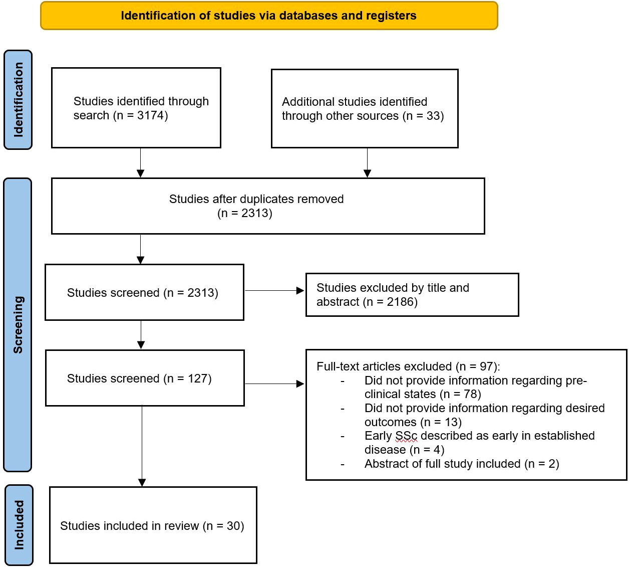
Our search strategy was developed with an experienced information specialist (Supplementary Material). We searched the databases EMBASE and MEDLINE with restrictions for the English language and included peer-reviewed manuscripts as well as conference abstracts. We sought to include studies which provided information regarding the role of adaptive and innate immune systems and the dysregulation of pathways which contributed to the development of classifiable SSc or SLE. Therefore, we included studies which explicitly studied individuals termed as UCTD-risk-SSc, Very early/early SSc, prescleroderma, pre-SLE, incomplete SLE, or lupus-like. Studies were excluded if they provided information regarding inflammatory pathways where patients with established disease were investigated. The search and inclusion of studies was performed by one reviewer (LMC) with review of included studies performed by both authors (LMC & JEP). Our search yielded 2313 manuscripts after duplicates were removed on August 10, 2021 and pertinent manuscripts have been included (Figure 1).
Systemic Sclerosis
Dysregulated Signalling Pathways and Autoimmunity
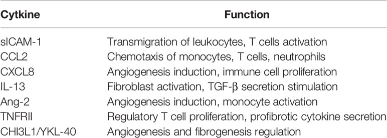
Progressive inflammation, vasculopathy and fibrosis orchestrated by aberrant cytokine production is a hallmark of SSc. Chemokines involved in extracellular matrix deposition, erroneous activation of fibroblasts, and anomalous immune system activation, including CCL2, MIP-1α/CCL3, CCL4, CCL7/MCP-3, and CXCL8, have been observed to be significantly upregulated in the serum of established SSc patients when compared to healthy controls (14–16). However, the presence of these chemokines is more nuanced in pre-clinical disease. Vettori et. al., compared the serum of UCTD-risk-SSc patients to fibromyalgia and/or osteoarthritis controls without RP, and definite SSc patients for soluble intercellular adhesion molecule-1 (sICAM-1), soluble vascular adhesion molecule-1 (sVCAM-1), CCL2, CXCL8, IL-13, IL-33, and transforming growth factor-β (TGF-β) (17). A significant increase was observed in sICAM-1, CCL2, CXCL8, and IL-13 along a disease spectrum gradient from UCTD-risk-SSc to limited cutaneous SSc (lcSSc) to diffuse cutaneous SSc (dcSSc). sICAM-1 is involved in the transmigration of leukocytes from vessels to endothelium and promotes inflammation through T cell activation and cytokine production (18, 19). CXCL8 and CCL2 are pro-fibrotic alter angiogenesis, and affect the migration of monocytes, T cells, and neutrophils (20–22). IL-13 contributes to fibrogenesis through fibroblast activation and TGF-β stimulation (23). Consequently, chemokines increase as disease severity worsens highlighting the progressive derangement of vasculature and autoimmune changes in SSc. Interestingly, higher IL-33 levels were found in UCTD-risk-SSc patients compared to controls and established SSc. IL-33 induces IL-4, IL-5, and IL-13 production leading to arterial vessel media hypertrophy and eosinophilic and mononuclear cell infiltration (24). Therefore, IL-33 functions as a very early mediator in the progression to established SSc, is involved in the fibrotic stage of SSc through IL-13 stimulation; and serves as a predictive marker to elucidate which patients will develop established disease (25).
Other cytokines are abnormal in UCTD-risk-SSc including soluble IL-2 receptor alpha (sIL-2Rα), aminoterminal propeptide of type III collagen (PIIINP), and CXCL4 (7, 26, 27). sIL-2Rα functions as a marker of T-cell activation, whereas PIIINP functions as a marker of collagen formation and fibroblast activation (28, 29). CXCL4 functions as a potent anti-angiogenic chemokine and serves to inhibit endothelial cell proliferation and migration (30). Additionally, CXCL4 has pro-fibrotic capabilities through inhibiting interferon-gamma (IFN-γ) expression and stimulating IL-13 and IL-4 production (31). CXCL4 levels, measured from serum, were higher in UCTD-risk-SSc than controls and were associated with anti-Scl 70 antibodies and sICAM-1 (27, 32). Furthermore, CXCL4 levels, drawn from non-platelet poor plasma, were reported to correlate with extent of skin fibrosis and were predictive of pulmonary arterial hypertension and lung and skin fibrosis progression in SSc (33).
Type I IFN represents another significant contributor to the pathogenesis of SSc through the upregulation of genes involved in the activation of the innate and adaptive immune systems. The increased expression of these type I IFN regulated genes, termed the type I IFN signature, has been previously observed in SLE and other autoimmune diseases (34, 35). Brkic et al., investigated the whole-blood samples of healthy controls without RP, patients with primary RP, UCTD-risk-SSc, and definite SSc patients to determine the expression of 11 type I IFN inducible genes (36). Authors report increased type I IFN related gene expression in UCTD-risk-SSc patients compared to healthy controls, but not in primary RP compared to controls. This finding eludes to the early contribution of the type I IFN pathway in the pathogenesis of SSc. Furthermore, the presence of polymorphisms of IFN regulated genes have been found to confer increased risk of SSc (37).
Vasculopathy and Fibrogenesis
Cossu et al. investigated angiogenetic and endothelial dysfunction markers involved in vasculopathy (38). Authors sampled the serum of healthy controls without RP, UCTD-risk-SSc, lcSSc, and dcSSc patients for angiopoietin-2 (ang-2), CXCL16, e-selectin, sICAM-1, CXCL8, sVCAM-1, and VEGF. There was a significant trend along a disease spectrum from controls to UCTD-risk-SSc to lcSSc and to dcSSc for ang-2, CXCL16, e-selectin, and sICAM-1. Authors also observed a significant difference in ang-2 between controls and UCTD-risk-SSc. Ang-2’s functioning is contextual as it facilitates angiogenesis if VEGF is present, but causes blood vessel regression if pro-angiogenic stimuli are absent (35). Clinically, ang-2 correlates with the extent of skin involvement in SSc as measured by the modified Rodnan skin score (mRSS), disease activity, and C-reactive protein (39). Tabata et. al., found that IGF-1, VEGF, and RANTES levels are significantly higher in mild established SSc compared to pre-clinical SSc (40).
Fibrogenic inflammatory pathways resulting from chronic inflammation and orchestrated through fibroblast dysfunction lead to excessive accumulation of extracellular matrix components, including hyaluronic acid, fibronectin, and proteoglycans, in SSc (41). Sera of healthy controls without RP, UCTD-risk-SSc, and non-fibrotic SSc patients were analyzed whereby elevated markers (CXCL10/IP-10, CXCL11/I-TAC, tumor necrosis factor receptor type II (TNFRII), and chitinase 3-like protein 1) were higher in UCTD-risk-SSc patients compared to controls (42). CXCL10 and CXCL11 are angiostatic and migration chemokines which drive smooth muscle cell proliferation, and recruit T cells, monocytes, and natural killer cells (43–45). Importantly, CXCL10 and CXCL11 levels are associated with UCTD-risk-SSc patients most at risk for developing established SSc (25, 46). Furthermore, CXCL10 and CXCL11 are observed to be correlated with type I IFN signature and decrease with type I IFN receptor blockade with anifrolumab (47).TNFRII has a role in the proliferation and activation of regulatory T cells (48). Additionally, TNFRII co-stimulated lymphocytes secrete pro-fibrotic cytokines in patients with SSc (49). Chitinase 3-like protein 1 has been implicated in regulating and stimulating angiogenesis and fibrogenesis through activation of Syndencan-1 and focal adhesion kinase (50). Furthermore, in SSc patients, chitinase 3-like protein 1 has been correlated with articular involvement and T cell activation (51). These findings highlight the interplay between the adaptive and innate immune systems alongside fibrogenesis.
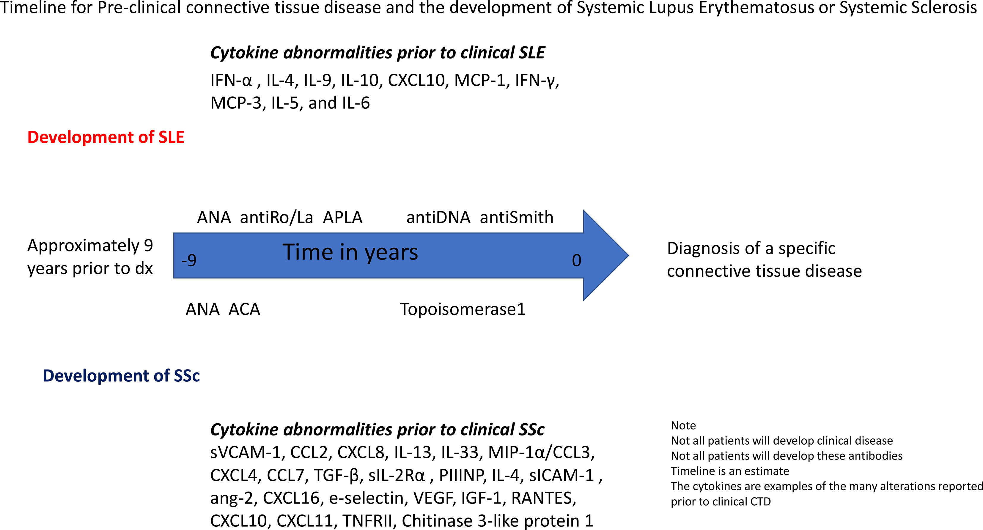
Alterations of natural killer (CD 56+) and natural killer T cells (CD56+ CD3+) in early SSc compared to controls, primary RP, and established SSc were found and thought to be related to differential Toll-like receptor (TLR) 1/2 stimulation (52). Early SSc demonstrated an intermediate activation pattern regarding CD56+ secretion of IL-6, TNF-a, and MIP-1α/CCL3 compared to controls with significant differences of IL-6 secretion. An increasing trend in CD56+ activation for TNF-α and CCL3 occurred between early SSc and controls. This pattern of elevated IL-6, TNF-α, and CCL3 alludes to the role of underlying innate immune mechanisms in prescleroderma or early SSc; which, may eventually lead to established SSc. The development of SSc is shown over time (Figure 2).

Figure 2 Timeline for Pre-clinical connective tissue disease and the development of Systemic Lupus Erythematosus or Systemic Sclerosis. Legend: SLE, Systemic lupus erythematosus; SSc, Systemic sclerosis; ANA, antinuclear antibody; APLA, antiphospholipid antibodies; ACA, anticentromere antibodies.
Systemic Erythematosus Lupus
Autoimmunity and Dysregulated Pathways
Antibodies predate the diagnosis of SLE by multiple years in a characteristic pattern evolving from non-specific ANA to more specific SLE antibodies prior to diagnosis. In a large serology study, a cohort of 130 military personnel who ultimately developed SLE were followed from first detection of ANA to diagnosis of SLE a median of 9.2 years later (11). Furthermore, anti-Ro, anti-La, anti-phospholipid, anti-double stranded DNA, anti-Smith, and anti-nuclear ribonucleoprotein (anti-RNP) antibodies were reported to have a time of first detection to diagnosis of 9.4 years, 8.1 years, 7.6 years, 9.3 years, 8.1 years, and 7.2 years, respectively. This observed pattern, corroborated by further studies, reflects progressive antibody evolution towards more specific SLE antibodies over time in patients ultimately diagnosed with SLE as ANA, anti-double stranded DNA, and anti-Smith antibodies have 86%, 94.7%, and 99% specificity, respectively (53–56). The presence and development of SLE specific antibodies can also serve as predictive makers of developing established disease. Munroe et. al., investigated unaffected blood relatives of SLE patients to identify risk factors of disease establishment (57). Relatives who developed SLE had elevated ANA and anti-Ro titers, and were likely to be anti-dsDNA and anti-RNP positive at baseline and follow up compared to those who did not transition. Anti-cardiolipin antibody positive patients also had more risk of developing SLE (58–60).
Cytokine changes in pre-clinical SLE have been studied (61). Interferon-α, IL-4, IL-9, IL-10, CXCL10 and monocyte chemotactic protein-1 (MCP-1/CCL2) were studied in sera of 35 patients prior to established SLE. CXCL10 was significantly higher in pre-clinical sera compared to controls and was correlated with interferon-α. One of the drivers of innate and adaptive immune dysregulation occurs through an up-regulation of interferon regulated genes, which is also known as the IFN signature of SLE (62). IFN-α, a type I IFN, stimulation leads to increased dendritic cell maturation, increased Th1 cell development and response, and enhanced NK, B, and T cell proliferation and survival (62). IFN-α correlates positively with IgG, and negatively with IgM autoantibodies (63). CXCL10 and IFN-α concentrations are higher in pre-clinical patients who are positive for any antibody compared to antibody negative patients.
Type II IFN (IFN-γ) is additionally implicated in SLE development (64). IFN-γ leads to production of IFN-α and the B-lymphocyte stimulator (BLyS) (65, 66). BLyS, otherwise known as B-cell activating factor of the TNF family (BAFF), is produced by innate immune cells and serves as a mediator of B cell proliferation and survival (67–69). BLyS induces a Th1 cellular response which coordinates both innate and cytotoxic immunity (65). Munroe et al., studied the timing and role of type I and II IFN, IFN-associated mediators, and antibody formation in pre-clinical patients who would later develop established SLE (70). Elevated IFN-γ, CXCL10, and MCP-3 levels occurred prior to IFN-α activity and antibodies. IFN-γ and MCP-3 are abnormal more than 4 years prior to the development of SLE. Therefore, though type I IFN is observed to be elevated in association with antibody positivity prior to SLE diagnosis, type II IFN and IFN-associated mediators seem to represent the pathogenetic intermediaries altering innate and adaptive immune system derangements through elevation of IFN-α and autoantibody formation. These findings agree with a finding that IFN-γ, IL-5, and IL-6 were elevated at least 3.5 years prior to classification (71). Importantly, these observed temporal differences may be secondary to the measurement techniques used in these studies and further investigations with direct measurements tools, such as single-molecule arrays, may further elucidate the temporal relationship between type I and II IFN. Figure 2 shows a timeline for the development of SLE and SSc.
Discussion
Understanding the immunological and inflammatory perturbations involved in the development of CTDs such as SSc and SLE provides clinicians with an opportunity to recognize pre-clinical patients that may benefit from close monitoring, investigations, and potentially early intervention to limit disease progression. Pre-clinical disease states, such as UCTD-risk-SSc, prescleroderma, and incomplete SLE, present with underlying aberrations, often years before clinical disease is present, of the innate and adaptive immune systems, and inflammatory pathways which drive pathogenesis and increase risk of developing established disease.
The pathogenic mechanisms present in UCTD-risk-SSc and prescleroderma include immune signal dysregulations, erroneous immune system recruitment, aberrant angiogenesis leading to vasculopathy, and inappropriate fibroblast activation leading to tissue fibrosis. Multiple cytokines are observed to increase along a disease spectrum from UCTD-risk-SSc to classified SSc and include sICAM-1, CCL2, CXCL8, ang-2, CXCL16, e-selectin, and IL-13 (Table 1). The mechanism of action of these cytokines includes transmigration of lymphocytes endothelium, innate immune cell activation and signal propagation, and extracellular matrix deposition. Furthermore, there are disease markers which are observed to be predictive of SSc and include sIL-2Rα, PIIINP, CXCL4, CXCL10, and CXCL11 (Table 2). Patients with SSc who have the limited cutaneous SSc subset frequently develop RP and anti-centromere antibody 8 years before other manifestations of SSc often followed by dilated nailfold capillaries, then puffy fingers or sclerodactyly and other features of SSc (2–4). The presence or absence of these features is significant in risk stratification where patients with RP but without antibodies or nailfold capillary changes are at 1.8% risk of definite SSc compared to 79.5% in those with RP and positive antibodies and nailfold capillary changes (5). At this point in time, other than treating RP to try to prevent ischemic changes, there is no specific treatment to change the natural history of future development of SSc. Also, 1/3 may develop SSc over the next 5 years (so 2/3 won’t) and this can lead to over-diagnosis, and patient anxiety. Interventions such as smoking cessation and reducing RP attacks and encouraging a healthy lifestyle including a diet high in omega3 fatty acids may be appropriate but this is speculation. Patients with diffuse cutaneous SSc do not develop RP until close to their diagnosis (often 1 to 2 years before or at the time of other signs and symptoms of SSc), so finding prescleroderma clinical features in the majority of these patients has not been possible.
Likewise, SLE development is rooted in aberrations of the innate and adaptive immune systems. Pre-clinical SLE is characterized by an evolving IFN signature and progressive SLE-specific antibody formation prior to disease classification. IFN-γ and IFN associated mediators can predate diagnoses by 3.5 years, and are present prior to and alongside antibody positivity. Throughout pre-clinical SLE, antibody formation occurs in a pattern that evolves from non-specific ANA to more specific SLE antibodies. Namely, ANA and anti-Ro formation can predate diagnosis by 9 years or more but are considered less specific. Whereas, the more specific anti-Smith and anti-dsDNA develop closer to disease onset. The development of SLE specific antibodies can function as predictive markers of transformation to clinical SLE.
Clinically, it is difficult to ascertain what to do with the findings. Other than close monitoring of patients at risk, it is not feasible to check cytokine panels (with high variability) and redoing antibodies is likely not cost effective. However, the changes in immune regulation that predate clinical CTD help in the understanding of pathogenesis and may in future provide targeted treatment for patients with a high probability of converting to chronic debilitating disease. It has been suggested that treating patients at risk for SLE with hydroxychloroquine may change the disease trajectory but large controlled studies are needed to determine if there is benefit in this approach (72); one such multicenter, randomized, placebo-controlled, double-blind clinical trial is currently underway (NCT0303118). Interestingly, there are already drug targets in clinically active SLE targeting signalling that has been shown to be abnormal prior to disease onset such as BlyS (belimumab) and type I interferon with anifrolumab. Intervening prior to clinical disease would not be appropriate with the knowledge we have but in future, personalized medicine may help to give a more robust prediction of who will develop chronic autoimmune CTD.
Conclusion
Ultimately, the coordinated dysregulation of the innate and adaptive immune systems, and inflammatory signalling pathways leads to the pathogenesis of connective tissue disease. Our improved understanding of these underlying aberrations in pre-clinical stages of disease will serve to better identify patients at increased risk.
Author Contributions
LM and JP were involved in study design, review of literature, and manuscript writing. All authors reviewed the manuscript and approved the final version.
Conflict of Interest
The authors declare that the research was conducted in the absence of any commercial or financial relationships that could be construed as a potential conflict of interest.
Publisher’s Note
All claims expressed in this article are solely those of the authors and do not necessarily represent those of their affiliated organizations, or those of the publisher, the editors and the reviewers. Any product that may be evaluated in this article, or claim that may be made by its manufacturer, is not guaranteed or endorsed by the publisher.
Acknowledgments
Assistance with the study: The authors thank Alla Iansavitchene for her contribution as information specialist.
Supplementary Material
The Supplementary Material for this article can be found online at: https://www.frontiersin.org/articles/10.3389/fimmu.2022.869172/full#supplementary-material
References
1. van den Hoogen F, Khanna D, Fransen J, Johnson SR, Baron M, Tyndall A, et al. Classification Criteria for Systemic Sclerosis: An American College of Rheumatology/European League Against Rheumatism Collaborative Initiative. Ann Rheum Dis (2013) 72(11):1747–55. doi: 10.1136/annrheumdis-2013-204424
2. Avouac J, Fransen J, Walker UA, Riccieri V, Smith V, Muller C, et al. Preliminary Criteria for the Very Early Diagnosis of Systemic Sclerosis: Results of a Delphi Consensus Study From EULAR Scleroderma Trials and Research Group. Ann Rheum Dis (2011) 70(3):476–81. doi: 10.1136/ard.2010.136929
3. Valentini G. Undifferentiated Connective Tissue Disease at Risk for Systemic Sclerosis (SSc) (So Far Referred to as Very Early/Early SSc or Pre-SSc). Autoimmun Rev (2015) 14(3):210–3. doi: 10.1016/j.autrev.2014.11.002
4. Matucci-Cerinic M, Bellando-Randone S, Lepri G, Bruni C, Guiducci S. Very Early Versus Early Disease: The Evolving Definition of the ‘Many Faces’ of Systemic Sclerosis. Ann Rheum Dis (2013) 72(3):319–21. doi: 10.1136/annrheumdis-2012-202295
5. Koenig M, Joyal F, Fritzler MJ, Roussin A, Abrahamowicz M, Boire G, et al. Autoantibodies and Microvascular Damage are Independent Predictive Factors for the Progression of Raynaud’s Phenomenon to Systemic Sclerosis: A Twenty-Year Prospective Study of 586 Patients, With Validation of Proposed Criteria for Early Systemic Sclerosis. Arthritis Rheumatol (2008) 58(12):3902–12. doi: 10.1002/art.24038
6. Valentini G, Marcoccia A, Cuomo G, Vettori S, Iudici M, Bondanini F, et al. Early Systemic Sclerosis: Analysis of the Disease Course in Patients With Marker Autoantibody and/or Capillaroscopic Positivity. Arthritis Care Res (Hoboken) (2014) 66(10):1520–7. doi: 10.1002/acr.22304
7. Valentini G, Pope JE. Undifferentiated Connective Tissue Disease at Risk for Systemic Sclerosis: Which Patients Might be Labeled Prescleroderma? Autoimmun Rev (2020) 19(11):102659. doi: 10.1016/j.autrev.2020.102659
8. Kiriakidou M, Ching CL. Systemic Lupus Erythematosus. Ann Intern Med (2020) 172(11):ITC81–96. doi: 10.7326/AITC202006020
9. Mosca M, Costenbader KH, Johnson SR, Lorenzoni V, Sebastiani GD, Hoyer BF, et al. Brief Report: How Do Patients With Newly Diagnosed Systemic Lupus Erythematosus Present? A Multicenter Cohort of Early Systemic Lupus Erythematosus to Inform the Development of New Classification Criteria. Arthritis Rheumatol (2019) 71(1):91–8. doi: 10.1002/art.40674
10. Lambers WM, Westra J, Bootsma H, de Leeuw K. From Incomplete to Complete Systemic Lupus Erythematosus; A Review of the Predictive Serological Immune Markers. Semin Arthritis Rheumatol (2021) 51(1):43–8. doi: 10.1016/j.semarthrit.2020.11.006
11. Arbuckle MR, McClain MT, Rubertone MV, Scofield RH, Dennis GJ, James JA, et al. Development of Autoantibodies Before the Clinical Onset of Systemic Lupus Erythematosus. N Engl J Med (2003) 349(16):1526–33. doi: 10.1056/NEJMoa021933
12. Lambers WM, Westra J, Jonkman MF, Bootsma H, de Leeuw K. Incomplete Systemic Lupus Erythematosus: What Remains After Application of American College of Rheumatology and Systemic Lupus International Collaborating Clinics Criteria? Arthritis Care Res (Hoboken) (2020) 72(5):607–14. doi: 10.1002/acr.23894
13. Eriksson C, Kokkonen H, Johansson M, Hallmans G, Wadell G, Rantapää-Dahlqvist S. Autoantibodies Predate the Onset of Systemic Lupus Erythematosus in Northern Sweden. Arthritis Res Ther (2011) 13(1):R30. doi: 10.1186/ar3258
14. Codullo V, Baldwin HM, Singh MD, Fraser AR, Wilson C, Gilmour A, et al. An Investigation of the Inflammatory Cytokine and Chemokine Network in Systemic Sclerosis. Ann Rheum Dis (2011) 70(6):1115–21. doi: 10.1136/ard.2010.137349
15. Distler JHW, Jüngel A, Caretto D, Schulze-Horsel U, Kowal-Bielecka O, Gay RE, et al. Monocyte Chemoattractant Protein 1 Released From Glycosaminoglycans Mediates its Profibrotic Effects in Systemic Sclerosis via the Release of Interleukin-4 From T Cells. Arthritis Rheumatol (2006) 54(1):214–25. doi: 10.1002/art.21497
16. Yanaba K, Komura K, Kodera M, Matsushita T, Hasegawa M, Takehara K, et al. Serum Levels of Monocyte Chemotactic Protein-3/CCL7 are Raised in Patients With Systemic Sclerosis: Association With Extent of Skin Sclerosis and Severity of Pulmonary Fibrosis. Ann Rheum Dis (2006) 65(1):124–6. doi: 10.1136/ard.2005.040782
17. Vettori S, Cuomo G, Iudici M, D’Abrosca V, Giacco V, Barra G, et al. Early Systemic Sclerosis: Serum Profiling of Factors Involved in Endothelial, T-Cell, and Fibroblast Interplay is Marked by Elevated Interleukin-33 Levels. J Clin Immunol (2014) 34(6):663–8. doi: 10.1007/s10875-014-0037-0
18. McCabe SM, Riddle L, Nakamura GR, Prashad H, Mehta A, Berman PW, et al. sICAM-1 Enhances Cytokine Production Stimulated by Alloantigen. Cell Immunol (1993) 150(2):364–75. doi: 10.1006/cimm.1993.1204
19. Lawson C, Wolf S. ICAM-1 Signaling in Endothelial Cells. Pharmacol Rep (2009) 61(1):22–32. doi: 10.1016/S1734-1140(09)70004-0
20. Russo RC, Garcia CC, Teixeira MM, Amaral FA. The CXCL8/IL-8 Chemokine Family and its Receptors in Inflammatory Diseases. Expert Rev Clin Immunol (2014) 10(5):593–619. doi: 10.1586/1744666X.2014.894886
21. Santos JC, de Brito CA, Futata EA, Azor MH, Orii NM, Maruta CW, et al. Up-Regulation of Chemokine C-C Ligand 2 (CCL2) and C-X-C Chemokine 8 (CXCL8) Expression by Monocytes in Chronic Idiopathic Urticaria. Clin Exp Immunol (2012) 167(1):129–36. doi: 10.1111/j.1365-2249.2011.04485.x
22. Moadab F, Khorramdelazad H, Abbasifard M. Role of CCL2/CCR2 Axis in the Immunopathogenesis of Rheumatoid Arthritis: Latest Evidence and Therapeutic Approaches. Life Sci (2021) 269:119034. doi: 10.1016/j.lfs.2021.119034
23. Fuschiotti P. Role of IL-13 in Systemic Sclerosis. Cytokine (2011) 56(3):544–9. doi: 10.1016/j.cyto.2011.08.030
24. Schmitz J, Owyang A, Oldham E, Song Y, Murphy E, McClanahan TK, et al. IL-33, an Interleukin-1-Like Cytokine That Signals via the IL-1 Receptor-Related Protein ST2 and Induces T Helper Type 2-Associated Cytokines. Immunity (2005) 23(5):479–90. doi: 10.1016/j.immuni.2005.09.015
25. Riccardi A, Borgia A, Fasano S, Messiniti V, Iraace R, Valentini G. Undifferentiated Connective Tissue Disease at Risk for SSc: Potential Role of Circulating CXCL-10, CXCL-11 and IL-33 in Predicting Disease Evolution. Arthritis Rheumatol (2019) 14(3):210–3.
26. Valentini G, Vettori S, Cuomo G, Iudici M, D’Abrosca V, Capocotta D, et al. Early Systemic Sclerosis: Short-Term Disease Evolution and Factors Predicting the Development of New Manifestations of Organ Involvement. Arthritis Res Ther (2012) 14(4):R188. doi: 10.1186/ar4019
27. Valentini G, Riccardi A, Vettori S, Irace R, Iudici M, Tolone S, et al. CXCL4 in Undifferentiated Connective Tissue Disease at Risk for Systemic Sclerosis (SSc) (Previously Referred to as Very Early SSc). Clin Exp Med (2017) 17(3):411–4. doi: 10.1007/s10238-016-0437-y
28. Gonzalez-Lopez L, Rocha-Muñoz AD, Olivas-Flores EM, Garcia-Gonzalez A, Peguero-Gómez AR, Flores-Navarro J, et al. Procollagen Type I and III Aminoterminal Propeptide Levels and Severity of Interstitial Lung Disease in Mexican Women With Progressive Systemic Sclerosis. Archivos Bronconeumología (English Edition) (2015) 51(9):440–8. doi: 10.1016/j.arbr.2014.06.027
29. Witkowska AM. On the Role of sIL-2r Measurements in Rheumatoid Arthritis and Cancers. Mediat Inflamm (2005) 2005(3):121–30. doi: 10.1155/MI.2005.121
30. Vandercappellen J, van Damme J, Struyf S. The Role of the CXC Chemokines Platelet Factor-4 (CXCL4/PF-4) and its Variant (CXCL4L1/PF-4var) in Inflammation, Angiogenesis and Cancer. Cytokine Growth Factor Rev (2011) 22(1):1–18. doi: 10.1016/j.cytogfr.2010.10.011
31. Romagnani P, Maggi L, Mazzinghi B, Cosmi L, Lasagni L, Liotta F, et al. CXCR3-Mediated Opposite Effects of CXCL10 and CXCL4 on T1 or T2 Cytokine Production. J Allergy Clin Immunol (2005) 116(6):1372–9. doi: 10.1016/j.jaci.2005.09.035
32. Valentini G, Riccardi A, Vettori S, Irace R, Ludici M, Tolone S, et al. Serum CXCL4 Increase in Patients With Undifferentiated Connective Tissue Disease at Risk for Systemic Sclerosis Is Associated With Anti-Scl70 Antibodies and ICAM-1, a Marker of Endothelial Activation. Arthritis Rheumatol (2015) 67:3606–7.
33. van Bon L, Affandi AJ, Broen J, Christmann RB, Marijnissen RJ, Stawski L, et al. Proteome-Wide Analysis and CXCL4 as a Biomarker in Systemic Sclerosis. New Engl J Med (2014) 370(5):433–43. doi: 10.1056/NEJMc1402401
34. Higgs BW, Liu Z, White B, Zhu W, White WI, Morehouse C, et al. Patients With Systemic Lupus Erythematosus, Myositis, Rheumatoid Arthritis and Scleroderma Share Activation of a Common Type I Interferon Pathway. Ann Rheum Dis (2011) 70(11):2029–36. doi: 10.1136/ard.2011.150326
35. Tan FK, Zhou X, Mayes MD, Gourh P, Guo X, Marcum C, et al. Signatures of Differentially Regulated Interferon Gene Expression and Vasculotrophism in the Peripheral Blood Cells of Systemic Sclerosis Patients. Rheumatology (2006) 45(6):694–702. doi: 10.1093/rheumatology/kei244
36. Brkic Z, van Bon L, Cossu M, van Helden-Meeuwsen CG, Vonk MC, Knaapen H, et al. The Interferon Type I Signature is Present in Systemic Sclerosis Before Overt Fibrosis and Might Contribute to its Pathogenesis Through High BAFF Gene Expression and High Collagen Synthesis. Ann Rheum Dis (2016) 75(8):1567–73. doi: 10.1136/annrheumdis-2015-207392
37. Skaug B, Assassi S. Type I Interferon Dysregulation in Systemic Sclerosis. Cytokine (2020) 132:154635. doi: 10.1016/j.cyto.2018.12.018
38. Cossu M, Andracco R, Santaniello A, Marchini M, Severino A, Caronni M, et al. Serum Levels of Vascular Dysfunction Markers Reflect Disease Severity and Stage in Systemic Sclerosis Patients. Rheumatology (2016) 55(6):1112–6. doi: 10.1093/rheumatology/kew017
39. Michalska-Jakubus M, Kowal-Bielecka O, Chodorowska G, Bielecki M, Krasowska D. Angiopoietins-1 and -2 are Differentially Expressed in the Sera of Patients With Systemic Sclerosis: High Angiopoietin-2 Levels are Associated With Greater Severity and Higher Activity of the Disease. Rheumatology (2011) 50(4):746–55. doi: 10.1093/rheumatology/keq392
40. Tabata K, Mikita N, Yasutake M, Matsumiya R, Tanaka K, Tani S, et al. Up-Regulation of IGF-1, RANTES and VEGF in Patients With Anti-Centromere Antibody-Positive Early/Mild Systemic Sclerosis. Modern Rheumatol (2021) 31(1):171–6. doi: 10.1080/14397595.2020.1726599
41. Ho YY, Lagares D, Tager AM, Kapoor M. Fibrosis—a Lethal Component of Systemic Sclerosis. Nat Rev Rheumatol (2014) 10(7):390–402. doi: 10.1038/nrrheum.2014.53
42. Cossu M, van Bon L, Preti C, Rossato M, Beretta L, Radstake TRDJ. Earliest Phase of Systemic Sclerosis Typified by Increased Levels of Inflammatory Proteins in the Serum. Arthritis Rheumatol (2017) 69(12):2359–69. doi: 10.1002/art.40243
43. Lacotte S, Brun S, Muller S, Dumortier H. CXCR3, Inflammation, and Autoimmune Diseases. Ann NY Acad Sci (2009) 1173(1):310–7. doi: 10.1111/j.1749-6632.2009.04813.x
44. Kuo PT, Zeng Z, Salim N, Mattarollo S, Wells JW, Leggatt GR. The Role of CXCR3 and Its Chemokine Ligands in Skin Disease and Cancer. Front Med (Lausanne) (2018) 5:271. doi: 10.3389/fmed.2018.00271
45. Bellando Randone S, George J, Mazzotta C, Guiducci S, Furst DE, Mor A, et al. Angiostatic and Angiogenic Chemokines in Systemic Sclerosis: An Overview. J Scleroderma Related Disord (2017) 2(1):1–10. doi: 10.5301/jsrd.5000226
46. Crescioli C, Corinaldesi C, Riccieri V, Raparelli V, Vasile M, del Galdo F, et al. Association of Circulating CXCL10 and CXCL11 With Systemic Sclerosis. Ann Rheum Dis (2018) 77(12):1845–6. doi: 10.1136/annrheumdis-2018-213257
47. Liu X, Mayes MD, Tan FK, Wu M, Reveille JD, Harper BE, et al. Correlation of Interferon-Inducible Chemokine Plasma Levels With Disease Severity in Systemic Sclerosis. Arthritis Rheumat (2013) 65(1):226–35. doi: 10.1002/art.37742
48. Ye L-L, Wei X-S, Zhang M, Niu Y-R, Zhou Q. The Significance of Tumor Necrosis Factor Receptor Type II in CD8+ Regulatory T Cells and CD8+ Effector T Cells. Front Immunol (2018) 9:583. doi: 10.3389/fimmu.2018.00583
49. Hügle T, O’Reilly S, Simpson R, Kraaij MD, Bigley V, Collin M, et al. Tumor Necrosis Factor-Costimulated T Lymphocytes From Patients With Systemic Sclerosis Trigger Collagen Production in Fibroblasts. Arthritis Rheumatol (2013) 65(2):481–91. doi: 10.1002/art.37738
50. Lee CG, da Silva CA, dela Cruz CS, Ahangari F, Ma B, Kang M-J, et al. Role of Chitin and Chitinase/Chitinase-Like Proteins in Inflammation, Tissue Remodeling, and Injury. Annu Rev Physiol (2011) 73:479–501. doi: 10.1146/annurev-physiol-012110-142250
51. la Montagna G, D’Angelo S, Valentini G. Cross-Sectional Evaluation of YKL-40 Serum Concentrations in Patients With Systemic Sclerosis. Relationship with Clinical and Serological Aspects of Disease. J Rheumatol (2003) 30(10):2147–51.
52. Cossu M, van Bon L, Nierkens S, Bellocchi C, Santaniello A, Dolstra H, et al. The Magnitude of Cytokine Production by Stimulated CD56+ Cells is Associated With Early Stages of Systemic Sclerosis. Clin Immunol (2016) 173:76–80. doi: 10.1016/j.clim.2016.09.004
53. Solomon DH, Kavanaugh AJ, Schur PH. American College of Rheumatology Ad Hoc Committee on Immunologic Testing Guidelines. Evidence-Based Guidelines for the Use of Immunologic Tests: Antinuclear Antibody Testing. Arthritis Rheumatol (2002) 47(4):434–44. doi: 10.1002/art.10561
54. Orme ME, Voreck A, Aksouh R, Ramsey-Goldman R, Schreurs MWJ. Systematic Review of anti-dsDNA Testing for Systemic Lupus Erythematosus: A Meta-Analysis of the Diagnostic Test Specificity of an anti-dsDNA Fluorescence Enzyme Immunoassay. Autoimmun Rev (2021) 20(11):102943. doi: 10.1016/j.autrev.2021.102943
55. Flechsig A, Rose T, Barkhudarova F, Strauss R, Klotsche J, Dähnrich C, et al. What is the Clinical Significance of Anti-Sm Antibodies in Systemic Lupus Erythematosus? A Comparison With anti-dsDNA Antibodies and C3. Clin Exp Rheumatol (2017) 35(4):598–606.
56. Slater CA, Davis RB, Shmerling RH. Antinuclear Antibody Testing. A study of clinical utility. Arch Intern Med (1996) 156(13):1421–5. doi: 10.1001/archinte.156.13.1421
57. Munroe ME, Young KA, Kamen DL, Guthridge JM, Niewold TB, Costenbader KH, et al. Discerning Risk of Disease Transition in Relatives of Systemic Lupus Erythematosus Patients Utilizing Soluble Mediators and Clinical Features. Arthritis Rheumatol (2017) 69(3):630–42. doi: 10.1002/art.40004
58. Hallengren CS, Nived O, Sturfelt G. Outcome of Incomplete Systemic Lupus Erythematosus After 10 Years. Lupus (2004) 13(2):85–8. doi: 10.1191/0961203304lu477oa
59. al Daabil M, Massarotti EM, Fine A, Tsao H, Ho P, Schur PH, et al. Development of SLE Among “Potential SLE” Patients Seen in Consultation: Long-Term Follow-Up. Int J Clin Pract (2014) 68(12):1508–13. doi: 10.1111/ijcp.12466
60. Vila L, Mayor A, Valent A, Garc M, Vila S. Clinical Outcome and Predictors of Disease Evolution in Patients With Incomplete Lupus Erythematosus. Lupus (2000) 9(2):110–5. doi: 10.1191/096120300678828073
61. Eriksson C, Rantapää-Dahlqvist S. Cytokines in Relation to Autoantibodies Before Onset of Symptoms for Systemic Lupus Erythematosus. Lupus (2014) 23(7):691–6. doi: 10.1177/0961203314523869
62. Bezalel S, Guri KM, Elbirt D, Asher I, Sthoeger ZM. Type I Interferon Signature in Systemic Lupus Erythematosus. Isr Med Assoc J (2014) 16(4):246–9.
63. Li Q-Z, Zhou J, Lian Y, Zhang B, Branch VK, Carr-Johnson F, et al. Interferon Signature Gene Expression is Correlated With Autoantibody Profiles in Patients With Incomplete Lupus Syndromes. Clin Exp Immunol (2010) 159(3):281–91. doi: 10.1111/j.1365-2249.2009.04057.x
64. Oke V, Gunnarsson I, Dorschner J, Eketjäll S, Zickert A, Niewold TB, et al. And Type III Associate With Distinct Clinical Features of Active Systemic Lupus Erythematosus. Arthritis Res Ther (2019) 21(1):107. doi: 10.1186/s13075-019-1878-y
65. Schroder K, Hertzog PJ, Ravasi T, Hume DA. Interferon-Gamma: An Overview of Signals, Mechanisms and Functions. J Leukoc Biol (2004) 75(2):163–89. doi: 10.1189/jlb.0603252
66. Harigai M, Kawamoto M, Hara M, Kubota T, Kamatani N, Miyasaka N. Excessive Production of IFN-Gamma in Patients With Systemic Lupus Erythematosus and its Contribution to Induction of B Lymphocyte Stimulator/B Cell-Activating Factor/TNF Ligand Superfamily-13B. J Immunol (2008) 181(3):2211–9. doi: 10.4049/jimmunol.181.3.2211
67. Scapini P, Nardelli B, Nadali G, Calzetti F, Pizzolo G, Montecucco C, et al. G-CSF–stimulated Neutrophils Are a Prominent Source of Functional BLyS. J Exp Med (2003) 197(3):297–302. doi: 10.1084/jem.20021343
68. Mackay F, Schneider P, Rennert P, Browning J. BAFF AND APRIL: A Tutorial on B Cell Survival. Annu Rev Immunol (2003) 21:231–64. doi: 10.1146/annurev.immunol.21.120601.141152
69. Schneider P, MacKay F, Steiner V, Hofmann K, Bodmer JL, Holler N, et al. BAFF, a Novel Ligand of the Tumor Necrosis Factor Family, Stimulates B Cell Growth. J Exp Med (1999) 189(11):1747–56. doi: 10.1084/jem.189.11.1747
70. Munroe ME, Lu R, Zhao YD, Fife DA, Robertson JM, Guthridge JM, et al. Altered Type II Interferon Precedes Autoantibody Accrual and Elevated Type I Interferon Activity Prior to Systemic Lupus Erythematosus Classification. Ann Rheum Dis (2016) 75(11):2014–21. doi: 10.1136/annrheumdis-2015-208140
71. Lu R, Munroe ME, Guthridge JM, Bean KM, Fife DA, Chen H, et al. Dysregulation of Innate and Adaptive Serum Mediators Precedes Systemic Lupus Erythematosus Classification and Improves Prognostic Accuracy of Autoantibodies. J Autoimmun (2016) 74:182–93. doi: 10.1016/j.jaut.2016.06.001
Keywords: systemic sclerosis, scleroderma, prescleroderma, pathogenesis, innate immunity, adaptive immunity, systemic lupus erythematosus, autoimmunity
Citation: Martin Calderon L and Pope JE (2022) Precursors to Systemic Sclerosis and Systemic Lupus Erythematosus: From Undifferentiated Connective Tissue Disease to the Development of Identifiable Connective Tissue Diseases. Front. Immunol. 13:869172. doi: 10.3389/fimmu.2022.869172
Received: 04 February 2022; Accepted: 05 April 2022;
Published: 05 May 2022.
Edited by:
David Karp, University of Texas Southwestern Medical Center, United StatesReviewed by:
Zsuzsanna McMahan, Johns Hopkins Medicine, United StatesShervin Assassi, University of Texas Health Science Center at Houston, United States
Copyright © 2022 Martin Calderon and Pope. This is an open-access article distributed under the terms of the Creative Commons Attribution License (CC BY). The use, distribution or reproduction in other forums is permitted, provided the original author(s) and the copyright owner(s) are credited and that the original publication in this journal is cited, in accordance with accepted academic practice. No use, distribution or reproduction is permitted which does not comply with these terms.
*Correspondence: Janet E. Pope, SmFuZXQuUG9wZUBzamhjLmxvbmRvbi5vbi5jYQ==
 Leonardo Martin Calderon
 Janet E. Pope

