 Weiwei Xiang1†
 Chong Xie1†
 Jiaying Luo1†
 Wei Zhang2
 Xinxin Zhao3
 Hong Yang4
 Yu Cai1
 Jie Ding1
 Yishu Wang1
 Yong Hao1Ying Zhang1
 Yangtai Guan1*
- 1Department of Neurology, Ren Ji Hospital, Shanghai Jiao Tong University School of Medicine, Shanghai, China
- 2Department of Ultrasound in Medicine, Shanghai Jiao Tong University Affiliated Sixth People’s Hospital, Shanghai, China
- 3Department of Radiology, Ren Ji Hospital, Shanghai Jiao Tong University School of Medicine, Shanghai, China
- 4Department of Neurology, The First Rehabilitation Hospital of Shanghai, Tongji University School of Medicine, Shanghai, China
A Corrigendum on
Low Frequency Ultrasound With Injection of NMO-IgG and Complement Produces Lesions Different From Experimental Autoimmune Encephalomyelitis Mice
By Xiang W, Xie C, Luo J, Zhang W, Zhao X, Yang H, Cai Y, Ding J, Wang Y, Hao Y, Zhang Y, Guan Y (2021). Front. Immunol. 12:4150. doi: 10.3389/fimmu.2021.727750
In the original article, there was a mistake in Figures 5 and 6 as published. The figures in their previous form do not best reflect the results. The corrected Figures 5 and 6 appear below.
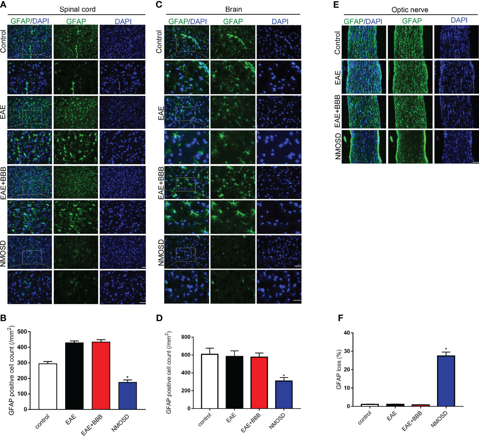
Figure 5 Loss of GFAP expression in the NMOSD model (A, B) Loss of GFAP expression in the spinal cord and statistic results, scale bar = 50μm; (C, D) Loss of GFAP expression in the brain and statistic results, scale bar = 50μm; (E, F) Loss of GFAP expression in the optic nerve and statistic results, scale bar = 50μm; Scale bar = 50μm. The experiment was repeated twice, with similar results. Data were presented as the mean ± SEM; *P < 0.05 vs control group; n = 6 in each group. LSD-t test was used.
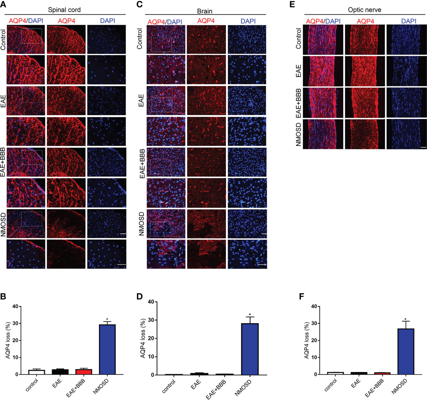
Figure 6 Loss of AQP4 expression in the NMOSD model (A, B) Loss of AQP4 expression in the spinal cord and statistic results, scale bar = 50μm; (C, D) Loss of AQP4 expression in the brain and statistic results, scale bar = 50μm; (E, F) Loss of AQP4 expression in the optic nerve and statistic results, scale bar = 50μm; The experiment was repeated twice, with similar results. Data were presented as the mean ± SEM; *P < 0.05 vs control group; n = 6 in each group. LSD-t test was used.
The authors apologize for this error and state that this does not change the scientific conclusions of the article in any way. The original article has been updated.
Publisher’s Note
All claims expressed in this article are solely those of the authors and do not necessarily represent those of their affiliated organizations, or those of the publisher, the editors and the reviewers. Any product that may be evaluated in this article, or claim that may be made by its manufacturer, is not guaranteed or endorsed by the publisher.
Keywords: neuromyelitis optica, mouse, Aquaporin-4, blood-brain barrier, low-frequency ultrasound
Citation: Xiang W, Xie C, Luo J, Zhang W, Zhao X, Yang H, Cai Y, Ding J, Wang Y, Hao Y, Zhang Y and Guan Y (2022) Corrigendum: Low Frequency Ultrasound With Injection of NMO-IgG and Complement Produces Lesions Different From Experimental Autoimmune Encephalomyelitis Mice. Front. Immunol. 13:842300. doi: 10.3389/fimmu.2022.842300
Received: 23 December 2021; Accepted: 04 January 2022;
Published: 25 January 2022.
Edited and reviewed by:
Sonja Hochmeister, Medical University of Graz, AustriaCopyright © 2022 Xiang, Xie, Luo, Zhang, Zhao, Yang, Cai, Ding, Wang, Hao, Zhang and Guan. This is an open-access article distributed under the terms of the Creative Commons Attribution License (CC BY). The use, distribution or reproduction in other forums is permitted, provided the original author(s) and the copyright owner(s) are credited and that the original publication in this journal is cited, in accordance with accepted academic practice. No use, distribution or reproduction is permitted which does not comply with these terms.
*Correspondence: Yangtai Guan, eWFuZ3RhaWd1YW5Ac2luYS5jb20=
†These authors have contributed equally to this work