 Michael Horckmans1
 Esteban Diaz Villamil
 Mariaelvy Bianchini
 Didier Communi
95% of researchers rate our articles as excellent or good
Learn more about the work of our research integrity team to safeguard the quality of each article we publish.
Find out more
CORRECTION article
Front. Immunol. , 28 October 2022
Sec. Inflammation
Volume 13 - 2022 | https://doi.org/10.3389/fimmu.2022.1061958
A Corrigendum on
Central role of PD-L1 in cardioprotection resulting from P2Y4 nucleotide receptor loss
By Horckmans M, Diaz Villamil E, Bianchini M, De Roeck L and Communi D (2022). 13:1006934. doi: 10.3389/fimmu.2022.1006934
In the published article, there was an error in the author list, and author Mariaelvy Bianchini was erroneously excluded. The corrected author list appears below.
Michael Horckmans1, Esteban Diaz Villamil1, Mariaelvy Bianchini2, Lucas De Roeck1, Didier Communi1
This author’s affiliation has been added: “Institute for Cardio-vascular Prevention, Ludwig-Maximilians-Universität (LMU), Munich, Germany”.
Accordingly, a correction has been made to Author contributions.
This sentence previously stated:
“MH and DC designed research study and analyzed data; MH, EDV, and LDR conducted experiments and acquired data, M.H. and D.C. wrote the manuscript.”
The corrected sentence appears below:
“MH and DC designed research study and analyzed data; MH, EDV, MB, and LDR conducted experiments and acquired data, MH and DC wrote the manuscript.”
A correction has also been made to Acknowledgments.
This sentence previously stated:
“The authors thank Mariaelvy Bianchini and Frédérick Libert for technical advice and help related to respectively, PD-L1 immunofluorescent reconstructions and RNA-sequencing experiments.”
The corrected sentence appears below:
“The authors thank Frédérick Libert for technical advice and help related to RNA-sequencing experiments.”
In the published article, there was also an error in the legend for Figure 2, panels E–F as published, due to errors in microscopy device and magnification.
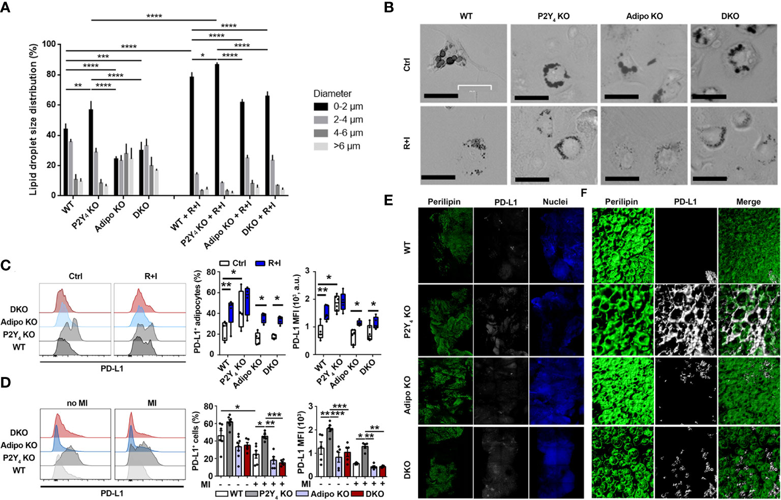
Figure 2 Identification of PD-L1 as a marker of beige adipocytes, upregulated in P2Y4 KO PAT. (A) Quantification of lipid droplet size in adipocytes generated from WT, P2Y4 KO, Adipo KO and DKO cADSCs in the presence or absence of R+I treatment. Quantification was performed using ImageJ software (n = 4). (B) Oil red O staining of cardiac adipose-derived stem cells (cADSC) cultures after adipogenic differentiation and after treatment with two adipocyte browning agents, rosiglitazone (1 µM) plus isoproterenol (100 µM) (R+I) (200x magnification). Adipogenic differentiation leads to big lipid droplet appearance in control (Ctrl) cADSC cultures. The additional presence of browning agents (R+I) in the differentiation medium leads to small lipid droplet appearance in cADSC cultures. (C) Higher PD-L1 expression in P2Y4 KO differentiated cADSCs. Density plots, percentage of PD-L1 positive cells and mean fluorescence intensities (MFI) obtained by flow cytometry quantification of PD-L1 expression in differentiated adipocytes isolated from PAT of WT, P2Y4 KO, Adipo KO and DKO mice, with or without R+I (n = 4-6). (D) Flow cytometry analysis showing increased level of PD-L1 expression in P2Y4 KO PAT. Density plots, percentage of PD-L1 positive cells and mean fluorescence intensities (MFI) detected by flow cytometry in PAT of sham (no MI) or ischemic (MI) WT, P2Y4 KO, Adipo KO and DKO mice, 24h post-MI (n = 5-7). (E, F). Loss of P2Y4 leads to increased PD-L1 immunohistological staining in ischemic PAT. (E) Immunofluorescent stainings of perilipin lipid droplet marker, and PD-L1 performed on optically cleared whole-mounted PAT of WT, P2Y4 KO, Adipo KO and DKO ischemic mice, 24h post-MI (5X magnification). (F) Representative confocal microscopy reconstructions of stained and optically cleared whole-mounted PAT (20X magnification).
The legend previously said: “E. Immunofluorescent stainings of perilipin lipid droplet marker, and PD-L1 performed on PAT sections of WT, P2Y4 KO, Adipo KO and DKO ischemic mice, 24h post-MI (10x magnification). F. Representative two-photon laser scanning microscopy reconstructions of stained PAT sections (100x magnification).”
The corrected legend appears below.
In the published article, there was an error in the Funding statement. A funding source was missing. The correct Funding statement appears below.
This work was supported by Research Project and Research Credit of the Fonds National de la Recherche Scientifique of Belgium (J.0060.18 CDR grant), by an ATIMI (Attract Brains for Brussels, Belgium) grant of Innoviris Brussels (2019-BFB-106 ATIMI grants), by the Fonds pour la Chirurgie Cardiaque, by the Fund Lokumo, King Baudouin Foundation, Belgium (2017-B7131100-207336 grant), by the Deutsche Forschungsgemeinschaft grants SFB1123/Z1 and INST409/150-1FUGG and by the Fonds et Crédit d’Encouragement à la Recherche (F.E.R./C.E.R., Free University of Brussels (U.L.B.)). MH was supported by ATIMI (Attract Brains for Brussels, Belgium) grants of Innoviris Brussels. EDV was supported by the F.R.I.A., Fonds National de la Recherche Scientifique, Belgium. LDR is supported by U.L.B., Belgium. DC is Senior Research Associate of the Fonds National de la Recherche Scientifique (F.N.R.S.). The funders had no role in study design, data collection and analysis, decision to publish, or preparation of the manuscript.
In the published article, there were errors in the used microscopy methodology related to PD-L1 and perilipin staining. Appropriate changes have been made in the microscopy device and method (two-photon laser scanning microscopy was replaced by confocal microscopy reconstructions) in a paragraph of the Materials and methods.
A correction has been made to Materials and methods, “Immunofluorescence experiments”, paragraph 1. These sentences previously stated:
“Frozen sections (5 µm) of mouse PAT were stained with antibodies against Programmed death ligand 1 (PD-L1) (BioLegend, San Diego, CA, USA) and perilipin (Sigma-Aldrich). For some experiments of PD-L1 and perilipin staining, two-photon laser scanning microscopy (TPLSC) was performed on a Leica SP5II MP equipped with Ti : Sa pulsed laser (Spectra-Physics, MKS Instruments, Andover, MA, USA) tuned at 800 nm.”
The corrected sentences appear below:
“Whole-mounted mouse PAT was fixed in 2% PFA, stained overnight with antibodies against Programmed death ligand 1 (PD-L1) (BioLegend, USA) and perilipin (Abcam, UK), then optically cleared with RapiClear 1.47 (SUNJin Lab, South Korea) for 6 hours. Samples were acquired as tile-scan overviews with a Leica Thunder Imager (Leica Microsystems, Germany). For higher optical resolution imaging, 3D image stacks were acquired using a Leica SP8 3X confocal microscope (Leica Microsystems, Germany) equipped with a tunable white light laser. Image 3D reconstructions were generated with Imaris 8 (Oxford Instruments, UK).”
In the published article, there were errors related to the used microscopy methodology (two-photon laser scanning microscopy was replaced by confocal microscopy reconstructions) related to PD-L1 and perilipin staining in the Results.
A correction has been made to Results, “PD-L1 is a marker of beige adipocytes up-regulated in the absence of P2Y4 receptor”, paragraph 2. These sentences previously stated:
“Immunohistological stainings of perilipin, a lipid droplet marker, and PD-L1 were then performed on PAT sections of WT, P2Y4 KO, adiponectin KO and DKO ischemic mice, 24h post-MI (Figure 2E). Two-photon laser scanning microscopy (TPLSM) reconstructions of perilipin and PD-L1 stainings were also made at higher magnification on PAT sections of ischemic mice, 24h post-MI (Figure 2F).”
The corrected sentences appear below:
“Immunohistological stainings of perilipin, a lipid droplet marker, and PD-L1 were then performed on whole-mounted PAT of WT, P2Y4 KO, adiponectin KO and DKO ischemic mice, 24h post-MI (Figure 2E). Confocal microscopy reconstructions of perilipin and PD-L1 stainings were also made at higher magnification on optically cleared whole mounted PAT of ischemic mice, 24h post-MI (Figure 2F).”
The authors apologize for these errors and state that they do not change the scientific conclusions of the article in any way. The original article has been updated.
All claims expressed in this article are solely those of the authors and do not necessarily represent those of their affiliated organizations, or those of the publisher, the editors and the reviewers. Any product that may be evaluated in this article, or claim that may be made by its manufacturer, is not guaranteed or endorsed by the publisher.
Keywords: cardioprotection, cardiac adipose tissue, PD-L1, P2Y receptor, adiponectin, ischemia
Citation: Horckmans M, Diaz Villamil E, Bianchini M, De Roeck L and Communi D (2022) Corrigendum: Central role of PD-L1 in cardioprotection resulting from P2Y4 nucleotide receptor loss. Front. Immunol. 13:1061958. doi: 10.3389/fimmu.2022.1061958
Received: 05 October 2022; Accepted: 18 October 2022;
Published: 28 October 2022.
Edited and Reviewed by:
Lisardo Bosca, Autonomous University of Madrid, SpainCopyright © 2022 Horckmans, Diaz Villamil, Bianchini, De Roeck and Communi. This is an open-access article distributed under the terms of the Creative Commons Attribution License (CC BY). The use, distribution or reproduction in other forums is permitted, provided the original author(s) and the copyright owner(s) are credited and that the original publication in this journal is cited, in accordance with accepted academic practice. No use, distribution or reproduction is permitted which does not comply with these terms.
*Correspondence: Didier Communi, RGlkaWVyLkNvbW11bmlAdWxiLmJl
Disclaimer: All claims expressed in this article are solely those of the authors and do not necessarily represent those of their affiliated organizations, or those of the publisher, the editors and the reviewers. Any product that may be evaluated in this article or claim that may be made by its manufacturer is not guaranteed or endorsed by the publisher.
Research integrity at Frontiers
Learn more about the work of our research integrity team to safeguard the quality of each article we publish.