 Dunja Bijelić1
 Marija Adžić1
 Mina Perić1
 Igor Jakovčevski2
 Eckart Förster2
 Melitta Schachner3*
 Pavle R. Andjus1*
- 1Centre for Laser Microscopy, Faculty of Biology, Institute of Physiology and Biochemistry “Jean Giaja”, University of Belgrade, Belgrade, Serbia
- 2Institut für Neuroanatomie und Molekulare Hirnforschung, Ruhr-Universität Bochum, Bochum, Germany
- 3Keck Center for Collaborative Neuroscience and Department of Cell Biology and Neuroscience, Rutgers University, Piscataway, NJ, United States
A Corrigendum on
Different Functions of Recombinantly Expressed Domains of Tenascin-C in Glial Scar Formation
Bijelić, D., Adžić, M., Perić, M., Jakovčevski, I., Förster, E., Schachner, M., and Andjus, P. R. (2021). Front. Immunol. 11(3944). doi: 10.3389/fimmu.2020.624612
In the original article, there was a mistake in Figure 2 as published. Instead of micrograph “+ FnA”, the micrograph for “+ Fn(D+A)” treatment in TnC +/+ genotype was duplicated by mistake. We have inserted the correct micrograph for “+ FnA”. The corrected Figure 2 appears below.
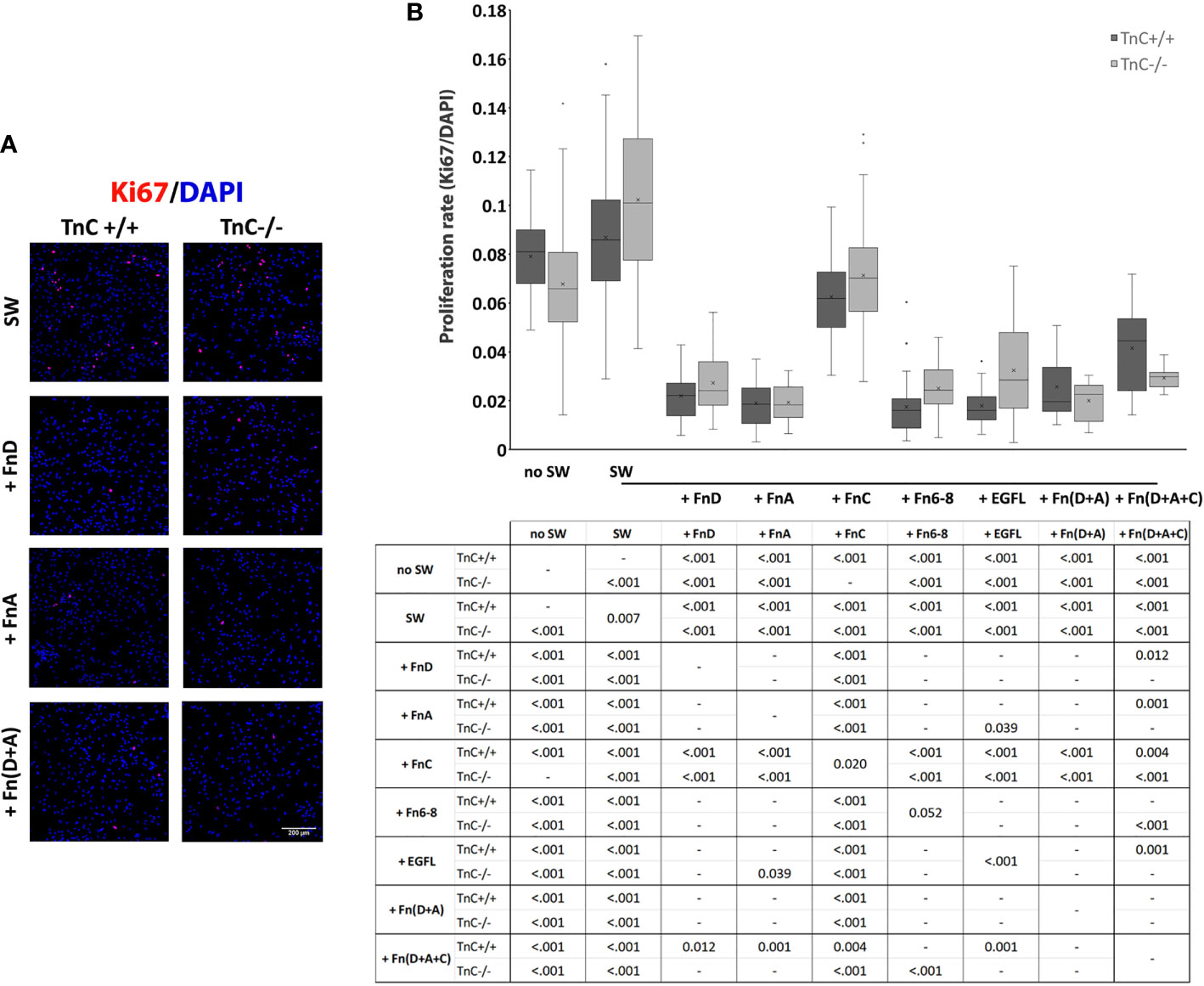
Figure 2 TnC fragments reduce proliferation in the astrocyte scratch wound assay. (A) Representative micrographs of Ki67+/DAPI+ immunofluorescence at 24 h after scratching in the control group and groups treated with FnA, FnD, and Fn(D+A); bar: 200 µm. (B) Proliferation was calculated as the number of Ki67+ nuclei compared to total DAPI+ nuclei. Results are presented as a box-and-whisker plot. Two-way ANOVA analysis shows a statistically significant interaction between the effects of genotype and treatment on cell proliferation rate (p = 0.005) with both the effects of genotype and treatment being significant (p = 0.040, p < 0.001, respectively). A statistically significant decrease in proliferation is seen in the presence of FnA, FnD, and Fn(D+A). All statistically significant pairwise comparisons are displayed below the box-and-whisker plot. n=3 independent astrocyte cultures.
The authors apologize for this error and state that this does not change the scientific conclusions of the article in any way. The original article has been updated.
Keywords: astrocyte, glial scar, microglia/macrophages, spinal cord injury, tenascin-C
Citation: Bijelić D, Adžić M, Perić M, Jakovčevski I, Förster E, Schachner M and Andjus PR (2021) Corrigendum: Different Functions of Recombinantly Expressed Domains of Tenascin-C in Glial Scar Formation. Front. Immunol. 12:672476. doi: 10.3389/fimmu.2021.672476
Received: 25 February 2021; Accepted: 08 March 2021;
Published: 16 March 2021.
Edited and reviewed by: Kyoko Imanaka-Yoshida, Mie University, Japan
Copyright © 2021 Bijelić, Adžić, Perić, Jakovčevski, Förster, Schachner and Andjus. This is an open-access article distributed under the terms of the Creative Commons Attribution License (CC BY). The use, distribution or reproduction in other forums is permitted, provided the original author(s) and the copyright owner(s) are credited and that the original publication in this journal is cited, in accordance with accepted academic practice. No use, distribution or reproduction is permitted which does not comply with these terms.
*Correspondence: Pavle R. Andjus, cGFuZGp1c0BiaW8uYmcuYWMucnM=; Melitta Schachner, c2NoYWNobmVyQGRscy5ydXRnZXJzLmVkdQ==