 Karen Toledo-Stuardo1Carolina H. Ribeiro1Andrea Canals2,3Marcela Morales1Valentina Gárate1Jose Rodríguez-Siza1Samantha Tello1Marco Bustamante4Ricardo Armisen5Douglas J. Matthies6Gerald Zapata-Torres6Patricio González-Hormazabal7María Carmen Molina1*
- 1Immunology Program, Faculty of Medicine, Institute of Biomedical Sciences (ICBM), University of Chile, Santiago, Chile
- 2Biostatistics Program, School of Public Health, University of Chile, Santiago, Chile
- 3Academic Direction, Clínica Santa María, Santiago, Chile
- 4Department of Surgery (Oriente), Hospital del Salvador, University of Chile, Santiago, Chile
- 5Center of Genetics and Genomics, Faculty of Medicine Clínica Alemana, Institute for Sciences and Innovations in Medicine (ICIM), Universidad del Desarrollo, Santiago, Chile
- 6Department of Inorganic and Analytical Chemistry, Faculty of Chemical and Pharmaceutical Sciences, University of Chile, Santiago, Chile
- 7Human Genetics Program, Institute of Biomedical Sciences (ICBM), University of Chile, Santiago, Chile
Gastric cancer (GC) is the fifth most prevalent type of cancer worldwide. Gastric tumor cells express MICA protein, a ligand to NKG2D receptor that triggers natural killer (NK) cells effector functions for early tumor elimination. MICA gene is highly polymorphic, thus originating alleles that encode protein variants with a controversial role in cancer. The main goal of this work was to study MICA gene polymorphisms and their relationship with the susceptibility and prognosis of GC. Fifty patients with GC and 50 healthy volunteers were included in this study. MICA alleles were identified using Sanger sequencing methods. The analysis of MICA gene sequence revealed 13 MICA sequences and 5 MICA-short tandem repeats (STR) alleles in the studied cohorts We identified MICA*002 (*A9) as the most frequent allele in both, patients and controls, followed by MICA*008 allele (*A5.1). MICA*009/049 allele was significantly associated with increased risk of GC (OR: 5.11 [95% CI: 1.39–18.74], p = 0.014). The analysis of MICA-STR alleles revealed a higher frequency of MICA*A5 in healthy individuals than GC patients (OR = 0.34 [95% CI: 0.12–0.98], p = 0.046). Survival analysis after gastrectomy showed that patients with MICA*002/002 or MICA*002/004 alleles had significantly higher survival rates than those patients bearing MICA*002/008 (p = 0.014) or MICA*002/009 (MICA*002/049) alleles (p = 0.040). The presence of threonine in the position MICA-181 (MICA*009/049 allele) was more frequent in GC patients than controls (p = 0.023). Molecular analysis of MICA-181 showed that the presence of threonine provides greater mobility to the protein than arginine in the same position (MICA*004), which could explain, at least in part, some immune evasion mechanisms developed by the tumor. In conclusion, our findings suggest that the study of MICA alleles is crucial to search for new therapeutic approaches and may be useful for the evaluation of risk and prognosis of GC and personalized therapy.
Introduction
Gastric cancer (GC) is the fifth most common neoplasm and the third leading cause of cancer-related death worldwide (1), accounting for 783,000 deaths in 2018 according to GLOBOCAN data.
The absence of early clinical tools for GC diagnosis decreases the success of therapies and survival of patients, which raises the question of the necessity to explore novel biological biomarkers of the disease (2–4). In this context, the major histocompatibility complex class I-related protein A (MICA) may provide relevant information about the pathological changes in the gastrointestinal mucosa. The healthy gastric mucosa express low levels of this protein, while the tumor tissue overexpresses it (5–7). MICA expression in the carcinogenesis process is regulated by transcriptional, translational and/or post-translational modification mechanisms (8, 9) that could be associated with Helicobacter pylori or Epstein Barr-virus infection (10, 11).
MICA is a ligand to natural killer group 2D (NKG2D), an activating receptor (12) that is important for the anti-tumor immune response (13). NKG2D is expressed by cytotoxic lymphocytes, including natural killer (NK) cells, γδ T cells, and CD8+ T cells. NK cells constitute the first line of defense against intracellular pathogens; they also contribute to the maintenance of mucosal homeostasis and the development of efficient immune responses against cancer (14). NKG2D binding to MICA on target cells triggers NK cell cytolytic activation, which results in target cell lysis through the release of granzyme and perforin by the effector cell (15). However, tumors have developed several strategies to evade the immune response, such as the proteolytic shedding of MICA from the cell membrane (13). The resulting soluble form of MICA induces the downregulation of NKG2D receptor on NK cells, compromising the immune response mediated by these and other cytolytic cells (16).
The human MICA gene is highly polymorphic; indeed, it has been reported more than 110 MICA alleles that encode over 100 protein variants (http://hla.alleles.org/data/mica.html). The human MICA gene is located in chromosome 6p21.3, 46 kb distant from HLA-B, and consists of six exons: exon 1–6 encode the leader peptide, the three extracellular protein domains (α1, α2, and α3), the transmembrane region, and the hydrophobic cytoplasmatic tail, respectively (17). Exon 5 contains a short tandem repeat (STR) with a variable number of GCT triplet repeats, which encode alanine (Ala). A second nomenclature for MICA alleles, MICA-STR (18), has thus emerged based on the presence of these repetitions; for instance, MICA*A5 consists of five repetitions of GCT. Such STR affects the length of the transmembrane region, as in the case of MICA*A9, which contains nine GCT repetitions, which consequently codifies nine residues of alanine (18). Interestingly, the MICA*A5.1 (prototype MICA*008) variant produces an insertion of guanine at position 952 (Chr6: 31380161-31380162 on Assembly GRCh37) in the transmembrane region, changing the reading frame to a pre-mature stop codon and generating a protein with a GPI anchor, which results in the recruitment of MICA to exosomes and release of the protein in the form of extracellular vesicles (EVs) (19, 20).
MICA polymorphisms have been previously studied in cancer, especially the MICA-129 residue, which is associated to the presence of methionine (Met) or valine (Val), where MICA-129 Met has shown a strong interaction with NKG2D, leading to the downregulation of the receptor more efficiently than MICA-129 Val (21). On the other hand, MICA-129 Val has been associated to an increase in soluble MICA (sMICA) levels in multiple myeloma, which induces NKG2D downregulation and contributes to immune evasion (16). Furthermore, several MICA transmembrane regions, based on the GCT repetitions, have been associated to higher susceptibility to certain types of cancers. For instance, MICA*A9 allele has been proposed to confer a risk for GC (22), while MICA*A5.1 allele may be associated with increased susceptibility to oral squamous cell carcinoma in Japanese patients (23). Since MICA alleles vary among human populations and generate proteins with different biological properties that may result in variable disease susceptibility, we decided to study MICA gene polymorphisms and susceptibility to gastric cancer and their relationship with the tumor progression.
Materials and Methods
Patients and Healthy Controls Samples
Gastric Cancer Patients Tissue Samples
During June 2011 and August 2015, a total of 50 patients (17 female, 33 male) aged 65.4 ± 10.3 years (range, 49–78 years) with confirmed diagnosis of gastric adenocarcinoma and treated at the Department of Gastrointestinal Surgery, Hospital del Salvador (Santiago, Chile), were enrolled in this study. Fresh primary gastric tumor tissue samples were obtained at the time of surgery and immediately processed for genomic DNA extraction.
Healthy Controls Blood Samples
Blood samples were obtained from 50 healthy donors (24 female, 26 male) aged 47.3 ± 15.6 years (range, 30–61 years) without prior gastrointestinal diseases or any type of cancer, who lived in Santiago, Chile. Blood samples were collected in 4 mL EDTA-containing vacutainer tubes (BD Biosciences, San José, CA, USA) for genomic DNA extraction.
Ethical Considerations
This study was approved by the Committee on Human Ethics Investigation of the Faculty of Medicine, University of Chile, and the Committee on Scientific Ethics of the Metropolitan Health Service of the Chilean Government. All patients and healthy volunteers signed a written Informed Consent for tissue and blood donation. Name and identification of patients and controls were omitted and rendered anonymous before data analysis.
Clinicopathological Data
The clinicopathological information included patient gender, age, primary tumor location, tumor size, Lauren's histological classification, Bormann classification (24), lymph node metastasis and tumor stage according to the American Joint Committee on Cancer, AJCC, 7th edition (25). Histopathological analysis was carried out by the team of pathologists from Hospital del Salvador (Santiago, Chile). The histological differentiation grade was based on the World Health Organization (WHO) classification (26). Patient survival was assessed for 36 months after surgery or until death due to tumor-specific disease.
Extraction and Purification of Genomic DNA
The genomic DNA was obtained from ~30 mg of tumor tissue using the E.Z.N.A Tissue DNA kit (Omega, Bio-tek, USA), following the manufacturer's instructions. The genomic DNA from blood samples was isolated using the salting out method previously described by Subbarayan PR and collaborators (27).
Measurement of DNA Purity and Integrity
The genomic DNA purity was assessed according to the mean 260/280 nm ratio in a Synergy™ microplate reader (Biotek, Winooski, VT, USA). The genomic DNA integrity was verified by electrophoresis using a 1% agarose gel (40 mg of analytical grade Agarose, 40 mL of Tris-borate-EDTA buffer) with 0.5 μL of ethidium bromide solution (Sigma-Aldrich/Merck KGaA, Darmstadt, Germany). The DNA bands were visualized using a UV transilluminator.
MICA Genotyping
To amplify the MICA gene of GC patients and healthy volunteers by PCR, we used two pairs of specific primers to 2-3 and 4-5 exons of the MICA gene, and MICA genotyping was carried out using bidirectional Sanger sequencing methods. Forward primer (2-3 exons): 5′ TGAAATCCTCGTTCTTGTCCCTTTGC 3′, Reverse primer (2-3 exons): 5′ AGGGTCCTCTACTTGCCCTGATTAC 3′; Forward primer (4-5 exons): 5′ TCAGCCAGAGTGAGAACAGTGAAGA 3′, Reverse primer (4-5 exons): 5′ TCATCCCCTGTTATGGAAGCCTTGTC 3′. The PCR reactions were analyzed by 0.8% agarose gel electrophoresis. The amplicons were purified using the Wizard SV Gel and PCR Clean-Up System Purification Kit (Promega, USA). The amplified products were sequenced on an ABI PRISM-3500 XL sequencer (Applied Biosystems, Foster City, CA). The genomic sequences of each sample were analyzed using Chromas 2.4 Viewer and were manually verified. These sequences were then compared with MICA allelic genotype sequences obtained from the IPD-IMGT/HLA database (28) (http://hla.alleles.org/data/mica.html). MICA allelic genotypes were obtained according to reference sequences of specific MICA alleles. The presence of heterozygous alleles in the sequences was assigned by Chromas and verified in the electropherogram. The exon 5 was carefully analyzed, manually, due to the presence of STR or microsatellites with different lengths, especially in heterozygous patients, as Sanger sequencing of this exon frequently results in base overlap. To validate our methodology, we analyzed two available cell lines with known MICA alleles, which included AGS ATCC CRL-1739 human gastric cancer cell line (which is homozygous for MICA*010) (29) and PC-3 ATCC CRL-1435 human prostate cancer cell line (which is heterozygous for MICA*008 and *012) (30). MICA*009 and MICA*049 alleles differ in one amino acid in the exon 6. Since the differential residue was not distinguished in this study, this allele was named as MICA*009/049; accordingly, allele MICA*002/009 was also named MICA*002/049.
Molecular Analysis
MICA protein sequence and its isoforms were retrieved from UniProtKB1 (31) (https://www.uniprot.org) in its FASTA format. A protein-protein BLAST (32) (http://blast.ncbi.nlm.nih.gov) was further performed to search for a protein template with the highest sequence identity to MICA, which resulted in PDB code 1HYR (17) with a 2.7 Å resolution. A sequence alignment with this template was then carried out using Clustal Omega server (33) (https://www.ebi.ac.uk). This target sequence alignment was fed on the Swiss-Model server (34) (https://swissmodel.expasy.org) to build the models of the proteins, which were then solvated in silico with water using a rectangular shape and neutralized with ions to a concentration of 0.15 M of NaCl to mimic physiological environment using the CHARMM-GUI server (35). Periodic boundary conditions were applied, and a minimization procedure was performed using a maximum number of 5,000 steps of conjugated gradient followed by 2,500 steps of steepest descent run. Next, the NVT ensemble was used for subsequent equilibration steps at 303.15 K using a timestep of 0.001 ps for 125 ps. NPT dynamic runs were used for production runs with a timestep of 0.004 ps for a period of 500 ns. The simulations were carried out using Amber14 (36) suite of programs.
Statistical Analysis
Descriptive analysis of demographic data and clinicopathological characteristics of cases and controls were performed using median and interquartile range for quantitative variables and absolute and percentage frequency distributions for categorical variables.
The distribution of MICA-sequence alleles and MICA-STR alleles among cases and controls, and the proportion of each allele among cases and controls were compared using Fisher's Exact Test. In addition, logistic regressions were adjusted for the prediction of GC by comparing each allele with the others, obtaining the odds ratio (OR) with their respective confidence intervals (95%, CI).
The distribution of genotypes of each MICA residue between cases and controls was also compared using Fisher's Exact Test.
Survival curves of GC patients, according to the presence of MICA alleles, were obtained using the Kaplan-Meier method and compared using the Log-rank test.
Statistical analysis was performed using Stata 14 software, and a p-value < 0.05 was considered significant.
Results
Clinicopathological Characteristics of GC Patients
Patients demographic characteristics and clinicopathological features of tumors are described in Table 1. Tumor size was given as the maximum tumor diameter measured on the freshly resected stomach. Most of GC patients (64%) presented with a tumor size higher than 5 cm. In 74.5% of patients, the tumors had cardia location. According to Lauren's histological classification, 27 out of 50 patients had intestinal type GC, 13 had diffuse type GC, and 10 patients presented with mixed type GC. Borrmann's classification showed that the gastric tumors were mainly at stage III (46%), followed by stage IV (11%), which is related to the TNM staging classification, where 41 patients were found at stage III-IV. Additionally, 46% of the patients showed Helicobacter pylori infection.
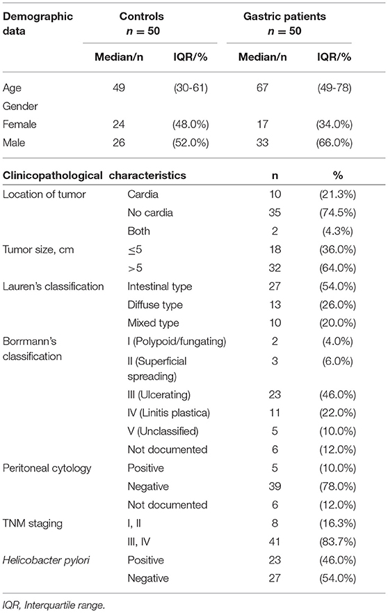
Table 1. Demographic data and clinicopathological characteristics of gastric cancer patients and controls.
MICA Allelic Frequency in GC Patients and Healthy Individuals
The analysis of MICA gene allowed us to identify 13 MICA-sequence alleles and 5 MICA-STR alleles in the studied population (Table 2). The distribution of MICA allelic frequencies identified in this study was different between GC patients and healthy volunteers (p = 0.024). We observed that the most frequent MICA allele found in both, patients and controls was the MICA*002 (*A9) allele, which was followed by the MICA*008 (*A5.1) allele. Together, both alleles represented more than 50 percent of all the alleles identified in our analysis. As shown in Supplementary Table 1, the MICA genotype frequency distribution in GC patients and healthy individuals did not show significant differences. The MICA*002/008 (*A9/A5.1) heterozygous genotype was the most common in both groups, representing 18.2 and 20% in GC patients and healthy controls, respectively.
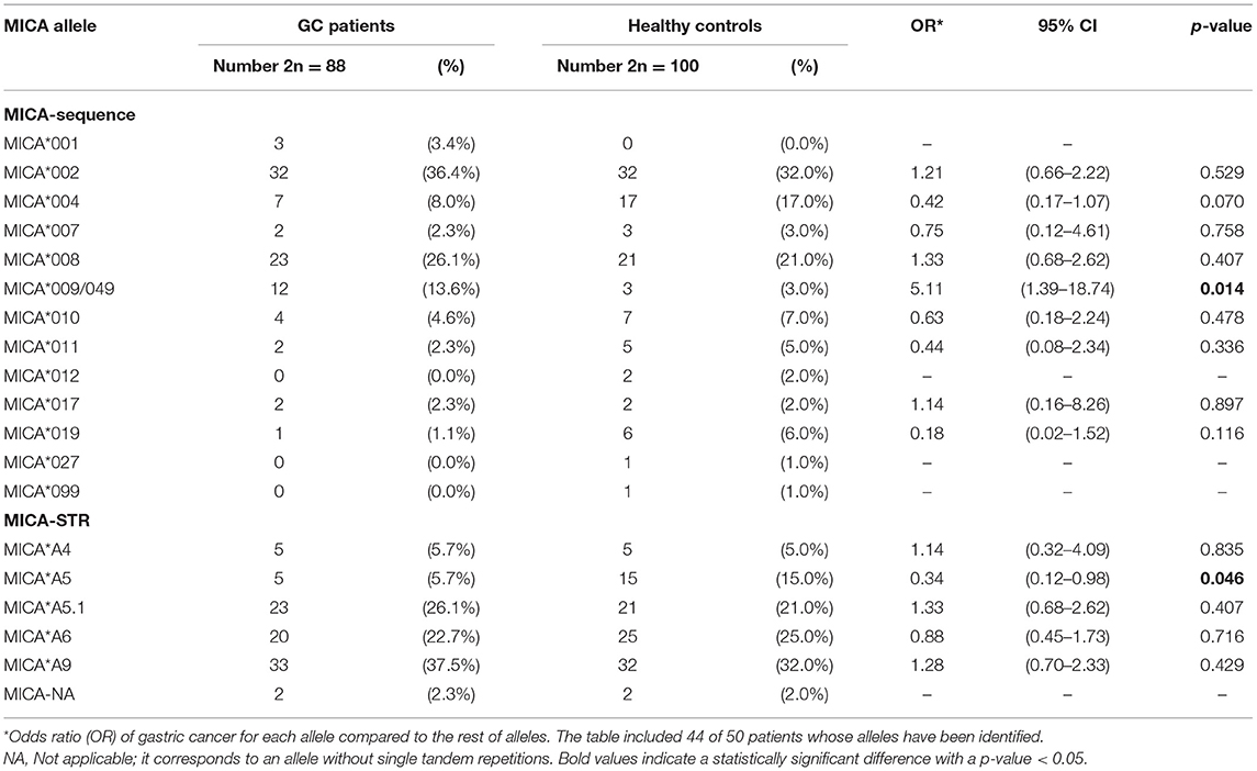
Table 2. MICA allelic frequency in gastric cancer patients and healthy controls.
We found that the MICA*009/049 allele frequency was significantly higher in GC patients than in healthy controls (p = 0.007), with a OR: 5.11 (95% CI: 1.39–18.74, p = 0.014). Additionally, we detected a higher frequency of MICA*A5 allele in healthy individuals than in GC patients, with a OR = 0.34 [95% CI: 0.12–0.98, p = 0.046) (Table 2). These results indicate that the presence of the MICA*A5 allele may be a protective factor in this type of tumor.
MICA Polymorphisms and Development of GC
We next analyzed the genotypes of the main amino-acids of MICA ectodomain in GC patients and healthy controls (Table 3). The results showed that the only amino-acid that varies between patients and controls was the residue in position 181 (p = 0.023), which corresponds to the substitution of threonine by arginine. This change defines the difference between MICA*004 allele (arginine) and MICA*009/049 allele (threonine). We observed that the majority of GC patients had a Thr/Thr genotype (42/50), while the Arg/Arg genotype was not identified in these patients. Patients with a Thr/Thr or Thr/Arg genotype did not show a significant difference in the tumor size or differentiation grade.
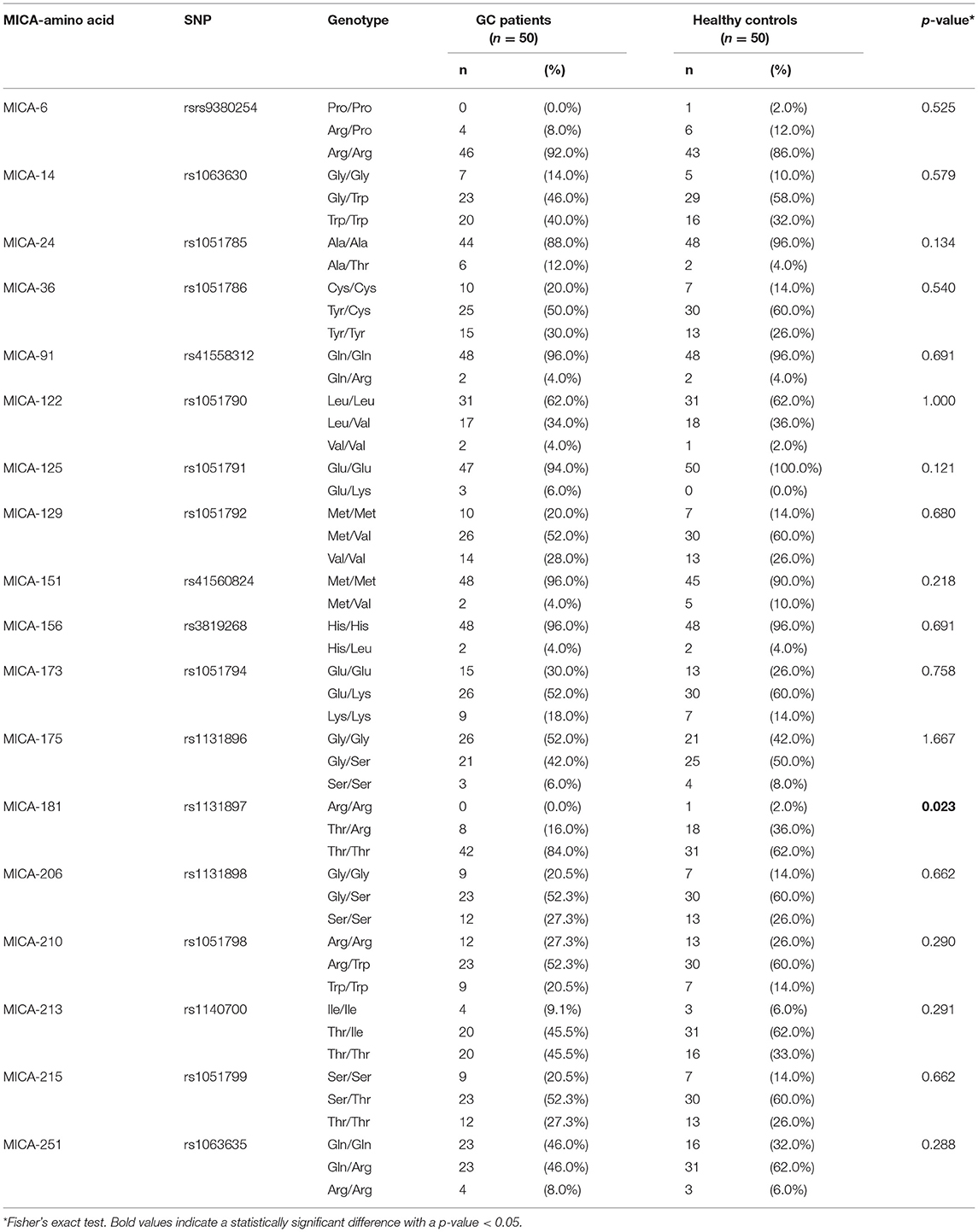
Table 3. MICA amino-acids genotypes of GC patients and healthy controls.
Additionally, we evaluated the possible association between clinical features related to the risk of developing GC and the MICA-129 polymorphism, since in position 129 resides one of the most studied residues that determine a variable affinity to NKG2D receptor. We found that MICA-129 single nucleotide polymorphism (SNP) (rs1051792) showed a strong linkage disequilibrium with other SNPs related to residues 36, 173, 206, 210, and 215. We did not identify associations between these genotypes and tumor size or differentiation grade. The presence of positive peritoneal cytology was identified in only five patients and, for this reason, it was not possible to classify the patients into groups according to this pathological characteristic, although positive peritoneal cytology was detected only in patients with Met/Val and Val/Val MICA-129 genotype (data no shown).
We studied the relevance of the MICA alleles, both MICA-sequence and MICA-STR, on the clinicopathological characteristics of the tumor. We observed that the majority of patients with the MICA*A9/A6 heterozygote allele showed mainly a poorly differentiated tumor (p = 0.031) (Supplementary Table 2), indicating that the presence of this genotype is related to an advanced stage of the tumor, as also observed previously (37). However, we did not find an association between the presence of the most frequent MICA-sequence alleles (*002, *008 and *009/049) and tumor size or differentiation grade (Supplementary Table 2).
Survival of GC Patients Based on MICA Alleles
We analyzed the overall survival of GC patients according to MICA alleles during 36 months after gastrectomy. We excluded the patients with a TNM stating I and II to avoid conflicting interpretations due to the possibility of a higher survival in patients with a lesser advanced disease. First, we classified the patients based on the main alleles found among them, which included patients homozygous for MICA*002 or MICA*008 and heterozygous for MICA*002/009 (MICA*002/049), MICA*002/004, or MICA*002/008. The Kaplan-Meier curves and Log-rank test showed significant differences among these groups of patients. MICA *002 and MICA*002/004 patients showed a higher survival rate than MICA*002/008 patients (p = 0.014) or MICA*002/009 (MICA*002/049) (p = 0.040). The survival distributions of homozygous patients for MICA*002 or heterozygous patients for MICA*002/004 were similar to that of homozygous patients for MICA*008 (p = 0.070). Likewise, no differences in the survival distributions of patients with MICA*002/008 compared to MICA*002/009 (MICA*002/049) or MICA*008/008 alleles could be detected (p = 0.611 and p = 0.909, respectively), neither between patients with MICA*002/009 (MICA*002/049) and those homozygous for MICA*008 (p = 0.883) (Figure 1A).
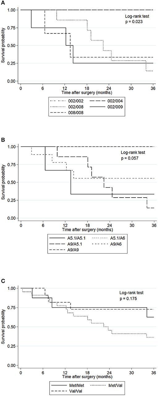
Figure 1. Kaplan-Meier curves for overall survival of GC patients with tumors at III and IV TNM staging, according to MICA-sequence, MICA-STR and MICA-129 genotype. The p-values were calculated by the Log-rank test. (A) The survival probability of GC patients with MICA*002/002 (n = 4) and MICA*002/004 (n = 4) alleles were significantly higher than that of MICA* 002/008 (n = 7) (p = 0.014) and MICA*002/009 (MICA*002/049) alleles (n = 4) (p = 0.040). The comparison between MICA*002/002 or MICA*002/004 and MICA*008/008 (n = 3) did not show significance (p = 0.070). (B) The survival probability of GC patients with MICA*A9/A9 (n = 4), MICA*A9/A5.1 (n = 7), MICA*A9/A6 (n = 9), MICA*A5.1/A6 (n = 3) and MICA*A5.1/A5.1 (n = 3) did not show significant differences (p = 0.057). (C) The survival probability of GC patients with the Met/Met genotype (n = 8), Met/Val (n = 22) and Val/Val (n = 11) did not show statistical differences (p = 0.175).
We also compared the survival curves of GC patients based on MICA-STR alleles (Figure 1B) and MICA-129 genotype (Figure 1C), which did not reach statistical significance (p = 0.057 and p = 0.175, respectively). Therefore, the MICA sequence, and not MICA-STR, may have a prognostic value in this type of cancer.
Molecular Analysis of MICA-181 Residue
Since we found that the MICA-181 residue showed differences between GC patients and controls (Table 3), and the fact that this residue defines the MICA*009/049 (Thr181) and MICA*004 (Arg181) alleles, we decided to perform a molecular analysis of these proteins to evaluate the impact of this amino-acid residue in the dynamic properties of MICA. Molecular dynamics simulations of both proteins displayed a different behavior during the simulation time of 500 ns. MICA*009/049 displayed greater mobility compared to MICA*004, which revealed a more restricted movement at the end of the simulation time, during which the change in the root mean square deviation (RMSD) values remained almost constant, as observed in Figure 2.
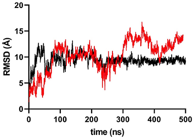
Figure 2. RMSD variation in MICA variant simulations. Black colored line represents MICA*004 (Arg181 variant), while red colored line represents MICA*009/049 (Thr181 variant). After 200 ns, MICA*004 remains constant, but MICA*009/049 displayed a less restrictive movement.
During the simulation, we noticed that the Arg181 residue established an electrostatic interaction with Asp29 (Figure 3), which, according to the simulations, is responsible for the flattening of the curve for MICA*004. Based on this observation, it was possible to measure a distance of Cα carbons of both residues (Arg181 vs. Thr181) with Asp29 in order to compare the importance of this interaction. We observed a higher distance for Thr181 than Arg181 around 300 ns (Figure 4). This implies that Thr181 does not accomplish a stabilizing interaction, probably due to its shorter side chain, as compared to Arg181. This confirmed that there is a greater movement in MICA*009/049, as Thr181 does not display the electrostatic interaction with Asp29.
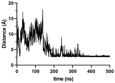
Figure 3. Distance, in Ångström, between Arg181 and Asp29 during simulation time of MICA. Distance was measured considering Nε of Arg181 and oxygen atoms of Asp29. The figure reveals that around 200 ns this hydrogen bond remains stable for the rest of the simulation.
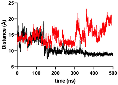
Figure 4. Distance, in Ångström, between Cα carbons of Thr181/Arg181 residues and Cα of Asp29 on MICA. Black colored line represents Arg181, while red colored line represents Thr181. Around 200 ns, the behavior of alleles differs due the hydrogen bond formation by the Arg181 and the Asp29.
Discussion
MICA, one of the main ligands to NKG2D receptor, has been considered an immunological target due to its participation in immune evasion mechanisms in cancer (38), including GC (5). MICA gene is highly polymorphic and, thus, originates different alleles based on its full-length sequence or STR (microsatellites) (MICA-STR). In this work, we identified MICA*002 (*A9) allele as the most frequent MICA variant in our cohort of GC patients and healthy controls, followed by MICA*008 (*A5.1) allele. In our study, both, patients as healthy controls shared the same geographic location; hence, the allelic frequencies described here are matched between cohorts. In this sense, the allele distribution found in our population differs from those reported in European, Asiatic, North American and Brazilian population (http://www.allelefrequencies.net), where the most frequent allele is MICA*008, followed by MICA*002 (39–43).
This difference could be explained by the ethnic diversity, which was not evaluated in the present work, and would certainly be recommended to be analyzed in future studies. Preliminary analysis of the rs67841474 in MICA gene, which corresponds to an indel that originates a frameshift variant (MICA*A5.1), has revealed that, in Chilean individuals from different ethnic origins, MICA*A5.1 is present in 20% of indigenous people from the North of Chile (Aymaras) in comparison to about 80% of indigenous people from the South of Chile (Mapuches) (44) (data available at http://genoma.med.uchile.cl:81/chilegenomico/). However, it is important to take into account that our cohorts are small when considering the variation that is exhibited by individuals in terms of MICA alleles; moreover, the frequencies would also be different depending on the population studied.
Even though it is clearly necessary to increase the cohort of study population for future investigations on NKG2D ligands (NKG2DL) polymorphisms, this time considering ethnical information and including human leukocyte antigen (HLA) analysis for a full characterization, we believe that our findings shed some light on the susceptibility and prognostic value of MICA polymorphisms in gastric cancer. Additionally, we also consider important to analyze the levels of soluble MICA in the serum of GC patients, which can be derived from the proteolytic shedding of MICA from the cell membrane, to evaluate whether these levels are related to MICA alleles, as some of the MICA variants could be more susceptible to this process. Such analysis would explain, at least in part, the broad interindividual variability of these levels in GC patients, which, in this particular type of cancer, appear to have a relatively low diagnostic impact (45); thus, MICA alleles could complement the diagnostic value of soluble MICA in this type of malignancy (46).
MICA polymorphic variants have been associated to several autoimmune and inflammatory diseases, as well as cancer (47, 48). Here, we observed that the presence of MICA*009/049 (MICA*A6) allele increases the susceptibility of developing GC, thus constituting a genetic risk factor for this type of cancer. In contrast, MICA*A5 allele was inversely associated with GC; therefore, this variant may be a protective factor for this disease, similar to findings previously reported for other types of cancer (49) and infectious diseases, such as tuberculosis (50).
MICA*009 allele has also been found in a high frequency in melanoma patients compared with controls (51). Additionally, both MICA*009 and MICA*004 (MICA*A6) have been associated with Behcet disease (18, 52–56) and rheumatoid arthritis (57). However, this variant was less frequent in patients with colorectal cancer (58), indicating that MICA alleles play a different role depending on the type of cancer, possibly due to cellular and molecular mechanisms that are particularly associated with different types of tumors. Accordingly, our results indicate a tendency for a higher frequency of MICA*004 among healthy individuals compared to GC patients. It is worth mentioning that MICA*009/049 and MICA*004 alleles produce a protein that differ at 181-residue, which makes it an interesting residue to analyze.
Based on the RMSD values and distance graphs described above, we hypothesize that different interactions between residues are achieved when both soluble MICA variants are simulated in a droplet of water. These results suggest that the interactions of MICA*004 are related to the physicochemical nature of Arg181, which bears a positive charge in its scaffold. This residue forms hydrogen bonds with neighboring residues, especially charged ones, as is the case of Asp29 in the simulation. Even though Thr181 residue can also form hydrogen bonds, this is a shorter and uncharged residue. A closer inspection of the interactions occurring for Arg181 reveals that not only is a coulombic interaction with Asp29 reinforced by hydrogen bonds, but it also displays a cation-π interaction with a terminal histidine residue (His3) at 4.5 Å. These interactions seem to be strong enough to keep the Arg181 stack in a crevice formed by these polar residues. In the case of Thr181 in MICA*009/049, these interactions are not observed during the simulation time, resulting in a more freely movement compared to Arg181. This observation implies that the change in only one amino acid can render a completely different dynamic behavior for both proteins. Figure 5 displays the Arg181 in the crevice formed by Asp29 and His3, which, in turn, displays a negative electrostatic potential surface, where Arg181 remains occluded. This is not observed with Thr181, which is not able to visit this site.
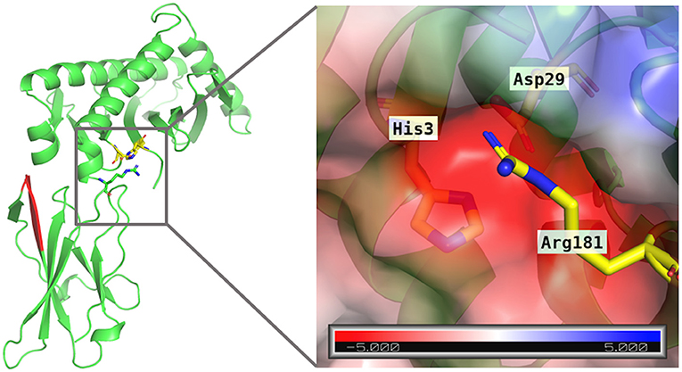
Figure 5. Electrostatic potential map for the crevice formed by residues His3 and Asp29 (yellow sticks) on MICA. The electrostatic potential map is given in kT/e units.
The MICA*009-HLA B*50, B*51 or B*52 haplotypes have been described in previous studies (59–61). Accordingly, HLA B*51 was found to be more frequent in Helicobacter pylori-positive pediatric patients with active gastritis and duodenal ulcer (62) and the HLA B*52 antigen has shown to be associated with lymph node metastasis in gastric cancer (63). Therefore, both HLA-B alleles and MICA*009/049 could to be factor risk in gastric cancer, so we suggest to consider this information for future studies.
When we analyzed the clinical characteristics of GC patients in relation to MICA polymorphisms, we observed that MICA-129, MICA-181 and MICA-sequence alleles were not related to tumor size, TNM stating or tumor differentiation grade in this type of cancer, whereas, tumors from MICA*A9/A6 heterozygote patients, who thus possess combinations of MICA*002 and MICA*009/049 or MICA*004, showed poorly differentiated tumors. These observations suggest that the transmembrane region of MICA may play a relevant role in GC progress. We suspect that the length of MICA transmembrane region, relative to the presence of six or more alanine residues, could implicate an increased susceptibility for proteolytic shedding of MICA by metalloproteases in the tumor microenvironment. If this is the case, the soluble levels of MICA would increase, which could negatively modulate the NKG2D receptor, favoring immune evasion mechanisms.
Here, we have also analyzed whether MICA variants affect the prognosis of GC. Our results showed that GC patients carrying the MICA*002 allele have better survival rates compared to GC patients carrying other MICA alleles. It has been previously demonstrated that the SNP rs9266825, which is part of MICA*002, *007, *018, *017, *001 alleles, is associated with increased survival rates in non-small cell lung cancer patients (64). Nevertheless, our study demonstrates, for the first time, the overall survival rates of GC patients based on the whole sequence of MICA gene.
We propose that certain MICA alleles have the potential to be more expressed on gastric tumor cells, depending on their cellular microenvironment, which may favor a better or worse immune response mediated by NK cells. According to our data, MICA*002 and MICA*004 alleles could be highly expressed on the surface of gastric tumor cells, which would trigger NKG2D receptor activation and an effective NK cell response. This is supported by other studies, which indicate that high-cell surface MICA/B expression in cancers of the digestive tract was associated with increased patient survival (65). In contrast, the levels of other protein variants, including MICA*009 and MICA*008, may be lower on tumor cell surface due to their shedding by metalloproteinases or release into the tumor milieu in extracellular vesicles, reducing target cell interaction with NK cells through the NKG2D receptor, thus affecting NK cell-mediated cytotoxicity. Support for this hypothesis comes from reports showing that patients with gastric tumors with high MICA expression had higher overall survival and disease-free-survival than patients bearing tumors with low MICA expression (66).
Our findings indicate that certain MICA alleles could have different effects on clinicopathological features of gastric tumor, such as the differentiation grade of tumor, as well as on patient overall survival after potentially curative gastrectomy, which may depend on the tumor microenvironment and regulation of MICA protein expression. In this sense, it will be interesting to analyze, in future studies, whether soluble MICA levels present any relationship with MICA alleles in GC patients.
In conclusion, MICA*009/049 allele increases the susceptibility to gastric cancer, whereas, MICA*A5 allele has a protective effect in this disease. MICA*002/002 and MICA*002/004 patients have higher survival rates than MICA*002/008 and MICA*002/009 (MICA*002/049) after surgery. Therefore, we consider that functional studies of MICA variants may help elucidate the mechanisms by which MICA confers protection or risk to GC development and prognosis, which may be a useful tool for the development of novel therapeutical approaches to treat this disease.
Data Availability Statement
The raw data supporting the conclusions of this article will be made available by the authors, without undue reservation, to any qualified researcher.
Ethics Statement
The studies involving human participants were reviewed and approved by Committee on Human Ethics Investigation of the Faculty of Medicine, University of Chile, and the Committee on Scientific Ethics of the Metropolitan Health Service of the Chilean Government. The patients/participants provided their written informed consent to participate in this study.
Author Contributions
KT-S, CHR, PG-H, GZ-T, and MCM interpreted the data and wrote the manuscript. AC analyzed the data. RA interpreted the data. GZ-T, DM, KT-S, MM, VG, JR-S, ST, and MB performed the bioinformatics analysis and laboratory experiments. MCM supervised the work. All authors contributed to manuscript revision and approved the submitted version.
Funding
This work was supported by the National Agency for Research and Development (ANID)/Scholarship Program/DOCTORADO BECAS CHILE/2017 Grant 21171812, ENLACE-VID ENL013/17 (University of Chile), Biomedical Sciences Institute (ICBM) Funding Grant 2020 (University of Chile) and REDES180146 UDECHILE from ANID and FONDECYT Grant 1171484.
Conflict of Interest
The authors declare that the research was conducted in the absence of any commercial or financial relationships that could be construed as a potential conflict of interest.
Acknowledgments
The authors would like to thank Mr. Bastián Jerez, Ms. Juana Orellana, Ms. Ruth Mora, and Ms. Nancy Fabres for their invaluable expert technical collaboration.
Supplementary Material
The Supplementary Material for this article can be found online at: https://www.frontiersin.org/articles/10.3389/fimmu.2021.645528/full#supplementary-material
References
1. Bray F, Ferlay J, Soerjomataram I, Siegel RL, Torre LA, Jemal A. Global cancer statistics 2018: GLOBOCAN estimates of incidence and mortality worldwide for 36 cancers in 185 countries. CA Cancer J Clin. (2018) 68:394–424. doi: 10.3322/caac.21492
2. Nakamura S, Kanda M, Kodera Y. Incorporating molecular biomarkers into clinical practice for gastric cancer. Expert Rev Anticancer Ther. (2019) 19:757–71. doi: 10.1080/14737140.2019.1659136
3. Nakamura S, Kanda M, Shimizu D, Sawaki K, Tanaka C, Hattori N, et al. STRA6 expression serves as a prognostic biomarker of gastric cancer. Cancer Genomics Proteomics. (2020) 17:509–16. doi: 10.21873/cgp.20207
4. Kanda M, Suh YS, Park DJ, Tanaka C, Ahn SH, Kong SH, et al. Serum levels of ANOS1 serve as a diagnostic biomarker of gastric cancer: a prospective multicenter observational study. Gastric Cancer. (2020) 23:203–11. doi: 10.1007/s10120-019-00995-z
5. Ribeiro CH, Kramm K, Galvez-Jiron F, Pola V, Bustamante M, Contreras HR, et al. Clinical significance of tumor expression of major histocompatibility complex class I-related chains A and B (MICA/B) in gastric cancer patients. Oncol Rep. (2016) 35:1309–17. doi: 10.3892/or.2015.4510
6. Groh V, Bahram S, Bauer S, Herman A, Beauchamp M, Spies T. Cell stress-regulated human major histocompatibility complex class I gene expressed in gastrointestinal epithelium. Proc Natl Acad Sci U S A. (1996) 93:12445–50. doi: 10.1073/pnas.93.22.12445
7. Allegretti YL, Bondar C, Guzman L, Cueto Rua E, Chopita N, Fuertes M, et al. Broad MICA/B expression in the small bowel mucosa: a link between cellular stress and celiac disease. PLoS ONE. (2013) 8:e73658. doi: 10.1371/journal.pone.0073658
8. Fattahi S, Golpour M, Amjadi-Moheb F, Sharifi-Pasandi M, Khodadadi P, Pilehchian-Langroudi M, et al. DNA methyltransferases and gastric cancer: insight into targeted therapy. Epigenomics. (2018)10:1477–97. doi: 10.2217/epi-2018-0096
9. Garrido-Tapia M, Hernandez CJ, Ascui G, Kramm K, Morales M, Ga Rate V, et al. STAT3 inhibition by STA21 increases cell surface expression of MICB and the release of soluble MICB by gastric adenocarcinoma cells. Immunobiology. (2017) 222:1043–51. doi: 10.1016/j.imbio.2017.05.009
10. Polakovicova I, Jerez S, Wichmann IA, Sandoval-Borquez A, Carrasco-Veliz N, Corvalan AH. Role of microRNAs and exosomes in helicobacter pylori and Epstein-Barr virus associated gastric cancers. Front Microbiol. (2018) 9:636. doi: 10.3389/fmicb.2018.00636
11. Wang R, Liu K, Chen XZ, group Sr. Associations between gastric cancer risk and virus infection other than Epstein-Barr virus: the protocol of a systematic review and meta-analysis based on epidemiological studies. Medicine (Baltimore). (2019) 98:e16708. doi: 10.1097/MD.0000000000016708
12. Risti M, Bicalho MD. MICA and NKG2D: is there an impact on kidney transplant outcome? Front Immunol. (2017) 8:179. doi: 10.3389/fimmu.2017.00179
13. Chitadze G, Bhat J, Lettau M, Janssen O, Kabelitz D. Generation of soluble NKG2D ligands: proteolytic cleavage, exosome secretion and functional implications. Scand J Immunol. (2013) 78:120–9. doi: 10.1111/sji.12072
14. Poggi A, Benelli R, Vene R, Costa D, Ferrari N, Tosetti F, et al. Human gut-associated natural killer cells in health and disease. Front Immunol. (2019) 10:961. doi: 10.3389/fimmu.2019.00961
15. Wensveen FM, Jelencic V, Polic B. NKG2D: A master regulator of immune cell responsiveness. Front Immunol. (2018) 9:441. doi: 10.3389/fimmu.2018.00441
16. Zingoni A, Vulpis E, Cecere F, Amendola MG, Fuerst D, Saribekyan T, et al. MICA-129 dimorphism and soluble MICA are associated with the progression of multiple myeloma. Front Immunol. (2018) 9:926. doi: 10.3389/fimmu.2018.00926
17. Li P, Morris DL, Willcox BE, Steinle A, Spies T, Strong RK. Complex structure of the activating immunoreceptor NKG2D and its MHC class I-like ligand MICA. Nat Immunol. (2001) 2:443–51. doi: 10.1038/87757
18. Mizuki N, Ota M, Kimura M, Ohno S, Ando H, Katsuyama Y, et al. Triplet repeat polymorphism in the transmembrane region of the MICA gene: a strong association of six GCT repetitions with Behcet disease. Proc Natl Acad Sci U S A. (1997) 94:1298–303. doi: 10.1073/pnas.94.4.1298
19. Ashiru O, Boutet P, Fernandez-Messina L, Aguera-Gonzalez S, Skepper JN, Vales-Gomez M, et al. Natural killer cell cytotoxicity is suppressed by exposure to the human NKG2D ligand MICA*008 that is shed by tumor cells in exosomes. Cancer Res. (2010) 70:481–9. doi: 10.1158/0008-5472.CAN-09-1688
20. Ashiru O, Lopez-Cobo S, Fernandez-Messina L, Pontes-Quero S, Pandolfi R, Reyburn HT, et al. A GPI anchor explains the unique biological features of the common NKG2D-ligand allele MICA*008. Biochem J. (2013) 454:295–302. doi: 10.1042/BJ20130194
21. Isernhagen A, Malzahn D, Bickeboller H, Dressel R. Impact of the MICA-129Met/Val Dimorphism on NKG2D-Mediated Biological Functions and Disease Risks. Front Immunol. (2016) 7:588. doi: 10.3389/fimmu.2016.00588
22. Lo SS, Lee YJ, Wu CW, Liu CJ, Huang JW, Lui WY. The increase of MICA gene A9 allele associated with gastric cancer and less schirrous change. Br J Cancer. (2004) 90:1809–13. doi: 10.1038/sj.bjc.6601750
23. Tamaki S, Sanefuzi N, Ohgi K, Imai Y, Kawakami M, Yamamoto K, et al. An association between the MICA-A5.1 allele and an increased susceptibility to oral squamous cell carcinoma in Japanese patients. J Oral Pathol Med. (2007) 36:351–6. doi: 10.1111/j.1600-0714.2007.00539.x
24. Dai X, Zhang X, Yu J. Clinicopathological features and Borrmann classification associated with HER2-positive in primary gastric cancer. Clin Exp Gastroenterol. (2019) 12:287–94. doi: 10.2147/CEG.S212895
25. Washington K. 7th edition of the AJCC cancer staging manual: stomach. Ann Surg Oncol. (2010) 17:3077–9. doi: 10.1245/s10434-010-1362-z
26. Watanabe H, Jass JR, Sobin LH, in collaboration with pathologists in 8 countries. Histological typing of oesophageal and gastric tumours, 2nd edition. In: WHO International Histological Classification of Tumors. Berlin: Springer-Verlag (1990).
27. Subbarayan PR, Sarkar M, Ardalan B. Isolation of genomic DNA from human whole blood. Biotechniques. (2002) 33:1231. doi: 10.2144/02336bm10
28. Robinson J, Barker DJ, Georgiou X, Cooper MA, Flicek P, Marsh SGE. IPD-IMGT/HLA database. Nucleic Acids Res. (2020) 48:D948–D55. doi: 10.1093/nar/gkz950
29. Li Z, Groh V, Strong RK, Spies T. A single amino acid substitution causes loss of expression of a MICA allele. Immunogenetics. (2000) 51:246–8. doi: 10.1007/s002510050039
30. Chitadze G, Lettau M, Bhat J, Wesch D, Steinle A, Furst D, et al. Shedding of endogenous MHC class I-related chain molecules A and B from different human tumor entities: heterogeneous involvement of the “a disintegrin and metalloproteases” 10 and 17. Int J Cancer. (2013) 133:1557–66. doi: 10.1002/ijc.28174
31. UniProt C. UniProt: a worldwide hub of protein knowledge. Nucleic Acids Res. (2019) 47:D506–D15. doi: 10.1093/nar/gky1049
32. Altschul SF, Gish W, Miller W, Myers EW, Lipman DJ. Basic local alignment search tool. J Mol Biol. (1990) 215:403–10. doi: 10.1016/S0022-2836(05)80360-2
33. Sievers F, Wilm A, Dineen D, Gibson TJ, Karplus K, Li W, et al. Fast, scalable generation of high-quality protein multiple sequence alignments using Clustal Omega. Mol Syst Biol. (2011) 7:539. doi: 10.1038/msb.2011.75
34. Guex N, Peitsch MC, Schwede T. Automated comparative protein structure modeling with SWISS-MODEL and Swiss-PdbViewer: a historical perspective. Electrophoresis. (2009) 30(Suppl. 1):S162–73. doi: 10.1002/elps.200900140
35. Jo S, Kim T, Iyer VG, Im W. CHARMM-GUI: a web-based graphical user interface for CHARMM. J Comput Chem. (2008) 29:1859–65. doi: 10.1002/jcc.20945
36. Case DA, Babin V, Berryman JT, Betz RM, Cai Q, Cerutti DS, et al. AMBER 14. San Francisco, CA: University of California (2014).
37. Feng F, Liu J, Wang F, Zheng G, Wang Q, Liu S, et al. Prognostic value of differentiation status in gastric cancer. BMC Cancer. (2018) 18:865. doi: 10.1186/s12885-018-4780-0
38. Schmiedel D, Mandelboim O. NKG2D ligands-critical targets for cancer immune escape and therapy. Front Immunol. (2018) 9:2040. doi: 10.3389/fimmu.2018.02040
39. Klussmeier A, Massalski C, Putke K, Schafer G, Sauter J, Schefzyk D, et al. High-throughput MICA/B genotyping of over two million samples: workflow and allele frequencies. Front Immunol. (2020) 11:314. doi: 10.3389/fimmu.2020.00314
40. Wang WY, Tian W, Zhu FM, Liu XX, Li LX, Wang F. MICA, MICB polymorphisms and linkage disequilibrium with HLA-B in a Chinese mongolian population. Scand J Immunol. (2016) 83:456–62. doi: 10.1111/sji.12437
41. Yamakawa RH, Saito PK, Gelmini GF, da Silva JS, Bicalho MDG, Borelli SD. MICA diversity and linkage disequilibrium with HLA-B alleles in renal-transplant candidates in southern Brazil. PLoS ONE. (2017) 12:e0176072. doi: 10.1371/journal.pone.0176072
42. Wenda S, Fae I, Sanchez-Mazas A, Nunes JM, Mayr WR, Fischer GF. The distribution of MICA alleles in an Austrian population: evidence for increasing polymorphism. Hum Immunol. (2013) 74:1295–9. doi: 10.1016/j.humimm.2013.06.013
43. Lucas D, Campillo JA, Lopez-Hernandez R, Martinez-Garcia P, Lopez-Sanchez M, Botella C, et al. Allelic diversity of MICA gene and MICA/HLA-B haplotypic variation in a population of the Murcia region in southeastern Spain. Hum Immunol. (2008) 69:655–60. doi: 10.1016/j.humimm.2008.07.011
44. Verdugo RA, Di Genova A, Herrera L, Moraga M, Acuna M, Berrios S, et al. Development of a small panel of SNPs to infer ancestry in Chileans that distinguishes Aymara and Mapuche components. Biol Res. (2020) 53:15. doi: 10.1186/s40659-020-00284-5
45. Jiang X, Huang JF, Huo Z, Zhang Q, Jiang Y, Wu X, et al. Elevation of soluble major histocompatibility complex class I related chain A protein in malignant and infectious diseases in Chinese patients. BMC Immunol. (2012) 13:62. doi: 10.1186/1471-2172-13-62
46. Zhao P, Chen D, Cheng H. Prognostic significance of soluble major histocompatibility complex class I-related chain A (sMICA) in gastric cancer. Br J Biomed Sci. (2018) 75:203–5. doi: 10.1080/09674845.2018.1505188
47. Wang Q, Zhou X. Associations of MICA polymorphisms with inflammatory rheumatic diseases. Open Rheumatol J. (2015) 9:94–100. doi: 10.2174/1874312901409010094
48. Onyeaghala G, Lane J, Pankratz N, Nelson HH, Thyagarajan B, Walcheck B, et al. Association between MICA polymorphisms, s-MICA levels, and pancreatic cancer risk in a population-based case-control study. PLoS ONE. (2019) 14:e0217868. doi: 10.1371/journal.pone.0217868
49. Ji M, Wang J, Yuan L, Zhang Y, Zhang J, Dong W, et al. MICA polymorphisms and cancer risk: a meta-analysis. Int J Clin Exp Med. (2015) 8:818–26.
50. Chen E, Chen C, Chen F, Yu P, Lin L. Positive association between MIC gene polymorphism and tuberculosis in Chinese population. Immunol Lett. (2019) 213:62–9. doi: 10.1016/j.imlet.2019.07.008
51. Campillo JA, Lopez-Hernandez R, Martinez-Banaclocha H, Bolarin JM, Gimeno L, Mrowiec A, et al. MHC class I chain-related gene a diversity in patients with cutaneous malignant melanoma from southeastern Spain. Dis Markers. (2015) 2015:831864. doi: 10.1155/2015/831864
52. Wallace GR, Verity DH, Delamaine LJ, Ohno S, Inoko H, Ota M, et al. MIC-A allele profiles and HLA class I associations in Behcet's disease. Immunogenetics. (1999) 49:613–7. doi: 10.1007/s002510050656
53. Carapito R, Shahram F, Michel S, Le Gentil M, Radosavljevic M, Meguro A, et al. On the genetics of the Silk Route: association analysis of HLA, IL10, and IL23R-IL12RB2 regions with Behcet's disease in an Iranian population. Immunogenetics. (2015) 67:289–93. doi: 10.1007/s00251-015-0841-6
54. Mizuki N, Meguro A, Tohnai I, Gul A, Ohno S, Mizuki N. Association of major histocompatibility complex class I chain-related gene A and HLA-B Alleles with Behcet's disease in Turkey. Jpn J Ophthalmol. (2007) 51:431–6. doi: 10.1007/s10384-007-0473-y
55. Lee YH, Song GG. Associations between major histocompatibility complex class I chain-related gene A polymorphisms and susceptibility to Behcet's disease. A meta-analysis. Z Rheumatol. (2015) 74:714–21. doi: 10.1007/s00393-014-1536-3
56. Eyerci N, Balkan E, Akdeniz N, Keles S. Association of MICA alleles and human leukocyte antigen B in Turkish patients diagnosed with Behcet's disease. Arch Rheumatol. (2018) 33:352–7. doi: 10.5606/ArchRheumatol.2018.6704
57. Wang Y, Li S, Chen C, Luo Q, Li Y, Liu L, et al. MICB*002 and MICB*014 protect against rheumatoid arthritis, whereas MICA*009 and MICA*A6 are associated with rheumatoid arthritis in a Hainan Han Chinese population. Int J Rheum Dis. (2019) 22:90–5. doi: 10.1111/1756-185X.13302
58. Ding W, Ma Y, Zhu W, Pu W, Zhang J, Qian F, et al. MICA (*)012:01 Allele facilitates the metastasis of KRAS-mutant colorectal cancer. Front Genet. (2020) 11:511. doi: 10.3389/fgene.2020.00511
59. Jarduli LR, Alves HV, de Souza VH, Uaska Sartori PV, Fava VM, de Souza FC, et al. Association of MICA and HLA-B alleles with leprosy in two endemic populations in Brazil. Int J Immunogenet. (2021) 48:25–35. doi: 10.1111/iji.12518
60. Cambra A, Munoz-Saa I, Crespi C, Serra A, Etxagibel A, Matamoros N, et al. MICA-HLA-B haplotype diversity and linkage disequilibrium in a population of Jewish descent from Majorca (the Balearic Islands). Hum Immunol. (2009) 70:513–7. doi: 10.1016/j.humimm.2009.04.005
61. Cha CH, Sohn YH, Oh HB, Ko SY, Cho MC, Kwon OJ. MICB polymorphisms and haplotypes with MICA and HLA alleles in Koreans. Tissue Antigens. (2011) 78:38–44. doi: 10.1111/j.1399-0039.2011.01694.x
62. Gonen S, Sari S, Kandur Y, Dalgic B, Soylemezoglu O. Evaluation of human leukocyte antigen class I and Ii antigens in helicobacter pylori-positive pediatric patients with active gastritis and duodenal ulcer. Arq Gastroenterol. (2017) 54:297–9. doi: 10.1590/s0004-2803.201700000-62
63. Ogoshi K, Tajima T, Mitomi T, Tsuji K. HLA antigens are candidate markers for prediction of lymph node metastasis in gastric cancer. Clin Exp Metastasis. (1996) 14:277–81.
64. Xu J, Tian S, Yin Z, Wu S, Liu L, Qian Y, et al. MicroRNA-binding site SNPs in deregulated genes are associated with clinical outcome of non-small cell lung cancer. Lung Cancer. (2014) 85:442–8. doi: 10.1016/j.lungcan.2014.06.010
65. Zhao Y, Chen N, Yu Y, Zhou L, Niu C, Liu Y, et al. Prognostic value of MICA/B in cancers: a systematic review and meta-analysis. Oncotarget. (2017) 8:96384–95. doi: 10.18632/oncotarget.21466
Keywords: gastric cancer, MICA gene, MICA polymorphism, MICA-129, MICA alleles
Citation: Toledo-Stuardo K, Ribeiro CH, Canals A, Morales M, Gárate V, Rodríguez-Siza J, Tello S, Bustamante M, Armisen R, Matthies DJ, Zapata-Torres G, González-Hormazabal P and Molina MC (2021) Major Histocompatibility Complex Class I-Related Chain A (MICA) Allelic Variants Associate With Susceptibility and Prognosis of Gastric Cancer. Front. Immunol. 12:645528. doi: 10.3389/fimmu.2021.645528
Received: 23 December 2020; Accepted: 23 February 2021;
Published: 31 March 2021.
Edited by:
Chanvit Leelayuwat, Khon Kaen University, ThailandReviewed by:
Steven Thomas Cox, Anthony Nolan, United KingdomJack D. Bui, University of California, San Diego, United States
Copyright © 2021 Toledo-Stuardo, Ribeiro, Canals, Morales, Gárate, Rodríguez-Siza, Tello, Bustamante, Armisen, Matthies, Zapata-Torres, González-Hormazabal and Molina. This is an open-access article distributed under the terms of the Creative Commons Attribution License (CC BY). The use, distribution or reproduction in other forums is permitted, provided the original author(s) and the copyright owner(s) are credited and that the original publication in this journal is cited, in accordance with accepted academic practice. No use, distribution or reproduction is permitted which does not comply with these terms.
*Correspondence: María Carmen Molina, mcmolina@med.uchile.cl