- 1Department of Medical Microbiology and Immunology, Medical School, University of Pécs, Pécs, Hungary
- 2Janos Szentagothai Research Centre, Pécs, Hungary
Immune checkpoint molecules, like CTLA-4, TIM-3, PD-1, are negative regulators of immune responses to avoid immune injury. Checkpoint regulators are thought to actively participate in the immune defense of infections, prevention of autoimmunity, transplantation, and tumor immune evasion. Maternal-fetal immunotolerance represents a real immunological challenge for the immune system of the mother: beside acceptance of the semiallogeneic fetus, the maternal immune system has to be prepared for immune defense mostly against infections. In this particular situation, the role of immune checkpoint molecules could be of special interest. In this review, we describe current knowledge on the role of immune checkpoint molecules in reproductive immunology.
Introduction
The activation of the immune system to eliminate harmful agents is usually followed by tissue damage at the site of the exposure. In order to keep this side effect of the immune response limited and localized, efficient immunoactivation of immune cells requires multiple incoming signals. Beside antigen recognition, co-stimulatory, survival, and proliferative signals, even environmental factors can determine the outcome of the immune response (1–4).
Immune checkpoint molecules are co-stimulatory receptors, occurring on the surface of several immune cells. After ligand binding, these regulators are capable of transducing inhibitory signals (5). CTLA-4, TIM-3, PD-1 are the most studied members from this group of cell surface receptors (5). The physiological role of immune checkpoints is to prevent a harmful immune attack against self-antigens during an immune response by negatively regulating the effector immune cells, e.g., by inducing T cell exhaustion (5, 6). Recent studies suggest that each checkpoint decreases immunoactivation through different intracellular signaling mechanisms (5, 7). Immune checkpoint regulators are thought to actively participate in the immune defense of infections, prevention of autoimmunity, transplantation, and tumor immune evasion (5, 7).
Pregnancy is a natural model of active immunotolerance, where maternal immune system simultaneously faces two challenges: beside acceptance of the semiallogeneic fetus, the maternal immune system has to be prepared for immune defense mostly against infections. In this particular situation, the role of immune checkpoint molecules could be of special interest. Therefore, this paper aims to review the literature presenting current knowledge about the role of immune checkpoint molecules in reproductive immunology.
CTLA-4
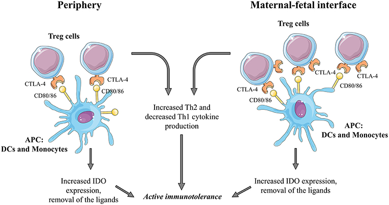
The first described inhibitory receptor CTLA-4 (Cytotoxic T-lymphocyte-associated protein 4) is predominantly and constitutively expressed intracellularly in regulatory T cells, and it is missing in naive conventional T cells (8, 9). Following activation, CTLA-4 is expressed on the cell surface of Tregs, but it can also be found on the cell surface of activated CD8+ or CD4+ T cells (10). The inhibitory effect of CTLA-4 results from the competition with the T cell activatory CD28 receptor to bind the B7 ligands CD80/CD86 present on the cell surface of antigen presenting cells (11). The ability of Treg cells to induce IDO expression in APCs through the CTLA-4-B7 binding was thought to be one of the major mechanism of immune suppression by these cells (12, 13). Interestingly, current thinking suggests, that the main function of CTLA-4 is not delivering negative signals through ligand binding but the removal of its ligands CD80/CD86 from the cell surface of APCs preventing thereby their binding to the costimulatory CD28 present on T cells (8, 14).
CTLA-4 in Murine Pregnancy
The significance of the CD80/86-CD28 activation pathway in T cells during fetal rejection was shown by blocking both ligands with mAb during the pregnancy of the abortion-prone murine model. The blockade resulted in the improvement of fetal survival with an increase of Th2 type cytokines at the maternal-fetal interface (MFI) and in the peripheral expansion of the CD4+ C25+ T cell population. Furthermore, CTLA-4 expression by T cells increased as well which was found to be significantly reduced at the MFI in abortion-prone matings (15, 16). Preventing binding of CD80/CD86 to CD28 is thought to be the way of action of CTLA-4 with similar beneficial effects in maternal-fetal tolerance. Blocking only CD86 using the same experimental setting resulted in the same observations (17). These findings support previous theories about CD80 might be the most functional ligand for CTLA-4. Blockade of the CD86 could turn off the co-stimulatory CD86/CD28 pathway while allowing a prolonged CD80/CTLA-4 interaction with all of the benefits (17–19).
Further evidence for the immunosuppressive capacity of CTLA-4 was delivered from experiments with the CTLA4Ig fusion protein. Using an adenoviral vector, CTLA4Ig was shown to be heavily expressed at the MFI. CTLA4Ig therapy of abortion-prone CBA/DBA matings could effectively improve pregnancy outcome by shifting serum cytokine levels toward Th2 bias and expanding regulatory T cell population at the periphery (20). Furthermore, the CTLA4Ig fusion protein significantly inhibited splenic lymphocyte proliferation and apoptosis of the fetoplacental unit (21). Interestingly, adoptive transfer of Treg with CTLA-4 blockade from normal pregnant mouse to CBA/DBA pregnancy didn't abolish the protective effect of Treg treatment without a blockade resulting in decreased abortion rates (22).
In another abortion-prone setting, in sonic stressed pregnant mice, decidual lymphocytes expressed decreased levels of CTLA-4, without any changes in CD28 expression suggesting the failure of the control of local immunoactivation. CTLA-4 expression by decidual lymphocytes of stressed animals could be enhanced by injections of the dipeptidyl peptidase IV inhibitor, a well-known terminator of T-cell activation (23, 24).
CTLA-4 in Human Pregnancy (Figure 1)
CTLA-4 at the Periphery
Although regulatory T cells increase in number in the periphery during early pregnancy, the enhanced CTLA-4 expression on the cell surface was not observed (10, 25). In contrast to these findings, the expression of one of the ligands of CTLA-4, namely CD86 showed an increased expression by peripheral DCs and monocytes in healthy pregnancy while CD80 expression patterns did not change (26).
CTLA-4Ig treatment of peripheral blood mononuclear cells resulted in a significantly higher IFN-γ secretion in normal pregnancy compared to non-pregnant condition (26). Despite the fact, that CTLA-4 is capable of inducing indoleamine 2,3 dioxygenase (IDO) expression in dendritic cells and monocytes through the induction of IFN-γ, there are conflicting data about whether CTLA-4Ig treatment could enhance IDO expression in DCs and monocytes in normal pregnancy (13, 26, 27).
CTLA-4 at the Maternal-Fetal Interface
Compared to the periphery, decidual Treg cells further increase in number, and the frequency of Treg expressing intracellular or surface CTLA-4 was also found to be elevated in the decidua (10, 27, 28). Interestingly, placental fibroblasts also express CTLA-4, but it is supposed to have non-immunological functions since fibroblasts are not directly in contact with maternal tissues (29). The CTLA-4 ligands, CD80, and CD86, are also present on decidual DCs and monocytes, and they show the same expression profile as in the periphery in normal pregnancy (26, 30). The decidual CTLA-4 expression is in a significantly positive correlation with decidual Th2 cytokine production and a negative correlation with decidual Th1 cytokine production suggesting remarkable immunosuppressive effects locally (30).
CTLA-4Ig treatment of decidual lymphocytes resulted in enhanced IFN-γ and IDO expression (10).
CTLA-4 in Pregnancy Complications
In the case of spontaneous abortion/miscarriage peripheral and decidual Tregs fail to increase to the levels observed in normal pregnancy (10). Data about CTLA-4 expression in these conditions are conflicting. In one hand, the overall ratio of CTLA-4+ peripheral and decidual lymphocytes as wells as the ratio of CTLA-4+ Tregs was found to be significantly reduced. Moreover, the ratios of CTLA-4+/CD28+ in regulatory T cells from miscarriage were significantly lower than that of normal pregnancy (30, 31). On the other hand, there was no significant difference in intracellular and cell surface expression of CTLA-4 on both peripheral and decidual Tregs when compared to non-pregnant and healthy pregnant controls (10). These controversy data may result from different patient inclusion criteria. From the two possible CTLA-4 ligands, only CD86 expression was found to be affected in miscarriage: peripheral monocytes, decidual monocytes, and DC showed significantly lower expression rates compared to those in normal pregnancy (26, 30). Response levels of IDO expression by both peripheral and decidual monocytes and DCs in spontaneous abortion with CTLA-4 treatments were lower compared to a healthy pregnancy (26).
Extensive research focused on the role of CTLA-4 gene polymorphism with different conclusions (32–37). The A/G polymorphism at position 49 in exon 1 of cytotoxic T lymphocyte antigen-4 (CTLA-4) gene may result in abnormal protein modification in the rough endoplasmic reticulum leading to reduced expression (38, 39). Further studies confirmed, that the 49 GG genotype was associated with a reduced inhibitory function of CTLA-4 whereas individuals with AA genotype had more expression of CTLA-4 both intracellular as on the cell surface of activated T cells (33, 40, 41). Further studies with larger sample sizes are needed to prove increased frequencies of G allele and GG genotype among patients with recurrent miscarriage.
Although preeclampsia is characterized by a diminished Treg frequency, a well-known alteration (42–46), little information is available about the possible role of CTLA-4 in the pathogenesis of the disease. CD80 and CD86 ligand expression levels on monocytes decrease in preeclampsia, while data about CTLA-4 expression of Treg are not conclusive, increased and unchanged expression patterns were reported as well. Therefore, it is difficult to determine the significance of the CTLA-4 pathway in preeclampsia (47–49). Two gene polymorphism studies of the exon-1 A49G region of the CTLA-4 gene revealed an increased frequency of the heterozygosity and GG phenotype in pre-eclamptic women (38, 50).
In women with successful IVF treatment, there is an increase in the peripheral Treg population compared to failed IVF attempts. Investigating CTLA-4 expression at the mRNA level, no differences could be observed in the two IVF patient group (51).
Heterozygous mutations in the immune checkpoint protein CTLA-4 leading to CTLA-4 deficiency results in different autoimmune clinical features, but no further information is available about pregnancy proceeding in these patients (52, 53).
TIM-3
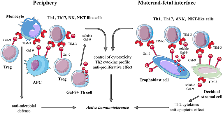
Extensive research has established that Tim-3/gal-9 pathway plays a significant role in the regulation of immune responses and induction of tolerance (54–58). TIM-3 was shown to be expressed by many types of immune cells, including Th1, Th17, NK and NKT-like cells, Tregs, and also on antigen-presenting immune cells (59). Interestingly, TIM-3 activity is thought to participate in both activation and inhibition of immune response (60, 61). In the case of a healthy pregnancy, expression of TIM-3 on Th1 cells may be a key element for reducing proinflammatory Th1-dependent T-cell response (57).
The ligand of TIM-3 receptor is galectin-9 (Gal-9), a β-galactose binding protein (62). Among other identified receptors of Gal-9, TIM-3 has been studied most intensively (54). Both in mice and humans, binding of TIM-3 to its ligand Gal-9 leads to the apoptosis of Th1 and Th17 cells and induce immunotolerance (63–65). Thus, the TIM-3/Gal-9 pathway may serve as a checkpoint regulator limiting the Th1- and Th17-driven immune response and modulating the Th1/Th2 cytokine balance (54).
TIM-3 in Murine Pregnancy
TIM-3 has been studied in detail in murine pregnancy models by several groups (66–71). First, immunofluorescence stainings revealed the presence of TIM-3 in midgestational uterus and flow cytometric analysis proved that this inhibitory molecule is expressed by a variety of immune cells residing locally in the uterus/decidua: uterine NK cells, γ/δ T cells, NKT-like cells, macrophages, dendritic cells (DC), and even by myeloid-derived (66–68). TIM-3 expression by these cells was shown to be dominant but variable throughout pregnancy, in the case of the most prevalent decidual immune cell type, NK cells upregulate TIM-3 during the first half of murine gestation (66, 67). Although TIM-3 expression of decidual NK cells and γ/δ T cells is similar to that in the periphery, their upregulated relative TIM-3 expression locally suggest that these cells are more mature and entirely functional (68, 72). However, the cytotoxic capacity of TIM-3+ decidual NK cells and γ/δ T cells was shown to be reduced when compared to the periphery; this might be due to the special local microenvironment at the MFI (68). In contrast to these findings, there is a smaller TIM-3+ NKT-like cell subset in the decidua with stronger lytic capacity. Therefore, separate action of TIM-3 on different immune cell types with varying functional outcomes could be concluded (68).
The TIM-3 ligand, galectin-9 is also present at the MFI at different sites. Both murine placental spongiotrophoblast and decidual regulatory T cells express galectin-9 and decidual Gal-9+ Th cells are the main source of the secreted, soluble form of Gal-9 (68). Since the presence of both the ligand, Gal-9 and its receptor, TIM-3 side by side, their binding interaction could be hypothesized, and the inhibitory signal derived from TIM-3 might contribute to maternal immunotolerance observed in murine pregnancy. This hypothesis is supported by the observation that TIM-3 blockade of allogeneic murine pregnancy resulted in litter size reduction, reduced live births, and an increased rate of resorption in vivo (66, 71).
Blocking TIM-3 with monoclonal antibodies (mAbs) provided further information about the possible function of this molecule at the MFI. Following inhibition, both apoptotic cells and macrophages accumulate locally, suggesting a deficiency of phagocytic clearance via failed recognition of phosphatidylserine through TIM-3 and enhanced pro-inflammatory cytokine production (66). Uterine granulocytes were also shown to increase in number and to enhance Th1 cytokine expression. These observations are in line with previous studies of experimental autoimmune/ischemic murine models where increased inflammation was due to macrophage and granulocyte activation following TIM-3 blockade (73, 74). Blocking TIM-3 on uterinal NK (uNK) cells affect both physiologic phenotype and function of these dominant cell population at the MFI (67). Although local accumulation and cytotoxic capacity of TIM-3+ uNK cells did not change, uNK cells upregulated the activation marker CD69, and their expression pattern of activating and inhibitory cell surface receptors was notably altered. Secretion of both proangiogenic (VEGF, IFN-γ) and immunosuppressive (IL-10) cytokines by TIM-3+ uNK cells were decreased. Additionally, TIM-3 inhibition resulted in reduced placental expression of the cytokines IL-15 and IL-9, which are important factors for NK cell survival and development (67, 75).
In abortion-prone mouse models, a reduced number of TIM-3+ dNK and CD4+ Th cells can be observed with predominantly Th1 cytokine profiles (69, 70).
All these data from murine pregnancy models suggest a protective role of TIM-3 present at the MFI.
TIM-3 in Human Pregnancy (Figure 2)
TIM-3 at the Periphery
In pregnant women, upregulation of TIM-3 expression by peripheral leukocytes throughout pregnancy was mainly observed on monocytes and NK cells (59, 76). The percentage of TIM-3+ Th, Tc, and NKT-like cells remained relatively constant (57). In the third trimester of a healthy pregnancy, among lymphocytes, ~80% of NK cells, 15% of CD8+ T cells express TIM-3, in the case of CD4+ T, and NKT-like cells, the ratio of TIM3+ cells was below 5% (77).
TIM-3+ CD8+ T and NK cells show increased cytotoxicity in the third trimester of pregnancy suggesting altered functional capacities toward the end of pregnancy. The increasing levels of soluble Gal-9 throughout pregnancy might have a counter-regulatory function to control enhanced cytotoxicity of TIM-3+ CD8+ T and NK cells (57, 78). These data are inconsistent with other findings where TIM-3+ NK cells were found to have a high capacity to secrete Th2 type cytokines and reduced cytotoxicity toward trophoblast cells as a possible consequence of galectin-9/TIM-3 interaction (76).
TIM-3 expression on monocytes is regulated by IL-4 (upregulation) and IFN-γ (downregulation) cytokines, and it is involved in the effective anti-microbial immune defense by synergizing with TLR signaling (59).
It has been demonstrated, that TGF-β1 can induce peripheral NK cells to form decidual NK-like phenotype (79, 80). TGF-β1 treatment upregulated TIM-3 expression on peripheral NK cells proposing an important function of this co-receptor at the MFI (81).
TIM-3 at the Maternal-Fetal Interface
Although TIM-3 expression at the MFI was shown on different decidual lymphocyte subsets, like CD8+, CD4+ T cells, NK cells (69–71, 81), little is known about their role in successful implantation and placentation. The majority of decidual NK (dNK) cells express TIM-3 (60–90%) (69, 81). According to CD117/CD94 expression, TIM-3+ dNK cells have a mature phenotype with Th2 cytokine profile (69, 81). Secretion of IL-4 could be further increased and secretion of TNF-α could be decreased by recombinant human Gal-9 treatment of LPS stimulated dNK cells suggesting regulatory function of TIM-3+ dNK cells on the exaggerated inflammatory response since trophoblast is capable of secreting a large amount of Gal-9, as well as decidual tissue showed high galectin-9 expression (69, 81). Interestingly, blocking TIM-3 signal with TIM-3 fusion protein resulted in the reduction of IFN-γ and TNF-α production of dNK cells (81). Immunohistochemical studies demonstrated, that the fetal part of the MFI, trophoblast cells of term placenta highly express galectin-9 as well (78).
Beside decidual immune cells, decidual stromal cells (DSCs) also express TIM-3, and TIM-3+ DSCs produce higher levels of Th2 cytokines suggesting immune activities of the decidual tissue itself (82). Furthermore, TIM-3 activation seems to be anti-apoptotic when DSCs were stressed through Toll-like receptor activation which is a new potential of this molecule since it acts pro-apoptotic on CD4+ T cells (64, 82, 83).
TIM-3 in Pregnancy Complications
The possible involvement of the TIM-3/Galectin-9 pathway in the pathogenesis of unexplained miscarriages, recurrent spontaneous abortion (RSA), and preeclampsia (PE) has been studied by several groups, both in the periphery as well as at the MFI. However, data should be interpreted cautiously since the inclusion and exclusion criteria for these clinical syndromes and recruitment of the patients involved may vary.
In RSA patient, reduced TIM-3 expression level of peripheral NK cells was observed which could be the result of lower serum TGF-β1 levels, a lack of stimulus for upregulation of TIM-3 (76, 81). Besides TIM-3 surface expression changes, there is an increase of soluble TIM-3 (sTIM-3) and a decrease of soluble galectin-9 in the sera of these patients assuming enhanced competitive binding of galectin-9 by sTIM-3 leading to failed inhibitory signals controlling inflammation (76, 84). Furthermore, TIM-3+ NK cells of RSA patients produce more pro-inflammatory and less anti-inflammatory cytokines suggesting functional deficiencies (76). The only genetic polymorphism analysis of the TIM-3 gene was carried out in RSA patients. TIM-3 polymorphism can affect ligand binding properties and may be involved in some immune-mediated diseases (85). However, analyzing polymorphism of the promoter region of the TIM-3 gene, no differences between the different genotype frequencies could be observed in healthy pregnant women and RSA patients (86). At the MFI, immunohistochemical studies revealed reduced expression of TIM-3 by decidual tissue of women with RSA. Furthermore, same findings were confirmed in the case of DSCs by flow cytometry (82). A decreased percentage of TIM-3 by dNK cells was also demonstrated, although in patients with unexplained miscarriage not with RSA (69). Conflicting data exist according to decidual TIM-3 expression, one study found upregulated TIM-3 and galectin-9 expression in decidua and chorionic villi, both at mRNA and at the protein level in patients with RSA. The authors interpret these findings as being reactive to downregulate Th1 responses observed in RSA (87).
There are few inconsistent data about the possible role of the TIM-3/Gal-9 pathway in the pathogenesis of preeclampsia. On the one hand, in preeclampsia, both TIM-3 and Gal-9 were found to be upregulated in decidual tissue, and TIM-3 expression of peripheral blood monocytes was shown to increase (4). On the other hand, a decreased ratio of TIM+3+ Th, NK, and Vdelta2+ T cells could be confirmed in the peripheral blood of preeclamptic patients (48, 77). Both findings suggest disturbed immune regulation of Th1 responses due to altered Gal-9 and TIM-3 interactions.
PD-1
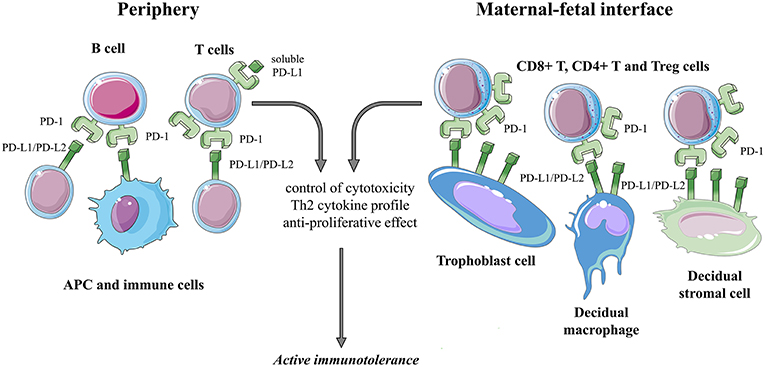
PD-1 is a transmembrane receptor expressed by e.g., T cells, B cells, natural killer (NK) cells, antigen presenting cells (5, 88). PD-1 generates a strong inhibitory signal upon binding to its ligands PD-L1 and PD-L2, resulting in down-regulation of pro-inflammatory T-cell activity (89). PD-L1 can be found on several immune cells (resting T cells, B cells, dendritic cells, macrophages), in various tissues, like placenta, heart, spleen (5, 90, 91). In contrast to that, PD-L2 expression is limited to dendritic cells and macrophages (92). Furthermore, ligand expression of PD-1 can be regulated, e.g., through the local cytokine environment. PD-L1 expression is increased by many pro-inflammatory factors (LPS, GM-CSF, VEGF) and cytokines (IFN-γ, TNF-α) (5, 93, 94).
PD-1 in Murine Pregnancy
In the allogeneic murine pregnancy model, surface expression of PD-1 on peripheral CD4+ and CD8+ T cells was not altered after conception and during gestation (1). PD-1 blockade in vivo was shown to enhance the proliferation of CD4+ and CD8+ T cells in unmated and pregnant mice and to erase the protective effect of Treg cells in Treg treated abortion-prone animals (1, 22).
There are only a few but very informative data about the local presence of PD-1 in murine pregnancy showing PD-1 expression by a broad spectrum of decidual lymphocyte subsets including CD4+ T cells, CD8+ T cells, T follicular helper cells, γδ T cells, NK, and NKT-like cells (1, 68, 95, 96). Furthermore, increased PD-1 expression by decidual NK, NKT-like, and γδ T cells was associated with the reduced cytolytic activity of these cells when compared to the periphery suggesting PD-1 dependent regulation of innate effector functions at the MFI (68).
Concerning the tissue distribution profile of the two ligands for PD-1, PD-L1, and PD-L2 at the MFI, both fetal and maternal compartments are involved: PD-L2 is expressed throughout the murine decidua, whereas PD-L1 expression is limited to the decidua basalis (97). Insufficient data exist about PD-L expression by the trophoblast suggesting PD-L1 expression by the syncytiotrophoblast but not by trophoblastic giant cells, which are next to the decidua basalis (97, 98). Therefore, PD-1 interaction with its ligands may occur in the decidua itself and is not affected by the fetal part. In vivo blockade of PD-1 ligands in allogenic murine pregnancy highlighted the functional role of the PD-1/PD-L1 pathway since anti-PD-L1 treatment resulted in increased fetal resorption rate and a reduction in the litter size, whereas PD-L2 blockade had no effect on fetal resorption (97). At the MFI, PD-L1 blockade resulted in infiltration of T cells, complement deposits, and higher levels of IFN-γ suggesting T cell-mediated rejection mechanisms locally. Another supportive report on the protective role of the PD-1/PD-L1 interaction in maternal-fetal tolerance was revealed by observations of the PD-L1 deficient pregnant mice, which showed similar results in fetal resorption rate, litter size, and a shift toward Th17 emphasizing the role of PD-L1 expressing regulatory T cells controlling fetal antigen-specific maternal T cell (99, 100). Yet, data are conflicting since in another experimental setting neither PD-L1 nor PD-1 deficient mice had significant alterations in gestational or in neonatal offspring parameters (101). These findings indicate doubt about the role of the PD-1/PD-L1 pathway in the survival of the fetal allograft in mice and further studies are needed to reconcile previous controversial results.
PD-1 in Human Pregnancy (Figure 3)
PD-1 at the Periphery
Although immunological acceptance of the fetus is primarily based on maternal tolerance mechanisms at the MFI locally, it exerts a significant impact on systemic immunity as well (102). Despite the fact that syncytiotrophoblast cells—which are bathed in maternal blood—express PD-L1 and PD-L2, data about possible changes in the PD-1 mediated systemic immune response during human pregnancy compared to healthy, non-pregnant controls are lacking (90, 103). The only information regarding this topic is that the frequency of PD-1 expressing T lymphocytes is elevated in the blood of healthy pregnant women compared to non-pregnant counterparts and soluble PD-L1 levels increase throughout gestation (78).
PD-1 at the Maternal-Fetal Interface
Immunofluorescent studies revealed a significantly higher PD-1 expression by decidual T lymphocytes similarly to non-pregnant endometrial T cells. This increase in PD-1 expression was demonstrated more in detail when compared to the periphery during pregnancy: decidual CD8+, CD4+, and regulatory T cells were shown to enhance PD-1 expression (104).
As already mentioned before, PD-L1 and PD-L2 are present in the placenta throughout pregnancy (90, 103, 105). In the first trimester, the major fetal source of PD-L1 is the villous syncytiotrophoblast and extravillous cytotrophoblast, while PD-L2 expression is much more restricted to villous cytotrophoblast (103, 105). On the maternal side, decidual stromal cells constitutively express both PD-L1 and PD-L2, but Th1 cytokines can further enhance their surface expression. PD-L1 expression by decidual macrophages is evident as well (104, 106).
PD-1 interaction with its ligand PD-L1 in different co-culture experiments resulted in reduced Th1 cytokine production by CD4+ T cells (104, 106). These findings suggest the contribution of the PD-1 mediated pathway to the establishment of a favorable Th2 type immune balance at the FMI in healthy human pregnancy.
PD-1 in Pregnancy Complications
Limited information is available about the involvement of the PD-1/PD-L pathway in pregnancy disorders. In preeclampsia, the percentage of PD-1 positive regulatory T cells was significantly higher than in healthy pregnancy with no difference in their PD-L1 expression. While the ratio of PD-1+ Th17 cells was not altered, the PDL1 expression by Th17 cells increased. PD-1 expression by CD3+, and CD4+ T cells did not significantly differ suggesting dysregulated PD-1/PD-L1 axis within the Treg/Th17 imbalance in the clinical phase of preeclampsia (47, 107).
In the case of RSA, decidual PD-L1 expression was significantly reduced on both mRNA as well as on protein level compared to healthy first-trimester decidua, while PD-1 expression by decidual lymphocytes showed no difference (108).
TIM-3 /PD-1 Co-expression
Effector cells of the immune system can be characterized by co-expression of different co-inhibitory molecules (109). In human pregnancy, TIM-3+PD-1+ CD8+ T cells preferentially accumulate in the decidua (71). Upregulation of both TIM-3 and PD-1 by decidual CD8+ T cells might be induced by embryonic trophoblast in an HLA-C dependent manner (71). These double positive cells display higher proliferative activity and produce more Th2 type cytokines than their TIM-3/PD-1 double negative CD8+ counterparts (71). Blocking both co-receptors increased cytotoxicity and decreased Th2 type cytokine production of TIM-3+PD-1+ CD8+ T cells suggesting a protective, anti-inflammatory role of TIM-3 and PD-1 co-expressing decidual CD8+ T lymphocytes at the MFI (71). This hypothesis is strengthened by further observations both in human as well as in mice. In murine pregnancy systemic blockade of both TIM-3 and PD-1 in vivo resulted in further reduction of fetal growth and litter size when compared to the blockade of either TIM-3 or PD-1 alone (71). In line with these findings, in patients with RSA, dual expression of TIM-3 and PD-1 by decidual CD8+ T cells was found to be significantly reduced and to be less proliferative in contrast to a healthy pregnancy (71).
Summary
Immune checkpoint molecules have a major impact on cellular immunity by limiting inflammatory immune response and thereby maintaining physiologic tissue conditions. From this point of view, maternal-fetal immunotolerance represents a real immunological challenge for the immune system of the mother where accurate, tight, and dynamic immune control is required for healthy pregnancy proceeding from the time of implantation on.
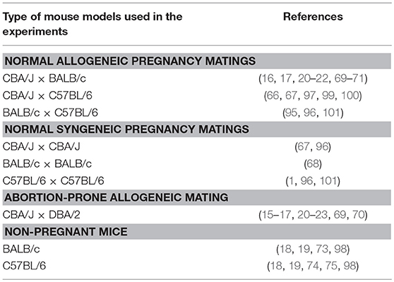
As presented in this review in detail, the possible role of immune checkpoint molecules in the establishment of maternal-fetal immunotolerance has been extensively studied. In the case of CTLA-4, TIM-3, and PD-1, the participation and possible role of these molecules in maternal immune response have been confirmed by different approaches, e.g., animal and human experiments, in vitro and in vivo studies. Table 1 shows the different mice strains used in animal experiments. However, data are sometimes conflicting and not comprehensive, which may be due to experimental setting differences, small sample size, and the highly complex and multilevel characteristics of immune cell activation. When considering the involvement of co-activatory molecules in maternofetal immune interactions, the co-signaling network is far more complicated (110).
Although there are no current clinical trials aiming at immune checkpoint molecules and interactions in pregnancy complications, some results of the studies discussed in this paper indicate the possible role of TIM-3 cell surface expression rate and serum levels of the soluble ligands PD-L1 and galectin-9 as potential biomarkers for screening during pregnancy (57, 76, 78).
Immune checkpoint inhibitors targeting the PD-1/PD-L1 and CTLA-4 pathways are revolutionary therapeutics in advanced malignancies and could be used in the treatment of chronic viral infections (HIV, HCV) as well in the future. Because of lacking a (68) adequate and well-controlled studies and based on the findings in mouse models, where blockade of the PD-1/PD-L1 pathway resulted in the adverse effect on pregnancy, checkpoint inhibitors are relatively contraindicated for the treatment of metastatic cancer in pregnant women requiring an individualized decision in each case (22, 97, 100, 111, 112).
Although intensive research and a large amount of information regarding the involvement of immune checkpoint molecules in reproductive immunology, the puzzle is not complete. Our current knowledge is quite deficient since there are several other immune checkpoint molecules described recently: Lymphocyte-activated gene-3 (LAG-3), T cell immunoreceptor with Ig and ITIM domains (TIGIT), B and T lymphocytes attenuator (BTLA), V-domain Ig suppressor of T cell activation (VISTA) are novel members of immune checkpoint molecules with proven immune regulatory activity (113). Until today, studies regarding this new generation of negative checkpoint regulators in the field of reproductive immunology are missing and urgently needed.
Author Contributions
EM writing, original draft preparation. MM and AB original draft preparation. KD table and figure preparation and editing. LS writing, review, and editing.
Funding
This work was supported by National Research, Development, and Innovation Office (NKFIH K119529 and PD112465), by the PTE ÁOK KA Research Grant (KA-2018-07 and KA-2018-18), by grants of GINOP-2.3.2-15-201600021, EFOP-3.6.3-VEKOP-16-2017-00009, and 20765-3/2018/FEKUTSTRAT.
Conflict of Interest Statement
The authors declare that the research was conducted in the absence of any commercial or financial relationships that could be construed as a potential conflict of interest.
References
1. Shepard MT, Bonney EA. PD-1 Regulates T cell proliferation in a tissue and subset-specific manner during normal mouse pregnancy. Immunol Invest. (2013) 42:385–408. doi: 10.3109/08820139.2013.782317
2. Matzinger P. Friendly and dangerous signals: is the tissue in control? Nat Immunol. (2007) 8:11–3. doi: 10.1038/ni0107-11
3. Haskins K, Kappler J, Marrack P. The major histocompatibility complex-restricted antigen receptor on T cells. Annu Rev Immunol. (1984) 2:51–66. doi: 10.1146/annurev.iy.02.040184.000411
4. Hao H, He M, Li J, Zhou Y, Dang J, Li F, et al. Upregulation of the Tim-3/Gal-9 pathway and correlation with the development of preeclampsia. Eur J Obstet Gynecol Reprod Biol. (2015) 194:85–91. doi: 10.1016/j.ejogrb.2015.08.022
5. Meggyes M, Szanto J, Lajko A, Farkas B, Varnagy A, Tamas P, et al. The possible role of CD8+/Vα7.2+/CD161++ T (MAIT) and CD8+/Vα7.2+/CD161 lo T (MAIT-like) cells in the pathogenesis of early-onset pre-eclampsia. Am J Reprod Immunol. (2018) 79:e12805. doi: 10.1111/aji.12805
6. Jiang Y, Li Y, Zhu B. T-cell exhaustion in the tumor microenvironment. Cell Death Dis. (2015) 6:e1792. doi: 10.1038/cddis.2015.162
7. Dai S, Jia R, Zhang X, Fang Q, Huang L. The PD-1/PD-Ls pathway and autoimmune diseases. Cell Immunol. (2014) 290:72–9. doi: 10.1016/j.cellimm.2014.05.006
8. Walker LSK. EFIS lecture: understanding the CTLA-4 checkpoint in the maintenance of immune homeostasis. Immunol Lett. (2017) 184:43–50. doi: 10.1016/j.imlet.2017.02.007
9. Saito S, Sasaki Y, Sakai M. CD4+CD25high regulatory T cells in human pregnancy. J Reprod Immunol. (2005) 65:111–20. doi: 10.1016/j.jri.2005.01.004
10. Sasaki Y. Decidual and peripheral blood CD4+CD25+ regulatory T cells in early pregnancy subjects and spontaneous abortion cases. Mol Hum Reprod. (2004) 10:347–53. doi: 10.1093/molehr/gah044
11. Chambers CA, Kuhns MS, Egen JG, Allison JP. C TLA−4-M ediated inhibition in regulation of T cell r esponses : mechanisms and manipulation in tumor immunotherapy. Annu Rev Immunol. (2001) 19:565–94. doi: 10.1146/annurev.immunol.19.1.565
12. Finger EB, Bluestone JA. When ligand becomes receptor—tolerance via B7 signaling on DCs. Nat Immunol. (2002) 3:1056–7. doi: 10.1038/ni1102-1056
13. Grohmann U, Orabona C, Fallarino F, Vacca C, Calcinaro F, Falorni A, et al. CTLA-4-Ig regulates tryptophan catabolism in vivo. Nat Immunol. (2002) 3:1097–101. doi: 10.1038/ni846
14. Walker LSK, Sansom DM. Confusing signals: recent progress in CTLA-4 biology. Trends Immunol. (2015) 36:63–70. doi: 10.1016/j.it.2014.12.001
15. Jin L-P, Zhou Y-H, Wang M-Y, Zhu X-Y, Li D-J. Blockade of CD80 and CD86 at the time of implantation inhibits maternal rejection to the allogeneic fetus in abortion-prone matings. J Reprod Immunol. (2005) 65:133–46. doi: 10.1016/j.jri.2004.08.009
16. Zhou WH, Dong L, Du MR, Zhu XY, Li DJ. Cyclosporin A improves murine pregnancy outcome in abortion-prone matings: involvement of CD80/86 and CD28/CTLA-44. Reproduction. (2008) 135:385–95. doi: 10.1530/REP-07-0063
17. Zhu X-Y, Zhou Y-H, Wang M-Y, Jin L-P, Yuan M-M, Li D-J. Blockade of CD86 signaling facilitates a Th2 bias at the maternal-fetal interface and expands peripheral CD4+CD25+ regulatory T cells to rescue abortion-prone fetuses1. Biol Reprod. (2005) 72:338–45. doi: 10.1095/biolreprod.104.034108
18. Judge TA, Wu Z, Zheng XG, Sharpe AH, Sayegh MH, Turka LA. The role of CD80, CD86, and CTLA4 in alloimmune responses and the induction of long-term allograft survival. J Immunol. (1999) 162:1947–51.
19. Yamada A, Kishimoto K, Dong VM, Sho M, Salama AD, Anosova NG, et al. CD28-independent costimulation of T cells in alloimmune responses. J Immunol. (2001) 167:140–6. doi: 10.4049/jimmunol.167.1.140
20. Li W, Li B, Fan W, Geng L, Li X, Li L, et al. CTLA4Ig gene transfer alleviates abortion in mice by expanding CD4+CD25+regulatory T cells and inducing indoleamine 2,3-dioxygenase. J Reprod Immunol. (2009) 80:1–11. doi: 10.1016/j.jri.2008.11.006
21. Li W, Li B, Li S. Adenovirus mediated CTLA4Ig transgene therapy alleviates abortion by inhibiting spleen lymphocyte proliferation and regulating apoptosis in the feto-placental unit. J Reprod Immunol. (2013) 97:167–74. doi: 10.1016/j.jri.2013.01.002
22. Wafula PO, Teles A, Schumacher A, Pohl K, Yagita H, Volk HD, et al. PD-1 but not CTLA-4 blockage abrogates the protective effect of regulatory T cells in a pregnancy murine model. Am J Reprod Immunol. (2009) 62:283–92. doi: 10.1111/j.1600-0897.2009.00737.x
23. Rüter J, Hoffmann T, Heiser U, Demuth HU, Arck PC, Klapp BF, et al. The expression of T-cell surface antigens CTLA-4, CD26, and CD28 is modulated by inhibition of dipeptidylpeptidase IV (DPP IV, CD26) activity in murine stress-induced abortions. Cell Immunol. (2002) 220:150–6. doi: 10.1016/S0008-8749(03)00028-5
24. Rüter J, Demuth H, Arck PC, Hofmann T, Klapp BF. Inhibition of dipeptidylpeptidase IV (DPP IV, CD26) activity modulates surface expression of CTLA-4 in stress-induced abortions. (2003)155–163.
25. Somerset DA, Zheng Y, Kilby MD, Sansom DM, Drayson MT. Normal human pregnancy is associated with an elevation in the immune suppressive CD25+ CD4+ regulatory T-cell subset. Immunology. (2004) 112:38–43. doi: 10.1111/j.1365-2567.2004.01869.x
26. Miwa N, Hayakawa S, Miyazaki S, Myojo S, Sasaki Y, Sakai M, et al. IDO expression on decidual and peripheral blood dendritic cells and monocytes/macrophages after treatment with CTLA-4 or interferon-γ increase in normal pregnancy but decrease in spontaneous abortion. Mol Hum Reprod. (2006) 11:865–70. doi: 10.1093/molehr/gah246
27. Heikkinen J, Möttönen M, Alanen A, Lassila O. Phenotypic characterization of regulatory T cells in the human decidua. Clin Exp Immunol. (2004) 136:373–8. doi: 10.1111/j.1365-2249.2004.02441.x
28. Dimova T, Nagaeva O, Stenqvist AC, Hedlund M, Kjellberg L, Strand M, et al. Maternal foxp3 expressing CD4+CD25+and CD4+CD25-regulatory T-cell populations are enriched in human early normal pregnancy decidua: a phenotypic study of paired decidual and peripheral blood samples. Am J Reprod Immunol. (2011) 66:44–56. doi: 10.1111/j.1600-0897.2011.01046.x
29. Kaufman KA, Bowen JA, Tsai AF, Bluestone JA, Hunt JS, Ober C. The CTLA-4 gene is expressed in placental fibroblasts. Mol Hum Reprod. (1999) 5:84–7. Available at: http://www.ncbi.nlm.nih.gov/pubmed/10050667 [Accessed January 9, 2019]
30. Jin LP, Fan DX, Zhang T, Guo PF, Li DJ. The costimulatory signal upregulation is associated with Th1 bias at the maternal-fetal interface in human miscarriage. Am J Reprod Immunol. (2011) 66:270–8. doi: 10.1111/j.1600-0897.2011.00997.x
31. Jin LP, Chen QY, Zhang T, Guo PF, Li DJ. The CD4+CD25brightregulatory T cells and CTLA-4 expression in peripheral and decidual lymphocytes are down-regulated in human miscarriage. Clin Immunol. (2009) 133:402–10. doi: 10.1016/j.clim.2009.08.009
32. Fan Q, Zhang J, Cui Y, Wang C, Xie Y, Wang Q, et al. The synergic effects of CTLA-4/Foxp3-related genotypes and chromosomal aberrations on the risk of recurrent spontaneous abortion among a Chinese Han population. J Hum Genet. (2018) 63:579–87. doi: 10.1038/s10038-018-0414-2
33. Gupta R, Prakash S, Parveen F, Agrawal S. Association of CTLA-4 and TNF-α polymorphism with recurrent miscarriage among North Indian women. Cytokine. (2012) 60:456–62. doi: 10.1016/j.cyto.2012.05.018
34. Misra MK, Mishra A, Phadke SR, Agrawal S. Association of functional genetic variants of CTLA4 with reduced serum CTLA4 protein levels and increased risk of idiopathic recurrent miscarriages. Fertil Steril. (2016) 106:1115–23.e6. doi: 10.1016/j.fertnstert.2016.06.011
35. Nasiri M, Rasti Z. CTLA-4 and IL-6 gene polymorphisms: risk factors for recurrent pregnancy loss. Hum Immunol. (2016) 77:1271–4. doi: 10.1016/j.humimm.2016.07.236
36. Wang X, Lin Q, Ma Z, Hong Y, Zhao A, Di W, et al. Association of the A/G polymorphism at position 49 in exon 1 of CTLA-4 with the susceptibility to unexplained recurrent spontaneous abortion in the Chinese population. Am J Reprod Immunol. (2005) 53:100–5. doi: 10.1111/j.1600-0897.2004.00251.x
37. Wang X, Ma Z, Hong Y, Lu P, Lin Q. Expression of CD28 and cytotoxic T lymphocyte antigen 4 at the maternal-fetal interface in women with unexplained pregnancy loss. Int J Gynecol Obstet. (2006) 93:123–9. doi: 10.1016/j.ijgo.2006.01.027
38. Jääskeläinen E, Toivonen S, Keski-Nisula L, Paattiniemi EL, Helisalmi S, Punnonen K, et al. CTLA-4 polymorphism 49A-G is associated with placental abruption and preeclampsia in Finnish women. Clin Chem Lab Med. (2008) 46:169–73. doi: 10.1515/CCLM.2008.034
39. Anjos S, Nguyen A, Ounissi-Benkalha H, Tessier M-C, Polychronakos C. A common autoimmunity predisposing signal peptide variant of the cytotoxic T-lymphocyte antigen 4 results in inefficient glycosylation of the susceptibility allele. J Biol Chem. (2002) 277:46478–86. doi: 10.1074/jbc.M206894200
40. Ligers A, Teleshova N, Masterman T, Huang W-X, Hillert J. CTLA-4 gene expression is influenced by promoter and exon 1 polymorphisms. Genes Immun. (2001) 2:145–152. doi: 10.1038/sj.gene.6363752
41. Chistiakov DA, Turakulov RI. CTLA-4 and its role in autoimmune thyroid disease. J Mol Endocrinol. (2003) 31:21–36. doi: 10.1677/jme.0.0310021
42. Toldi G, Švec P, Vásárhelyi B, Mészáros G, Rigó J, Tulassay T, et al. Decreased number of FoxP3+ regulatory T cells in preeclampsia. Acta Obstet Gynecol Scand. (2008) 87:1229–33. doi: 10.1080/00016340802389470
43. Steinborn A, Haensch GM, Mahnke K, Schmitt E, Toermer A, Meuer S, et al. Distinct subsets of regulatory T cells during pregnancy: is the imbalance of these subsets involved in the pathogenesis of preeclampsia? Clin Immunol. (2008) 129:401–12. doi: 10.1016/j.clim.2008.07.032
44. Prins JR, Boelens HM, Heimweg J, Van der Heide S, Dubois AE, Van Oosterhout AJ, et al. Preeclampsia is associated with lower percentages of regulatory T cells in maternal blood. Hypertens Pregnancy. (2009) 28:300–11. doi: 10.1080/10641950802601237
45. Darmochwal-Kolarz D, Saito S, Rolinski J, Tabarkiewicz J, Kolarz B, Leszczynska-Gorzelak B, et al. Activated T lymphocytes in pre-eclampsia. Am J Reprod Immunol. (2007) 58:39–45. doi: 10.1111/j.1600-0897.2007.00489.x
46. Sasaki Y, Darmochwal-Kolarz D, Suzuki D, Sakai M, Ito M, Shima T, et al. Proportion of peripheral blood and decidual CD4+ CD25bright regulatory T cells in pre-eclampsia. Clin Exp Immunol. (2007) 149:139–45. doi: 10.1111/j.1365-2249.2007.03397.x
47. Toldi G, Vásárhelyi B, Biró E, Fügedi G, Rigó J, Molvarec A. B7 costimulation and intracellular indoleamine-2,3-dioxygenase expression in peripheral blood of healthy pregnant and pre-eclamptic women. Am J Reprod Immunol. (2013) 69:264–71. doi: 10.1111/aji.12069
48. Miko E, Szereday L, Barakonyi A, Jarkovich A, Varga P, Szekeres-Bartho J. Immunoactivation in preeclampsia: Vδ2+ and regulatory T cells during the inflammatory stage of disease. J Reprod Immunol. (2009) 80:100–8. doi: 10.1016/j.jri.2009.01.003
49. Boij R, Mjösberg J, Svensson-Arvelund J, Hjorth M, Berg G, Matthiesen L, et al. Regulatory T-cell subpopulations in severe or early-onset preeclampsia. Am J Reprod Immunol. (2015) 74:368–78. doi: 10.1111/aji.12410
50. Samsami Dehaghani A, Doroudchi M, Kalantari T, Pezeshki AM, Ghaderi A. Heterozygosity in CTLA-4 gene and severe preeclampsia. Int J Gynecol Obstet. (2005) 88:19–24. doi: 10.1016/j.ijgo.2004.09.007
51. Zhou J, Wang Z, Zhao X, Wang J, Sun H, Hu Y. An increase of treg cells in the peripheral blood is associated with a better in vitro fertilization treatment outcome. Am J Reprod Immunol. (2012) 68:100–6. doi: 10.1111/j.1600-0897.2012.01153.x
52. Verma N, Burns SO, Walker LSK, Sansom DM. Immune deficiency and autoimmunity in patients with CTLA-4 (CD152) mutations. Clin Exp Immunol. (2017) 190:1–7. doi: 10.1111/cei.12997
53. Lo B, Abdel-Motal UM. Lessons from CTLA-4 deficiency and checkpoint inhibition. Curr Opin Immunol. (2017) 49:14–9. doi: 10.1016/j.coi.2017.07.014
54. Lajko A, Meggyes M, Polgar B, Szereday L. The immunological effect of Galectin-9/TIM-3 pathway after low dose Mifepristone treatment in mice at 14.5 day of pregnancy. PLoS ONE. (2018) 13:e0194870. doi: 10.1371/journal.pone.0194870
55. Chabtini L, Mfarrej B, Mounayar M, Zhu B, Batal I, Dakle PJ, et al. TIM-3 regulates innate immune cells to induce fetomaternal tolerance. J Immunol. (2013) 190:88–96. doi: 10.4049/jimmunol.1202176
56. Sánchez-Fueyo A, Tian J, Picarella D, Domenig C, Zheng XX, Sabatos CA, et al. Tim-3 inhibits T helper type 1–mediated auto- and alloimmune responses and promotes immunological tolerance. Nat Immunol. (2003) 4:1093–101. doi: 10.1038/ni987
57. Meggyes M, Miko E, Polgar B, Bogar B, Farkas B, Illes Z, et al. Peripheral blood TIM-3 positive NK and CD8+ T cells throughout pregnancy: TIM-3/galectin-9 interaction and its possible role during pregnancy. PLoS ONE. (2014) 9:e92371. doi: 10.1371/journal.pone.0092371
58. Hastings WD, Anderson DE, Kassam N, Koguchi K, Greenfield EA, Kent SC, et al. TIM-3 is expressed on activated human CD4+ T cells and regulates Th1 and Th17 cytokines. Eur J Immunol. (2009) 39:2492–501. doi: 10.1002/eji.200939274
59. Zhao J, Lei Z, Liu Y, Li B, Zhang L, Fang H, et al. Human pregnancy up-regulates Tim-3 in innate immune cells for systemic immunity. J Immunol. (2009) 182:6618–24. doi: 10.4049/jimmunol.0803876
60. van de Weyer PS, Muehlfeit M, Klose C, Bonventre JV, Walz G, Kuehn EW. A highly conserved tyrosine of Tim-3 is phosphorylated upon stimulation by its ligand galectin-9. Biochem Biophys Res Commun. (2006) 351:571–6. doi: 10.1016/j.bbrc.2006.10.079
61. Lee J, Su EW, Zhu C, Hainline S, Phuah J, Moroco JA, et al. Phosphotyrosine-dependent coupling of Tim-3 to T-cell receptor signaling pathways. Mol Cell Biol. (2011) 31:3963–74. doi: 10.1128/MCB.05297-11
62. von Wolff M, Wang X, Gabius H-J, Strowitzki T. Galectin fingerprinting in human endometrium and decidua during the menstrual cycle and in early gestation. MHR Basic Sci Reprod Med. (2004) 11:189–94. doi: 10.1093/molehr/gah144
63. Seki M, Oomizu S, Sakata K, Sakata A, Arikawa T, Watanabe K, et al. Galectin-9 suppresses the generation of Th17, promotes the induction of regulatory T cells, and regulates experimental autoimmune arthritis. Clin Immunol. (2008) 127:78–88. doi: 10.1016/j.clim.2008.01.006
64. Zhu C, Anderson AC, Schubart A, Xiong H, Imitola J, Khoury SJ, et al. The Tim-3 ligand galectin-9 negatively regulates T helper type 1 immunity. Nat Immunol. (2005) 6:1245–52. doi: 10.1038/ni1271
65. Sabatos CA, Chakravarti S, Cha E, Schubart A, Sánchez-Fueyo A, Zheng XX, et al. Interaction of Tim-3 and Tim-3 ligand regulates T helper type 1 responses and induction of peripheral tolerance. Nat Immunol. (2003) 4:1102–10. doi: 10.1038/ni988
66. Chabtini L, Mfarrej B, Mounayar M, Zhu B, Batal I, Dakle PJ, et al. TIM-3 regulates innate immune cells to induce fetomaternal tolerance. J Immunol. (2013) 190:88–96. doi: 10.4049/jimmunol.1202176
67. Tripathi S, Chabtini L, Dakle PJ, Smith B, Akiba H, Yagita H, et al. Effect of TIM-3 blockade on the immunophenotype and cytokine profile of murine uterine NK cells. PLoS ONE. (2015) 10:e0123439. doi: 10.1371/journal.pone.0123439
68. Meggyes M, Lajko A, Palkovics T, Totsimon A, Illes Z, Szereday L, et al. Feto-maternal immune regulation by TIM-3/galectin-9 pathway and PD-1 molecule in mice at day 14.5 of pregnancy. Placenta. (2015) 36:1153–60. doi: 10.1016/j.placenta.2015.07.124
69. Li Y-H, Zhou W-H, Tao Y, Wang S-C, Jiang Y-L, Zhang D, et al. The galectin-9/Tim-3 pathway is involved in the regulation of NK cell function at the maternal–fetal interface in early pregnancy. Cell Mol Immunol. (2016) 13:73–81. doi: 10.1038/cmi.2014.126
70. Wang S, Zhu X, Xu Y, Zhang D, Li Y, Tao Y, et al. Programmed cell death-1 (PD-1) and T-cell immunoglobulin mucin-3 (Tim-3) regulate CD4+ T cells to induce Type 2 helper T cell (Th2) bias at the maternal-fetal interface. Hum Reprod. (2016) 31:700–11. doi: 10.1093/humrep/dew019
71. Wang S-C, Li Y-H, Piao H-L, Hong X-W, Zhang D, Xu Y-Y, et al. PD-1 and Tim-3 pathways are associated with regulatory CD8+ T-cell function in decidua and maintenance of normal pregnancy. Cell Death Dis. (2015) 6:e1738. doi: 10.1038/cddis.2015.112
72. Ndhlovu LC, Lopez-Verges S, Barbour JD, Jones RB, Jha AR, Long BR, et al. Tim-3 marks human natural killer cell maturation and suppresses cell-mediated cytotoxicity. Blood. (2012) 119:3734–43. doi: 10.1182/blood-2011-11-392951
73. Monney L, Sabatos CA, Gaglia JL, Ryu A, Waldner H, Chernova T, et al. Th1-specific cell surface protein Tim-3 regulates macrophage activation and severity of an autoimmune disease. Nature. (2002) 415:536–41. doi: 10.1038/415536a
74. Uchida Y, Ke B, Freitas MCS, Yagita H, Akiba H, Busuttil RW, et al. T-Cell immunoglobulin mucin-3 determines severity of liver ischemia/reperfusion injury in mice in a TLR4-dependent manner. Gastroenterology. (2010) 139:2195–206. doi: 10.1053/j.gastro.2010.07.003
75. Ashkar AA, Black GP, Wei Q, He H, Liang L, Head JR, et al. Assessment of requirements for IL-15 and IFN regulatory factors in uterine NK cell differentiation and function during pregnancy. J Immunol. (2003) 171:2937–44. doi: 10.4049/jimmunol.171.6.2937
76. Li Y, Zhang J, Zhang D, Hong X, Tao Y, Wang S, et al. Tim-3 signaling in peripheral NK cells promotes maternal-fetal immune tolerance and alleviates pregnancy loss. Sci Signal. (2017) 10:eaah4323. doi: 10.1126/scisignal.aah4323
77. Miko E, Meggyes M, Bogar B, Schmitz N, Barakonyi A, Varnagy A, et al. Involvement of galectin-9/TIM-3 pathway in the systemic inflammatory response in early-onset preeclampsia. PLoS ONE. (2013) 8:e71811. doi: 10.1371/journal.pone.0071811
78. Enninga EAL, Harrington SM, Creedon DJ, Ruano R, Markovic SN, Dong H, et al. Immune checkpoint molecules soluble program death ligand 1 and galectin-9 are increased in pregnancy. Am J Reprod Immunol. (2018) 79:e12795. doi: 10.1111/aji.12795
79. Keskin DB, Allan DSJ, Rybalov B, Andzelm MM, Stern JNH, Kopcow HD, et al. TGFbeta promotes conversion of CD16+ peripheral blood NK cells into CD16- NK cells with similarities to decidual NK cells. Proc Natl Acad Sci USA. (2007) 104:3378–83. doi: 10.1073/pnas.0611098104
80. Eriksson M, Meadows SK, Wira CR, Sentman CL. Unique phenotype of human uterine NK cells and their regulation by endogenous TGF-β. J Leukoc Biol. (2004) 76:667–75. doi: 10.1189/jlb.0204090
81. Sun J, Yang M, Ban Y, Gao W, Song B, Wang Y, et al. Tim-3 is upregulated in NK cells during early pregnancy and inhibits NK cytotoxicity toward trophoblast in galectin-9 dependent pathway. PLoS ONE. (2016) 11:e0147186. doi: 10.1371/journal.pone.0147186
82. Wang S, Cao C, Piao H, Li Y, Tao Y, Zhang X, et al. Tim-3 protects decidual stromal cells from toll-like receptor-mediated apoptosis and inflammatory reactions and promotes Th2 bias at the maternal-fetal interface. Sci Rep. (2015) 5:9013. doi: 10.1038/srep09013
83. Kuchroo VK, Meyers JH, Umetsu DT, DeKruyff RH. TIM family of genes in immunity and tolerance. in Frederick W. Alt, editor. Advances in Immunology. (2006) Academic press. p. 227–249. doi: 10.1016/S0065-2776(06)91006-2
84. Wu M, Zhu Y, Zhao J, Ai H, Gong Q, Zhang J, et al. Soluble costimulatory molecule sTim3 regulates the differentiation of Th1 and Th2 in patients with unexplained recurrent spontaneous abortion. Int J Clin Exp Med. (2015) 8:8812–9.
85. Xu J, Yang Y, Liu X, Wang Y. The −1541 C>T and +4259 G>T of TIM-3 polymorphisms are associated with rheumatoid arthritis susceptibility in a Chinese Hui population. Int J Immunogenet. (2011) 38:513–8. doi: 10.1111/j.1744-313X.2011.01046.x
86. Shen Y, Wang C, Hong D, Zeng B, Fang C, Yuan C, et al. The relationship between polymorphisms in the promoter region of Tim-3 and unexplained recurrent spontaneous abortion in Han Chinese women. Reprod Biol Endocrinol. (2013) 11:104. doi: 10.1186/1477-7827-11-104
87. Li J, Li F, Zuo W, Zhou Y, Hao H, Dang J, et al. Up-regulated expression of Tim-3/Gal-9 at maternal-fetal interface in pregnant woman with recurrent spontaneous abortion. J Huazhong Univ Sci Technol. (2014) 34:586–90. doi: 10.1007/s11596-014-1320-2
88. Keir ME, Butte MJ, Freeman GJ, Sharpe AH. PD-1 and its ligands in tolerance and immunity. Annu Rev Immunol. (2008) 26:677–704. doi: 10.1146/annurev.immunol.26.021607.090331
89. Chikuma S. Basics of PD-1 in self-tolerance, infection, and cancer immunity. Int J Clin Oncol. (2016) 21:448–55. doi: 10.1007/s10147-016-0958-0
90. Petroff MG, Chen L, Phillips TA, Azzola D, Sedlmayr P, Hunt JS. B7 family molecules are favorably positioned at the human maternal-fetal interface1. Biol Reprod. (2003) 68:1496–504. doi: 10.1095/biolreprod.102.010058
91. Veras E, Kurman RJ, Wang T-L, Shih I-M. PD-L1 Expression in human placentas and gestational trophoblastic diseases. Int J Gynecol Pathol. (2016) 36:146–53. doi: 10.1097/PGP.0000000000000305
92. Lewkowich IP, Lajoie S, Stoffers SL, Suzuki Y, Richgels PK, Dienger K, et al. PD-L2 modulates asthma severity by directly decreasing dendritic cell IL-12 production. Mucosal Immunol. (2013) 6:728–39. doi: 10.1038/mi.2012.111
93. Radvanyi L, Pilon-Thomas S, Peng W, Sarnaik A, Mulé JJ, Weber J, et al. Antagonist antibodies to PD-1 and B7-H1 (PD-L1) in the treatment of advanced human cancer–letter. Clin Cancer Res. (2013) 19:5541. doi: 10.1158/1078-0432.CCR-13-1054
94. Kondo A, Yamashita T, Tamura H, Zhao W, Tsuji T, Shimizu M, et al. Interferon-gamma and tumor necrosis factor-alpha induce an immunoinhibitory molecule, B7-H1, via nuclear factor-kappaB activation in blasts in myelodysplastic syndromes. Blood. (2010) 116:1124–31. doi: 10.1182/blood-2009-12-255125
95. Zeng W, Liu Z, Zhang S, Ren J, Ma X, Qin C, et al. Characterization of T follicular helper cells in allogeneic normal pregnancy and PDL1 blockage-induced abortion. Sci Rep. (2016) 6:36560. doi: 10.1038/srep36560
96. Barton BM, Xu R, Wherry EJ, Porrett PM. Pregnancy promotes tolerance to future offspring by programming selective dysfunction in long-lived maternal T cells. J Leukoc Biol. (2017) 101:975–87. doi: 10.1189/jlb.1A0316-135R
97. Guleria I, Khosroshahi A, Ansari MJ, Habicht A, Azuma M, Yagita H, et al. A critical role for the programmed death ligand 1 in fetomaternal tolerance. J Exp Med. (2005) 202:231–7. doi: 10.1084/jem.20050019
98. Liang SC, Latchman YE, Buhlmann JE, Tomczak MF, Horwitz BH, Freeman GJ, et al. Regulation of PD-1, PD-L1, and PD-L2 expression during normal and autoimmune responses. Eur J Immunol. (2003) 33:2706–16. doi: 10.1002/eji.200324228
99. D'Addio F, Riella LV, Mfarrej BG, Chabtini L, Adams LT, Yeung M, et al. The link between the PDL1 costimulatory pathway and Th17 in fetomaternal tolerance. J Immunol. (2011) 187:4530–41. doi: 10.4049/jimmunol.1002031
100. Habicht A, Dada S, Jurewicz M, Fife BT, Yagita H, Azuma M, et al. A link between PDL1 and T regulatory cells in fetomaternal tolerance. J Immunol. (2007) 179:5211–9. doi: 10.4049/jimmunol.179.8.5211
101. Taglauer ES, Yankee TM, Petroff MG. Maternal PD-1 regulates accumulation of fetal antigen-specific CD8+ T cells in pregnancy. J Reprod Immunol. (2009) 80:12–21. doi: 10.1016/j.jri.2008.12.001
102. Borzychowski AM, Croy BA, Chan WL, Redman CWG, Sargent IL. Changes in systemic type 1 and type 2 immunity in normal pregnancy and pre-eclampsia may be mediated by natural killer cells. Eur J Immunol. (2005) 35:3054–63. doi: 10.1002/eji.200425929
103. Petroff MG, Kharatyan E, Torry DS, Holets L. The immunomodulatory proteins B7-DC, B7-H2, and B7-H3 are differentially expressed across gestation in the human placenta. Am J Pathol. (2005) 167:465–73. doi: 10.1016/S0002-9440(10)62990-2
104. Taglauer ES, Trikhacheva AS, Slusser JG, Petroff MG. Expression and function of PDCD1 at the human maternal-fetal interface. Biol Reprod. (2008) 79:562–9. doi: 10.1095/biolreprod.107.066324
105. Petroff MG, Chen L, Phillips TA, Hunt JS. B7 family molecules: novel immunomodulators at the maternal-fetal interface. Placenta. (2002) 23:S95–101. doi: 10.1053/plac.2002.0813
106. Nagamatsu T, Schust DJ, Sugimoto J, Barrier BF. Human decidual stromal cells suppress cytokine secretion by allogenic CD4+ T cells via PD-1 ligand interactions. Hum Reprod. (2009) 24:3160–71. doi: 10.1093/humrep/dep308
107. Tian M, Zhang Y, Liu Z, Sun G, Mor G, Liao A. The PD-1/PD-L1 inhibitory pathway is altered in pre-eclampsia and regulates T cell responses in pre-eclamptic rats. Sci Rep. (2016) 6:27683. doi: 10.1038/srep27683
108. Li G, Lu C, Gao J, Wang X, Wu H, Lee C, et al. Association between PD-1/PD-L1 and T regulate cells in early recurrent miscarriage. Int J Clin Exp Pathol. (2015) 8:6512–8.
109. Blackburn SD, Shin H, Haining WN, Zou T, Workman CJ, Polley A, et al. Coregulation of CD8+ T cell exhaustion by multiple inhibitory receptors during chronic viral infection. Nat Immunol. (2009) 10:29–37. doi: 10.1038/ni.1679
110. Xu Y-Y, Wang S-C, Li D-J, Du M-R. Co-signaling molecules in maternal-fetal immunity. Trends Mol Med. (2017) 23:46–58. doi: 10.1016/j.molmed.2016.11.001
111. Poulet FM, Wolf JJ, Herzyk DJ, DeGeorge JJ. An evaluation of the impact of PD-1 pathway blockade on reproductive safety of therapeutic PD-1 inhibitors. Birth Defects Res Part B Dev Reprod Toxicol. (2016) 107:108–19. doi: 10.1002/bdrb.21176
112. Johnson DB, Sullivan RJ, Menzies AM. Immune checkpoint inhibitors in challenging populations. Cancer. (2017) 123:1904–11. doi: 10.1002/cncr.30642
Keywords: immune checkpoint molecule, reproductive immunology, pregnancy, immunotolerance, CTLA-4, TIM-3, PD-1
Citation: Miko E, Meggyes M, Doba K, Barakonyi A and Szereday L (2019) Immune Checkpoint Molecules in Reproductive Immunology. Front. Immunol. 10:846. doi: 10.3389/fimmu.2019.00846
Received: 23 January 2019; Accepted: 01 April 2019;
Published: 18 April 2019.
Edited by:
Nandor Gabor Than, Hungarian Academy of Sciences (MTA), HungaryReviewed by:
Jolan Eszter Walter, University of South Florida, United StatesAlexander Steinkasserer, University Hospital Erlangen, Germany
Attila Kumanovics, Mayo Clinic, United States
Copyright © 2019 Miko, Meggyes, Doba, Barakonyi and Szereday. This is an open-access article distributed under the terms of the Creative Commons Attribution License (CC BY). The use, distribution or reproduction in other forums is permitted, provided the original author(s) and the copyright owner(s) are credited and that the original publication in this journal is cited, in accordance with accepted academic practice. No use, distribution or reproduction is permitted which does not comply with these terms.
*Correspondence: Eva Miko, bWlrby5ldmFAcHRlLmh1
 Eva Miko
 Matyas Meggyes1,2
 Laszlo Szereday


