
94% of researchers rate our articles as excellent or good
Learn more about the work of our research integrity team to safeguard the quality of each article we publish.
Find out more
ORIGINAL RESEARCH article
Front. Genet., 19 October 2023
Sec. Livestock Genomics
Volume 14 - 2023 | https://doi.org/10.3389/fgene.2023.1113411
This article is part of the Research TopicApplication of Genomics in Livestock Populations under Selection or ConservationView all 20 articles
 Changchun Lin1,2
 Weimin Wang3
 Deyin Zhang3
 Kai Huang3Yukun Zhang3Xiaolong Li3
 Yuan Zhao3Liming Zhao3Jianghui Wang1Bubo Zhou1
 Jiangbo Cheng3Dan Xu1Wenxin Li1
 Xiaoxue Zhang1*Wenxin Zheng2*
Feed efficiency (FE), an important economic trait in sheep production, is indirectly assessed by residual feed intake (RFI). However, RFI in sheep is varied, and the molecular processes that regulate RFI are unclear. It is thus vital to investigate the molecular mechanism of RFI to developing a feed-efficient sheep. The miRNA-sequencing (RNA-Seq) was utilized to investigate miRNAs in liver tissue of 6 out of 137 sheep with extreme RFI phenotypic values. In these animals, as a typical metric of FE, RFI was used to distinguish differentially expressed miRNAs (DE_miRNAs) between animals with high (n = 3) and low (n = 3) phenotypic values. A total of 247 miRNAs were discovered in sheep, with four differentially expressed miRNAs (DE_miRNAs) detected. Among these DE_miRNAs, three were found to be upregulated and one was downregulated in animals with low residual feed intake (Low_RFI) compared to those with high residual feed intake (High_RFI). The target genes of DE_miRNAs were primarily associated with metabolic processes and biosynthetic process regulation. Furthermore, they were also considerably enriched in the FE related to glycolysis, protein synthesis and degradation, and amino acid biosynthesis pathways. Six genes were identified by co-expression analysis of DE_miRNAs target with DE_mRNAs. These results provide a theoretical basis for us to understand the sheep liver miRNAs in RFI molecular regulation.
Feed efficiency (FE), an important economic trait in sheep production, is indirectly assessed by residual feed intake (RFI) and feed conversion ratio (FCR) (Carberry et al., 2012; Zhang et al., 2017a; Claffey et al., 2018; McGovern et al., 2018). RFI is defined as the discrepancy between the amount of feed actually consumed and amount anticipated to be needed for maintenance and growth (Mebratie et al., 2019). Improved FE has the potential to reduce meat production costs, with feed and feeding-related expenses accounting for 75% of total variable production costs in beef cattle farming (Ahola and Hill, 2012). Zhang et al. (2017b) showed in indoor sheep husbandry, feed expenditures account for 65%–70% of overall costs. In addition, research has been shown that enhancing ruminant FE may effectively mitigate greenhouse gas emissions and provide positive environmental outcomes (Nkrumah et al., 2006; Hegarty et al., 2007; Deng et al., 2018). Thus, livestock producers have a keen interested in the domain of genetic selection and breeding, particularly with regard to enhancing FE of their animals. However, the precise definition of FE in animals is currently being disputed, due to imperfect quality of ratios such as FCR (Gunsett, 1984). Therefore, RFI serves only as a metric for evaluating FE within the context of animal production. RFI is influenced by a variety of internal and external environmental variables such as body composition, nutrition digestion and metabolism, energy expenditure, physical activity, and control of body temperature (Zhang et al., 2017b). Recently, there has been a growing interest in the subject of FE within the context of livestock and poultry production, and researches on RFI-related genes have mainly focused on swine (Do et al., 2014; Jing et al., 2015; Horodyska et al., 2017; Messad et al., 2019), cattle (Santana et al., 2014; Salleh et al., 2018) and poultry (Yi et al., 2015). Animals with low_RFI exhibit reduced feed intake, and resulting in decreasedless waste and generation, which do not affect the body size, productivity, or weight of the animals (Koch et al., 1963). Therefore, studying the mechanisms of Low_RFI animals will not only reduce costs but also benefit the environment. The liver, being a vital digestive gland and metabolic organ (Xing et al., 2019), plays an important part in the metabolism of lipids, carbohydrates, and glucose metabolism, and has crucial physiological roles in oxidation, metabolism and reduction (El-Badawy et al., 2019; Cigrovski Berkovic et al., 2020; Ndiaye et al., 2020). Given the essential function played by the liver in the metabolic processes of livestock and poultry, it was selected as the sample in this present research.
MicroRNAs (miRNAs) are a class of small (∼22 nucleotides) endogenous non-coding RNAs that exhibit a high degree of conservation across different species (Nelson et al., 2011). The miRNAs have been discovered in several physiological fluids, tissues, and cell types, where they play a crucial role in regulating gene expression at the post-transcriptional level, and they are associated with a wide range of important biological processes (Halushka et al., 2018). Previous studies has shown that miRNA exert control over gene expression by their binding to particular messenger RNA (mRNA), which ultimately leads to the subsequent destruction or inhibition of the targeted transcript. miRNA is associated with the regulation of almost all cellular and developmental processes in eukaryotes (Nejad et al., 2018). For instance, it has been shown that miR-1, miR-133a, miR-133b, and miR-206 exhibit increased expression throughout the advanced phases of human of human fetal muscle development (Koutsoulidou et al., 2011). The miR-33 has inhibitory effects on the process of fatty acid breakdown by targeting several genes involved in fatty acid β-oxidation (Gerin et al., 2010). Moreover, miRNAs has been demonstrated to be essential for the development of brain structures and to support critical systems that, if disturbed, may lead to or cause neurodevelopmental disorders (Hollins et al., 2014). Previous studies have shown that a number of miRNAs in the liver tissues of various livestock play an important role in influencing FE. For instance, miR-338 influences fatty acid synthase (Xing et al., 2019), miR-185 affects glucose and lipid metabolism (Li et al., 2016), and miR-545-3p in pig liver affects fat deposition (Chu et al., 2017). In the liver of cattle, miR-19b regulates lipid metabolism of fat, miR-122-3p influences hepatic cholesterol and lipid metabolism, and miR-143 affects insulin signaling and glucose homeostasis (Al-Husseini et al., 2016). However, the precise processes via which miRNAs regulate RFI in sheep have yet to be fully unclear.
Thus, the present study aimed to identify candidate miRNAs that regulate FE to breed a Low_RFI Hu sheep population. We used sequencing to determine transcription differences in liver tissue of sheep with extreme RFI phenotypes.
The animal studies were done in accordance with the regulations and guidelines set out by the government of Gansu Province, as well as with the approval of the Animal Health and Ethics Committee of Gansu Agricultural University (Animal Experimentation License No. 2012-2-159).
The experimental animals used in this study have been comprehensively detailed, together with their corresponding management regimens, in previous publications (Zhang et al., 2017b; Zhang et al., 2019). To put it simply, a total of 137 male Hu sheep were obtained from Jinchang Zhongtian Sheep Co., Ltd. (Jinchang China) and transported to Minqin Zhongtian Sheep Co., Ltd. (Minqin China) during the same time frame for the purpose of breeding. The process of weaning was established when the lambs reached 56 days of age. Subsequently, during the initial phases of the experimental study, only lambs displaying resilient development and overall outstanding health were selected as candidates. The lambs were supplied with nourishment in a standardized single pen (0.8 × 1 m), whereby they access to fresh water and food every day until the end of the experiment (180 days of age). The lambs attained an ideal age of 80 days for the start of the official experiment, which was recorded as day one. The performance experiment is concluded when the lambs reach 180 days, hence rendering the official duration of the trial as 100 days. Consequently, all lambs participating in this study had a 2-week transitional period prior before a 10 days pre-feeding phase. Throughout the transitional phase, a regular percentage adjustment was made to the form of feed utilized each day. Additionally, the whole pelleted feed was consumed on the initial day of the pre-fed phase. During the first 10-day pre-fed period and ensuing 100-day official trial, the pellet feed was purchased from Gansu Sanyang Jinyuan Animal Husbandry Co., Ltd., (Gansu China).
Lambs were treated to a feeding regimen structured in 20-day cycles until the completion of the feeding experiment (180 days of age), with their initial weight being measured on the first day of the specified period (80 days of age). The lambs underwent daily weighing before to feeding, using a calibrated electronic scale. No modifications were made to the participants or the equipment utilized over the whole period of the experiment. Furthermore, each sheep was weighed for remaining feed before each weighing period, which was used to calculate feed intake and RFI. For the computational model used in this study, the main reference was the formula of Zhang et al. (2017b). The specific formula used in this particular instance was as follows:
where Yk represents the average daily feed intake of the ith individual; β0 regres-sion intercept; β1 regression coefficient for mid-test metabolic body weight (MBW); β2 regression coefficient for average daily gain (ADG); ek represents uncontrolled error of the ith individual; BWf represents final body weight; BWi represents initial body weight; and N, experimental period (days).
The methodologies used for the collection and processing methods of the tissues were cited from previous scholarly investigations (Zhang et al., 2017b). The methodology may be concisely summarized in the following manner: all lambs are uniformly transferred to a professional slaughterhouse after the end of the measurement. The RFI values of the six sheep used in this study are provided fully, concerning prior research conducted by our research group. The lambs were subjected to a 24 h of fasting period before to being weighed and then executed in standard procedures. Each liver sample was immediately collected after slaughter process and then preserved in liquid nitrogen for temporary storage. Following the process of slaughter, it was transferred to −80°C for long-term storage until RNA was extracted. As described in previous study, we selected 3 High_RFI and 3 Low_RFI sheep from 137 male Hu lambs for total RNA extraction (Zhang et al., 2017b; Zhang et al., 2019). The total RNA extraction was performed using the TRIzol Reagent (Invitrogen, Waltham, MA, United States) method as per the provided instructions. The NanoPhotometer® spectrophotometer (IMPLEN, CA, United States) was used to quantify the purity of RNA, while the integrity of RNA was performed using the Agilent Bioanalyzer 2,100 system’s RNA Nano 6000 Assay Kit (Agilent Technologies, CA, United States).
The small RNA library preparation kits were used to produce small RNA sequencing libraries (Galina-Pantoja et al., 2006). There are many steps that involved, as seen below: firstly, whole RNA molecule was used as a template to directly connect the 3′ and 5′ ends of the small RNA with the synthetic adaptors. The synthesis cDNA from total RNA was conducted with M-MuLV reverse transcriptase (NEB, United States), followed by amplification of the resulting cDNA in accordance with the recommended protocols provided by Illumina. The PCR products underwent purification and recovery processes using 8% polyacrylamide gels. These gels exhibited the capability to effectively separate DNA fragments with sizes 140 to 160 base pairs. Following that, the DNA fragments that had undergone purification were dissolved in 8 μL of elution solution. In the end, the evaluation of the library’s integrity on the Agilent Bioanalyzer 2,100 system may be conducted by using DNA high-sensitivity chip. Once the library has undergone qualification, the product was subjected to sequencing on the Illumina HiSeq 2,500 platform, resulting in the generation of a 50 bp single-ended read.
The raw data collected by the sequencing equipment was then given a Base Calling analysis with the intention of producing FASTQ files. The acquisition of clean data included the elimination of extraneous information from the raw data via the use of a customized Perl script. Concurrently, Q20, Q30, and GC-contents of the raw data were obtained. Then, for all subsequent analysis, choose certain range of length from clean reads. The Bowtie (Langmead et al., 2009) method was used for the purpose of aligning small RNA tags with the reference sequence, facilitating the assessment of their expression levels on the reference. Then used miRbase 20.0 as a reference to mapping known miRNAs. In order to exclude protein-coding genes, repetitive sequences, rRNA, tRNA, snRNA, and snoRNA, custom scripts are used to extract miRNAs of predetermined length. Following, the software tools miREvo (Wen et al., 2012) and mirdeep2 (Friedländer et al., 2012) were used to map sequences with the sheep reference genome (Oar_v1.0) to provide predictions about novel miRNAs. The use of the whole rRNA ratio served as an indicator of sample quality. In animal samples, this ratio should ideally be below 40%. Additionally, the cumulative p values for RNA folding were used as a metric for output measure.
The miRNA expression levels were estimated by TPM (transcript per million) (Zhou et al., 2010). The processing procedure differs according on the sample’s quality. To perform differential expression analysis on Low_RFI and High_RFI samples with biological recurrence, use the DESeq R package (version 1.8.3). The filtering conditions for differential expression of miRNAs in this study were p-value <0.05 and |log2 (foldchange)|≤ 0.5 (Tang et al., 2007). The threshold for substantial differential expression is set to the default value. The target gene of miRNA was then predicted for animals using miRanda (Enright et al., 2003). Statistics calculations were carried out to analyze the expression levels of the most prevalent known miRNAs and novel miRNAs in both experimental groups. Additionally, the most common miRNA or DE_miRNA expression was estimated in relation to the anticipated mRNA expression of its target gene.
The target gene candidates of DE_miRNAs were analyzed using Gene Ontology (GO) enrichment analysis (“target gene candidates” in the follows). For GO enrichment analysis, a GOseq-based Wallenius non-central hyper-geometric distribution (Young et al., 2010) was used, which might correct for gene length bias. Kyoto Encyclopedia of Genes and Genomes (KEGG) (Kanehisa et al., 2008) was a database used to understand the advanced functions and benefits of biological systems based on molecular-level information, especially genome sequencing and other highly experimental technologies (http://www.genome.jp/kegg/). To assess the statistical enrichment of target gene candidates in KEGG pathways, we employed the KOBAS (Mao et al., 2005) tool.
Investigate the possible association between DE_miRNA found in this study and Zhang et al. describes DE_mRNA (Cigrovski Berkovic et al., 2020). The construction of a miRNA-mRNA interaction network was facilitated by using Cytoscape software (Shannon et al., 2003). This network was established by including DE_miRNAs and DE_mRNAs based on their specific roles. Specifically, mRNAs exhibiting discernible association with miRNAs were integrated into the miRNA-mRNA interaction network. The same shape with various colors shows types that are up-or downregulated in DE_miRNAs and DE_mRNAs.
3.2 Illumina sequencing generated more than 10 M (million) high quality raw reads for each of the two groups of Hu sheep (except for High_RFI No.1 Hu sheep) (Table 1). The raw data obtained from sequencing is given to a filtering process so as to generate clean reads. Reads with more than 10% N content were removed first. An average of 124 reads was removed in the Low_RFI group, less than 0.01%, and similarly an average of 133 reads was removed in the High_RFI group, less than 0.01% (Supplementary Table S1). Further, after removing low-quality readings, the Low_RFI group averaged 0.32% and the High_RFI group 0.35% being removed (Supplementary Table S1). The number of deletions resulting from the existence of 5 adapter contamination and the presence of ployA/T/G/C was low, with an average 0.00% and 0.03% in the Low_RFI group 0.00% and 0.06% in the High_RFI group, respectively (Supplementary Table S1). More reads were deleted due to the absence of 3 adapter null or insert null, about 0.97% and 0.95%. All samples were filtered to retain clean reads of 98% or more. Finally, the clean reads of each sample were screened for sRNAs within a certain length range (18∼35 nt) for subsequent analysis (Table 1). The length-screened sRNAs were localized to the Ovis aries reference genome to analyze the distribution of small RNAs (Table 1). From the clean data, a total of 10,278,758, 10,014,172, 9,533,396, 8,699,934, 9,391,798, and 13,765,901 mapped reads from the LR1 (Low_RFI No.1 Hu sheep), LR2 (Low_RFI No.2 Hu sheep), LR3 (Low_RFI No.3 Hu sheep), HR1 (High_RFI No.1 Hu sheep), HR2 (High_RFI No.2 Hu sheep), and HR3 (High_RFI No.3 Hu sheep) libraries were retrieved, with over 90% mapping to the Ovis aries reference genome (Table 1). In terms of miRNA expression level, our research results indicate that genes with a TPM <60 retrieved from RNA-seq accounted for about 75% of the total, whereas high-expressed genes, that is, genes with TPM >60, accounted for around 25% (Table 2). However, HR2 accounted for only 7.81%. Screening miRNAs ranged from 18 to 35 nt in length (Figures 1A, B). Furthermore, a variety of non-coding RNAs (ncRNAs) were discovered, including transfer RNAs (tRNAs), snRNAs and miRNAs (Figures 1C, D). Among the identified ncRNAs, a minute fraction constituted recently discovered miRNAs.
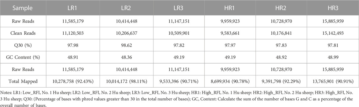
TABLE 1. Summary of clean reads mapped to the Ovis aries reference genome.
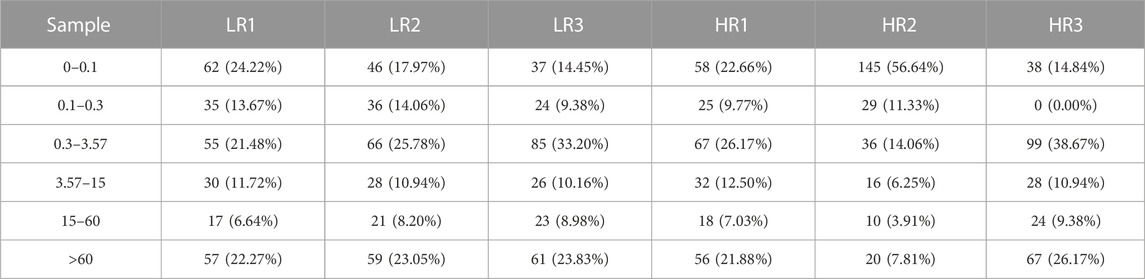
TABLE 2. Analysis of miRNA expression levels.
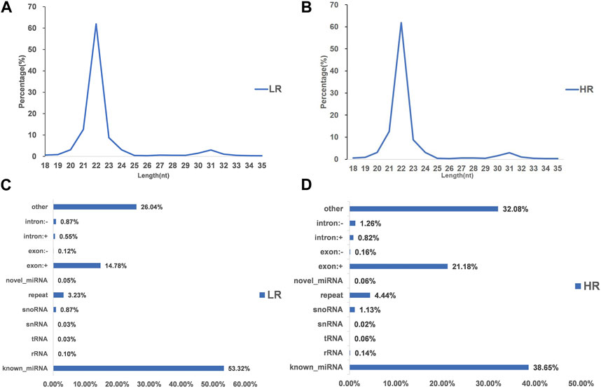
FIGURE 1. Characterization of microRNA (miRNA) profiling and the percentage of detected miRNAs from ncRNAs. (A, B) Length distribution of clean reads from identified miRNA fragments. (C, D) Categories of identified non-coding RNAs (ncRNAs) via sequencing in Low_FRI and High_FRI. Note “LR: Low_RFI, HR: High_RFI” (the following figures are identical).
We identified 121, 120, and 122 known miRNAs in the High_RFI samples, and 119, 83, and 128 known miRNAs in the Low_RFI samples (Table 3). In all of these reads, approximately 59% of known miRNAs were detected in all samples (Supplementary Table S2). The miRNA the highest abundance across all samples was oar-miR-148a, with mean paired reads from HR1, HR2, HR3, LR1, LR2, and LR3 of 2,885,255, 1,771,013, 2,178,809, 3,049,001, 7,921,051, and 1,323,994, respectively. Among the top 20 expressed miRNAs in each group, the expression levels of 10 miRNAs such as oar-miR-148a, oar-let-7f, oar-miR-143, oar-miR-30a-5p, oar-miR-26a, oar-miR-21, oar-let-7g, oar-let-7i, oar-miR-30d and oar-miR-99a accounted for an average of more than 94% (Supplementary Table S3). The expression analysis shows the top 20 highly expressed miRNAs in the study samples from each respective group (Supplementary Figure S1).

TABLE 3. Number and ratio of identified miRNA matrices.
The hairpin structure that is characteristic of miRNA precursors may be used as a means to forecast the existence of novel miRNAs. We identified 73, 90, and 97 novel miRNAs in the High_RFI samples, and 79, 28, and 90 novel miRNAs in the Low_RFI samples (Table 3). Among the novel that were identified, only 23 were identified in all samples (Supplementary Table S3). Of the 117 unique novel miRNAs, novel_31, novel_50 and novel_41were the most expressed in the samples with an average of 1,826, 1,246 and 1,173 reads aligned to these miRNAs, respectively (Supplementary Table S3). All detected new miRNAs were supplied (Supplementary Table S3), and the top 20 expressed novel miRNAs for each group were presented (Supplementary Table S4).
The sequencing data has been submitted to the NCBI Sequence Read Archive (SRA) database under the biological project PRJNA813431. Differential miRNA expression analysis between Low_RFI and High_RFI groups from the same population of sheep with different phenotypic values. The relevant phenotypic data for the selected sheep were detailed in previous studies (Zhang et al., 2022). Phenotypic differences were not significant except for FCR, RFI and feed intake (FI). In total, an average of 11.62 million raw read were obtained from each sample. A total of 247 miRNAs were detected in 6 liver samples, of which four miRNAs (one known miRNA and three novel miRNAs) were identified as differentially expressed (p < 0.05) (Table 4). The Venn diagrams show miRNAs that are uniquely expressed or co-expressed in different groups, with 205 miRNAs co-expressed in both groups (Figure 2A). Four miRNAs were differentially expressed in the Low_RFI group compared to the High_RFI group, including three upregulated and a downregulated (p < 0.05) (Table 4) (Figure 2B). Of all the DE_miRNAs identified, novel_41 and novel_115 were expressed in all samples. The novel_41 was upregulated in Low_RFI group, while novel_115 showed downregulated compared to the High_RFI group (Table 4).
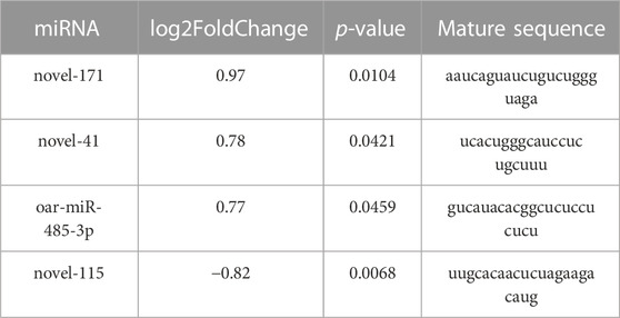
TABLE 4. Four differentially expressed miRNAs in Hu sheep with Low and High_RFI.
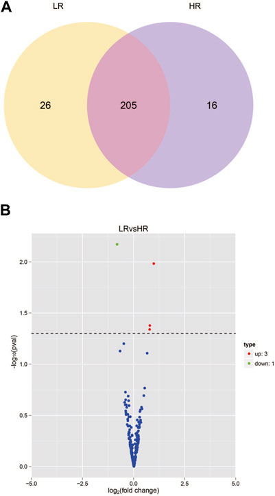
FIGURE 2. miRNA expression in the liver of sheep with different RFI. (A) Venn diagram showing the total number of miRNAs expressed in each group individually and in both. (B) Volcano map of differentially expressed miRNAs. In the volcano plot, significant downregulated genes are indicated by “green” dots, while significant upregulated genes are indicated by “red” dots.
The target genes of ten most highly expressed miRNAs (seven known and three novel) were predicted in the two groups for further functional analysis (Supplementary Table S5). The majority of these target genes mainly involved in various biological processes, including glycolysis, protein synthesis and catabolism, cell growth and proliferation, scavenging of free radicals, as well as cell death and survival (Supplementary Figure S2). In terms of glycolytic processes, the target genes were mainly involved in nucleoside diphosphate kinase activity, nucleoside kinase activity, adenylate kinase activity, hexokinase activity, and other glucose binding activities. For protein synthesis and catabolism, target genes were involved in the positive regulation of protein secretion, protein polymerization and protein deubiquitination, among other roles. For cell growth and proliferation, target genes were involved in platelet alpha granulation, cell cortex proliferation, and the proliferation and development of microtubule cell ribosomes (Supplementary Figure S2).
To further understand the biological functions and roles of these four DE_miRNAs, target genes were identified (miRDB) for highly differentiated miRNAs between Low_RFI and High_RFI gruops. Upregulated miRNAs in Low_RFI group were integrated to several target genes: top ranking genes were DZANK1, CYP26B1, IDH3G, TRAC, IL36RN, UBE2Z, ZMYND12, PDGFD, VSIG2, and ELK4, whereas top-ranking target genes with downregulated miRNAs were CFAP221, HLA-DOB, TOP2B, ACSL4, FRYL, RAB3GAP2, TSPAN9, ILKAP, USP13, and PRR36. The provided diagram illustrates the top 20 anticipated target genes for the four DE_miRNAs (Figure 3). To further elucidate the functions of the DE_miRNAs, we performed enrichment analysis of their candidate target genes. GO enrichment results showed that these target genes were mainly related to metabolism and binding: Biological processes: metabolism, organic metabolism, primary metabolism and macromolecular metabolism. Cellular components: intracellular, intracellular fractions, organelles and membrane-bound organelles. Molecular functions: binding, protein binding, catalytic activity and Hydrolytic enzyme activity (Figure 4A). It was shown that the metabolism in the liver plays an important role in the efficiency of animal feed. KEGG pathway showed DE_miRNAs target genes were mainly enriched in transcriptional dysregulation in herpes simplex virus infection, leishmaniasis, and biosynthesis of amino acids (Figure 4B).
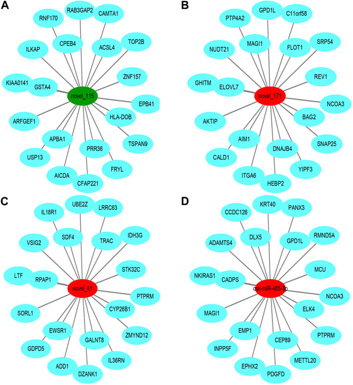
FIGURE 3. Top ranked (based on total target score of miRDB) DE_miRNAs for target genes. In the network plot, significant downregulated genes are indicated by green, while significant upregulated genes are indicated by red. The target gene predicted by psRobottar is shown in blue.
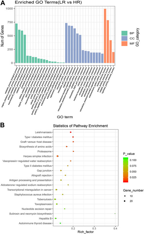
FIGURE 4. Enrichment analysis of high and Low_RFI differential miRNAs. (A) Using GO (Gene Ontology) enrichment analysis, BP indicates biological process, CC indicates cellular component, MF indicates molecular function. (B) Kyoto Encyclopedia of Genes and Genomes (KEGG) differentially expressed miRNAs target gene enrichment.
To fully understand the potential RFI effects of miRNA, we used DE_miRNA and their targets genes to create an interactive population network. A total of 1423 DE_miRNAs target genes were identified. We previously discovered 101 DE_mRNAs between the low and high RFI sheep groups using the same objective as the present investigation (Zhang et al., 2022). Among these, seven miRNAs were co-expressed (Figure 5A). However, some target genes were predicted to be the target genes of a single miRNA, and it was observed that upregulation of DE_miRNAs did not always result in downregulation of DE_mRNAs in liver tissue obtained from animals with Low_RFI. Most DE_miRNAs were predicted to primarily target a single differential target gene. As shown in figure, the upregulated miRNA novel_171 targets the upregulated target gene RTP4, and the upregulated miRNA novel_41 targets the downregulated target gene CD274 (Figure 5B). However, the upregulated miRNA oar-miR-485-3p targeted three different genes, the downregulated target gene OAS1 and the two upregulated target genes SHISA3 and PLEKHH2.
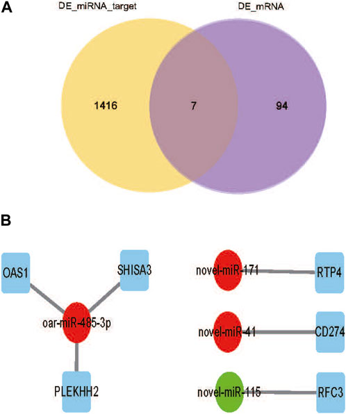
FIGURE 5. (A) Venn diagram showing the number of differential mRNA target genes in yellow, the number of differential miRNA candidate target genes in purple, and the overlapping part indicates the co-expressed part. (B) DE_miRNAs are indicated by circles, significant downregulated genes are indicated by green, while significant upregulated genes are indicated by red. And squares indicate DE_miRNAs co-expressing target genes with DE_mRNA.s.
RNA sequencing can serve as a powerful miRNAs expression profiling tool to identify the DE_miRNAs(Motameny et al., 2010), even at low expression levels in all cells, as well as allows for the parallel analysis of known miRNAs and the identification of miRNAs (Pritchard et al., 2012). Along with the simultaneous analysis of known miRNAs, the examination of novel miRNAs also becomes feasible. Furthermore, the use of mature miRNA sequences may facilitate the identification of prospective target genes for both known and undiscovered miRNAs. In this study, miRNAs sequencing was used to identify miRNA expression profiles in liver tissue from 6 Hu sheep with extreme RFI from the same farm. Sequencing results showed that sequencing data were of high quality with an average Q30 value of 94%. In addition, after quality control processing of raw sequencing data, the read sequences was an average length of 22 bp, while the majority of reads f were ranged between 20 and 24 bp length from both Low_RFI and High_RFI gruops, providing a high quality and reliable data for subsequent analysis (Friedländer et al., 2012) (Figures 1A, B). The observed average alignment rate, above 90%, indicates a strong agreement between the identified miRNAs and the liver samples. Furthermore, roughly 83% of these miRNAs were found to be expressed in all liver samples, which aligns with the findings of a previous research on miRNAs in bovine liver (Mukiibi et al., 2018). This observation implies that miRNAs exhibit a high degree of conservation within a given population. Among the miRNAs that have been identified, ten highly expressed miRNAs, including oar-miR-148a, oar-let-7f, oar-miR-143, oar-miR-30a-5p, oar-miR-26a, oar-miR-21, oar-let-7g, oar-let-7i, oar-miR-30d and oar-miR-99a, accounted for an average of 92.14% and 95.92% of the total aligned sequence reads in the High and Low_RFI groups, respectively. According to a publication, let-7 miRNA has been detected in various animal species, including humans (Lee et al., 2016). In the present study, it was shown that oar-let-7f, oar-let-7g and oar-let-7i which are members of the let-7 family in sheep, had highly expressed levels in the liver of both groups of FRI sheep. This finding suggests that let-7 family of miRNAs was substantially conserved. The description was consistent with the previously reported results (Friedman et al., 2009). Therefore, from this we speculate that let-7 family miRNAs have the same trend in the same species. Interestingly, oar-miR-148a was the most highly expressed miRNA in all samples, and it belongs to the miR-148/152 family, whose homologous members are involved in a variety of biological functions and diseases in different species. For example, it has been reported that overexpression of miR-148 significantly promotes myogenic differentiation in C2C12-derived myoblasts and primary myoblasts (Zhang et al., 2012). In sheep, miR-148a have been reported to accelerate lipogenic differentiation of sheep preadipocytes and inhibit the proliferation of sheep preadipocytes by inhibiting PTEN expression (Jin et al., 2021). Furthermore, it had an inhibitory effect on the proliferation of Hu sheep hair papilla cells and was associated with hair follicle growth and development (Lv et al., 2019).
To explore the biological significance of sheep-associated DE_miRNAs with varying RFI characteristics. We performed target gene prediction for ten miRNAs that were highly expressed in two groups of RFI sheep. Among these target genes, the main biological functions involved include: negative term regulation of the apoptotic process, cell growth and proliferation, apoptosis and survival, and adipocyte differentiation. Some miRNAs with higher abundance have been discovered as significant regulators of animal cell proliferation and development, apoptosis, and regeneration, which is consistent with our results (Wang et al., 2009). As an example, the second highest expressed miRNA in our research, namely, oar-miR-30a-5p, has been previously associated to lipid and insulin metabolism in mice (Sud et al., 2017; Kim et al., 2019). miR-26a and miR-143 are involved in the regulation of mouse hepatocyte proliferation, a significant aspect in liver tissue regeneration (Geng et al., 2016; Zhou et al., 2019). miR-99a and miR-148a (Gailhouste et al., 2013) were identified as regulators hepatic detoxification in liver tissues of mice and human animals. Based on the observed of miRNA-mRNA interactions across mammalian species and our results of our study, we hypothesized that these miRNAs highly expressed in sheep liver may perform similar biological functions to other species. Moreover, since these highly expressed miRNAs are in a state of continuous self-regeneration or regeneration, it explains their involvement in proliferation as well as apoptosis and regeneration of different cells.
The liver, being the biggest internal organ, plays crucial functions in several physiological metabolic processes, including detoxification (He et al., 2020; Wang et al., 2020). It also serves as a central regulator of energy metabolism, with glycation as a fundamental feature, and is an important coordinator of metabolism and a key site for maintaining metabolic homeostasis (He et al., 2017; Matz et al., 2017; Xue et al., 2019; Moscoso and Steer, 2020). It has been reported that miRNAs are involved in almost every aspect of cell biology (Chen and Verfaillie, 2014). miRNAs play crucial biological roles in cell differentiation, proliferation, metabolism and apoptosis, as well as in viral infection (Kim et al., 2009). Hence, the variable expression of liver miRNAs in Low_RFI and High_RFI sheep might potentially lead to molecular differences in FE. In this study, one known and three novel miRNAs were identified between Low_RFI and High_RFI gruops. However, most of the detected DE_miRNAs (50%) were conservative, which is consistent with the conclusion that miRNAs are conservative (Friedman et al., 2009). For this study to validate the present findings, it would be necessary to conduct more investigations including bigger cohorts of sheep and more broad range of phenotypic animal populations, given that a lower threshold of DE_miRNA screening (p < 0.05) was used. In the present study, more than 75% of the DE_miRNAs were upregulated in Low_RFI animals, which was consistent with the results of differential expression analysis of miRNAs in beef cattle with different FE phenotypes (Mukiibi et al., 2020). Thus, this suggests that reduced expression of target genes for these miRNAs is expected.
To investigate the potential biological role of RFI-associated DE_miRNAs in sheep, we predicted their target genes. The target genes are associated with many crucial biological activities, such as metabolic processes, organic metabolism, cell assembly and structure, lipid metabolism, protein breakdown, protein binding, protein metabolism, catalytic activity, and hydrolytic enzyme activity. Among these functions, lipid metabolism and protein synthesis have been reported to be relevant in other species (Chen et al., 2011; Alexandre et al., 2015; Tizioto et al., 2015; Mukiibi et al., 2018). To further understand how DE_miRNAs interact with the 101 DE_mRNAs identified in previous studies (Zhang et al., 2019). Only seven DE_mRNAs (annotated as RTP4, CD274, OAS1, PLEKHH2, SHISA3, and RFC3) were identified as target genes for DE_miRNAs. These target DE_mRNAs play important roles in innate antiviral response, protein-coupled receptor trafficking, immunity, cell mobility, intercellular signaling and connections, cell death, cell development, differentiation, and gene regulation. Meanwhile, the RTP4 gene has been shown to be associated with RFI in sheep (Zhang et al., 2022). Certain DE_miRNAs have the potential to impact hepatic functional efficiency FE via their distinct regulatory effects on several biological processes inside the liver. According to the DE_miRNAs- mRNAs interaction network (Figure 5), some single miRNAs were predicted to be targets of single or multiple DE_mRNAs. Because a single miRNA using its seed region may bind to multiple sites in the 3′-UTR of distinct genes (mRNAs), and one target can have multiple binding sites for one or more miRNAs, miRNAs can modulate multiple biological processes even if they are few in number compared to mRNAs they regulate (Ambros, 2004; Bartel, 2004; Brennecke et al., 2005).
Overall, the comparison of DE_miRNAs and DE_mRNAs expression patterns in liver tissue that we identified was consistent with expectation. This may be attributed to the fact that miRNAs accelerate degradation of target genes by promoting the deadenylation of their target transcripts (Stroynowska-Czerwinska et al., 2014). Consequently, we have observe different patterns of DE_miRNA targeting of DE_mRNAs, perhaps attributable to variations in the regulatory mechanisms governing mRNA degradation. To better understand the relationship between miRNAs and mRNAs, further studies at the cellular level are needed to verify these interactions.
In the present study, we employed RNA-seq to analyze liver miRNAs in sheep populations. Among these miRNAs, oar-miR-148a, oar-let-7f, oar-miR-143, oar-miR-30a-5p, oar-miR-26a, oar-miR-21, oar-let-7g, oar-let-7i, oar-miR-30d and oar miR-99a had the highest expression levels in all samples. By differential miR-mRNA expression analysis, four miRNAs associated to RFI were discovered, including three novel miRNAs (novel_41, novel_115, and novel_171). Only two miRNAs (novel_41 and novel_115) were expressed in all samples, indicating that most DE_miRNAs were distinct. The predicted target genes of identified DE_miRNAs are involved in a variety of cellular and molecular functions. In addition, only 6.30% of the identified common target genes were found in the liver tissue of the same subjects. These target genes primarily regulate lipid metabolism, molecular transport, intercellular communication and connections, cell death, and survival. These results provide a theoretical basis for us to understand miRNA expression profile and the molecular mechanisms of miRNA related to FE in sheep liver.
The datasets presented in this study can be found in online repositories. The names of the repository/repositories and accession number(s) can be found in the article/Supplementary Material
All animal experiments were conducted out in compliance with the rules and recommendations of Gansu Province’s NPC government and were authorized by Gansu Agricultural University’s Animal Health and Ethics Committee. The study was conducted in accordance with the local legislation and institutional requirements.
XZ, CL, and WZ designed the study. XL, YuZ, JW, JC, DX, WL, BZ, and LZ involved in animal husbandry and liver sample collection. YkZ, DZ, KH, and WW correct the manuscript. CL and XZ analyzed the data and wrote the manuscript. All authors contributed to the article and approved the submitted version.
This work was supported by the National Key R&D Program of China (2022YFD1302000), the National for joint research on improved breeds of livestock and poultry (19210365), the West Light Foundation of the Chinese Academy of Sciences (CN), and the China Agriculture Research System (CARS-39).
The authors declare that the research was conducted in the absence of any commercial or financial relationships that could be construed as a potential conflict of interest.
All claims expressed in this article are solely those of the authors and do not necessarily represent those of their affiliated organizations, or those of the publisher, the editors and the reviewers. Any product that may be evaluated in this article, or claim that may be made by its manufacturer, is not guaranteed or endorsed by the publisher.
The Supplementary Material for this article can be found online at: https://www.frontiersin.org/articles/10.3389/fgene.2023.1113411/full#supplementary-material
SUPPLEMENTARY Table S1 | Raw data filtering information.
SUPPLEMENTARY Table S2 | Identification of all known miRNAs information.
SUPPLEMENTARY Table S3 | Identification of all novel miRNAs information.
SUPPLEMENTARY Table S4 | Top 20 expressed novel miRNAs information.
SUPPLEMENTARY Table S5 | Top 10 miRNAs target gene prediction.
SUPPLEMENTARY FIGURE S1 | Top 20 expressed known miRNAs information.
SUPPLEMENTARY FIGURE S2 | Top 10 miRNAs target gene GO enrichment.
Ahola, J. K., and Hill, R. A. (2012). Input factors affecting profitability: a changing paradigm and a challenging time: feed efficiency in the beef industry.
Alexandre, P. A., Kogelman, L. J., Santana, M. H., Passarelli, D., Pulz, L. H., Fantinato-Neto, P., et al. (2015). Liver transcriptomic networks reveal main biological processes associated with feed efficiency in beef cattle. BMC genomics 16, 1073. doi:10.1186/s12864-015-2292-8
Al-Husseini, W., Chen, Y., Gondro, C., Herd, R. M., Gibson, J. P., and Arthur, P. F. (2016). Characterization and profiling of liver microRNAs by RNA-sequencing in cattle divergently selected for residual feed intake. Asian-Australasian J. animal Sci. 29 (10), 1371–1382. doi:10.5713/ajas.15.0605
Ambros, V. (2004). The functions of animal microRNAs. Nature 431 (7006), 350–355. doi:10.1038/nature02871
Bartel, D. P. (2004). MicroRNAs: genomics, biogenesis, mechanism, and function. Cell. 116 (2), 281–297. doi:10.1016/s0092-8674(04)00045-5
Brennecke, J., Stark, A., Russell, R. B., and Cohen, S. M. (2005). Principles of microRNA-target recognition. PLoS Biol. 3 (3), e85. doi:10.1371/journal.pbio.0030085
Carberry, C. A., Kenny, D. A., Han, S., McCabe, M. S., and Waters, S. M. (2012). Effect of phenotypic residual feed intake and dietary forage content on the rumen microbial community of beef cattle. Appl. Environ. Microbiol. 78 (14), 4949–4958. doi:10.1128/AEM.07759-11
Chen, Y., and Verfaillie, C. M. (2014). MicroRNAs: the fine modulators of liver development and function. Liver Int. 34 (7), 976–990. official journal of the International Association for the Study of the Liver. doi:10.1111/liv.12496
Chen, Y., Gondro, C., Quinn, K., Herd, R. M., Parnell, P. F., and Vanselow, B. (2011). Global gene expression profiling reveals genes expressed differentially in cattle with high and low residual feed intake. Anim. Genet. 42 (5), 475–490. doi:10.1111/j.1365-2052.2011.02182.x
Chu, A. Y., Deng, X., Fisher, V. A., Drong, A., Zhang, Y., Feitosa, M. F., et al. (2017). Multiethnic genome-wide meta-analysis of ectopic fat depots identifies loci associated with adipocyte development and differentiation. Nat. Genet. 49 (1), 125–130. doi:10.1038/ng.3738
Cigrovski Berkovic, M., Virovic-Jukic, L., Bilic-Curcic, I., and Mrzljak, A. (2020). Post-transplant diabetes mellitus and preexisting liver disease - a bidirectional relationship affecting treatment and management. World J. Gastroenterol. 26 (21), 2740–2757. doi:10.3748/wjg.v26.i21.2740
Claffey, N. A., Fahey, A. G., Gkarane, V., Moloney, A. P., Monahan, F. J., and Diskin, M. G. (2018). Effect of breed and castration on production and carcass traits of male lambs following an intensive finishing period. Transl. animal Sci. 2 (4), 407–418. doi:10.1093/tas/txy070
Deng, K. D., Xiao, Y., Ma, T., Tu, Y., Diao, Q. Y., Chen, Y. H., et al. (2018). Ruminal fermentation, nutrient metabolism, and methane emissions of sheep in response to dietary supplementation with Bacillus licheniformis. Animal Feed Sci. Technol., S0377840117313950. doi:10.1016/j.anifeedsci.2018.04.014
Do, D. N., Strathe, A. B., Ostersen, T., Pant, S. D., and Kadarmideen, H. N. (2014). Genome-wide association and pathway analysis of feed efficiency in pigs reveal candidate genes and pathways for residual feed intake. Front. Genet. 5, 307. doi:10.3389/fgene.2014.00307
El-Badawy, R. E., Ibrahim, K. A., Hassan, N. S., and El-Sayed, W. M. (2019). Pterocarpus santalinus ameliorates streptozotocin-induced diabetes mellitus via anti-inflammatory pathways and enhancement of insulin function. Iran. J. basic Med. Sci. 22 (8), 932–939. doi:10.22038/ijbms.2019.34998.8325
Enright, A., John, B., Gaul, U., Tuschl, T., Biology, CSJG, and Marks, D. S. (2003). MicroRNA targets in Drosophila. MicroRNA targets Drosophila 5 (11), R1. doi:10.1186/gb-2003-5-1-r1
Friedländer, M. R., Mackowiak, S. D., Li, N., Chen, W., and Rajewsky, N. (2012). miRDeep2 accurately identifies known and hundreds of novel microRNA genes in seven animal clades. Nucleic acids Res. 40 (1), 37–52. doi:10.1093/nar/gkr688
Friedman, R. C., Farh, K. K., Burge, C. B., and Bartel, D. P. (2009). Most mammalian mRNAs are conserved targets of microRNAs. Genome Res. 19 (1), 92–105. doi:10.1101/gr.082701.108
Gailhouste, L., Gomez-Santos, L., Hagiwara, K., Hatada, I., Kitagawa, N., Kawaharada, K., et al. (2013). miR-148a plays a pivotal role in the liver by promoting the hepatospecific phenotype and suppressing the invasiveness of transformed cells. Hepatol. Baltim. Md) 58 (3), 1153–1165. doi:10.1002/hep.26422
Galina-Pantoja, L., Mellencamp, M. A., Bastiaansen, J., Cabrera, R., Solano-Aguilar, G., and Lunney, J. K. (2006). Relationship between immune cell phenotypes and pig growth in a commercial farm. Anim. Biotechnol. 17 (1), 81–98. doi:10.1080/10495390500461146
Geng, X., Chang, C., Zang, X., Sun, J., Li, P., Guo, J., et al. (2016). Integrative proteomic and microRNA analysis of the priming phase during rat liver regeneration. Gene 575 (2), 224–232. doi:10.1016/j.gene.2015.08.066
Gerin, I., Clerbaux, L. A., Haumont, O., Lanthier, N., Das, A. K., Burant, C. F., et al. (2010). Expression of miR-33 from an SREBP2 intron inhibits cholesterol export and fatty acid oxidation. J. Biol. Chem. 285 (44), 33652–33661. doi:10.1074/jbc.M110.152090
Gunsett, F. C. (1984). Linear index selection to improve traits defined as ratios. J. animal Sci. 59 (5), 1185–1193. doi:10.2527/jas1984.5951185x
Halushka, M. K., Fromm, B., Peterson, K. J., and McCall, M. N. (2018). Big strides in cellular MicroRNA expression. Trends Genet. TIG 34 (3), 165–167. doi:10.1016/j.tig.2017.12.015
He, X., Li, L., Fang, Y., Shi, W., Li, X., and Ma, H. (2017). In vivo imaging of leucine aminopeptidase activity in drug-induced liver injury and liver cancer via a near-infrared fluorescent probe. Chem. Sci. 8 (5), 3479–3483. doi:10.1039/c6sc05712h
He, B., Shi, J., Wang, X., Jiang, H., and Zhu, H. J. (2020). Genome-wide pQTL analysis of protein expression regulatory networks in the human liver. BMC Biol. 18 (1), 97. doi:10.1186/s12915-020-00830-3
Hegarty, R. S., Goopy, J. P., Herd, R. M., and McCorkell, B. (2007). Cattle selected for lower residual feed intake have reduced daily methane production. J. animal Sci. 85 (6), 1479–1486. doi:10.2527/jas.2006-236
Hollins, S. L., Goldie, B. J., Carroll, A. P., Mason, E. A., Walker, F. R., Eyles, D. W., et al. (2014). Ontogeny of small RNA in the regulation of mammalian brain development. BMC genomics 15 (1), 777. doi:10.1186/1471-2164-15-777
Horodyska, J., Hamill, R. M., Varley, P. F., Reyer, H., and Wimmers, K. (2017). Genome-wide association analysis and functional annotation of positional candidate genes for feed conversion efficiency and growth rate in pigs. PloS one 12 (6), e0173482. doi:10.1371/journal.pone.0173482
Jin, X., Hao, Z., Zhao, M., Shen, J., Ke, N., Song, Y., et al. (2021). MicroRNA-148a regulates the proliferation and differentiation of ovine preadipocytes by targeting PTEN. Animals: an open access. J. MDPI 11 (3), 820. doi:10.3390/ani11030820
Jing, L., Hou, Y., Wu, H., Miao, Y., Li, X., Cao, J., et al. (2015). Transcriptome analysis of mRNA and miRNA in skeletal muscle indicates an important network for differential Residual Feed Intake in pigs. Sci. Rep. 5, 11953. doi:10.1038/srep11953
Kanehisa, M., Araki, M., Goto, S., Hattori, M., Hirakawa, M., Itoh, M., et al. (2008). KEGG for linking genomes to life and the environment. Nucleic Acids Res. 36, D480–D484. doi:10.1093/nar/gkm882
Kim, V. N., Han, J., and Siomi, M. C. (2009). Biogenesis of small RNAs in animals. Nat. Rev. Mol. Cell. Biol. 10 (2), 126–139. doi:10.1038/nrm2632
Kim, J. Y., Jun, J. H., Park, S. Y., Yang, S. W., Bae, S. H., and Kim, G. J. (2019). Dynamic regulation of miRNA expression by functionally enhanced placental mesenchymal stem cells PromotesHepatic regeneration in a rat model with bile duct ligation. Int. J. Mol. Sci. 20 (21), 5299. doi:10.3390/ijms20215299
Koch, R. M., Swiger, L. A., Chambers, D., and Gregory, K. E. (1963). Efficiency of feed use in beef cattle. J. animal Sci. 22 (2), 486–494. doi:10.2527/jas1963.222486x
Koutsoulidou, A., Mastroyiannopoulos, N. P., Furling, D., Uney, J. B., and Phylactou, L. A. (2011). Expression of miR-1, miR-133a, miR-133b and miR-206 increases during development of human skeletal muscle. BMC Dev. Biol. 11, 34. doi:10.1186/1471-213X-11-34
Langmead, B., Trapnell, C., Pop, M., and Salzberg, S. L. (2009). Ultrafast and memory-efficient alignment of short DNA sequences to the human genome. Genome Biol. 10 (3), R25. doi:10.1186/gb-2009-10-3-r25
Lee, H., Han, S., Kwon, C. S., and Lee, D. (2016). Biogenesis and regulation of the let-7 miRNAs and their functional implications. Protein and Cell. 7 (2), 100–113. doi:10.1007/s13238-015-0212-y
Li, Y., Li, X., Sun, W. K., Cheng, C., Chen, Y. H., Zeng, K., et al. (2016). Comparison of liver microRNA transcriptomes of Tibetan and Yorkshire pigs by deep sequencing. Gene 577 (2), 244–250. doi:10.1016/j.gene.2015.12.003
Li, P., Fan, C., Cai, Y., Fang, S., Zeng, Y., Zhang, Y., et al. (2020). Transplantation of brown adipose tissue up-regulates miR-99a to ameliorate liver metabolic disorders in diabetic mice by targeting NOX4. Adipocyte 9 (1), 57–67. doi:10.1080/21623945.2020.1721970
Lv, X., Gao, W., Jin, C., Wang, L., Wang, Y., Chen, W., et al. (2019). Preliminary study on microR-148a and microR-10a in dermal papilla cells of Hu sheep. BMC Genet. 20 (1), 70. doi:10.1186/s12863-019-0770-8
Mao, X., Cai, T., Olyarchuk, J. G., and Wei, L. (2005). Automated genome annotation and pathway identification using the KEGG Orthology (KO) as a controlled vocabulary. Bioinformatics 21 (19), 3787–3793. doi:10.1093/bioinformatics/bti430
Matz, P., Wruck, W., Fauler, B., Herebian, D., Mielke, T., and Adjaye, J. (2017). Footprint-free human fetal foreskin derived iPSCs: a tool for modeling hepatogenesis associated gene regulatory networks. Sci. Rep. 7 (1), 6294. doi:10.1038/s41598-017-06546-9
McGovern, E., Kenny, D. A., McCabe, M. S., Fitzsimons, C., McGee, M., Kelly, A. K., et al. (2018). 16S rRNA sequencing reveals relationship between potent cellulolytic genera and feed efficiency in the rumen of bulls. Front. Microbiol. 9, 1842. doi:10.3389/fmicb.2018.01842
Mebratie, W., Madsen, P., Hawken, R., Romé, H., Marois, D., Henshall, J., et al. (2019). Genetic parameters for body weight and different definitions of residual feed intake in broiler chickens. Genet. Sel. Evol. 51 (1), 53. doi:10.1186/s12711-019-0494-2
Messad, F., Louveau, I., Koffi, B., Gilbert, H., and Gondret, F. (2019). Investigation of muscle transcriptomes using gradient boosting machine learning identifies molecular predictors of feed efficiency in growing pigs. BMC genomics 20 (1), 659. doi:10.1186/s12864-019-6010-9
Moscoso, C. G., and Steer, C. J. (2020). The evolution of gene therapy in the treatment of metabolic liver diseases. Genes 11 (8), 915. doi:10.3390/genes11080915
Motameny, S., Wolters, S., Nürnberg, P., and Schumacher, B. (2010). Next generation sequencing of miRNAs - strategies, resources and methods. Genes 1 (1), 70–84. doi:10.3390/genes1010070
Mukiibi, R., Vinsky, M., Keogh, K. A., Fitzsimmons, C., Stothard, P., Waters, S. M., et al. (2018). Transcriptome analyses reveal reduced hepatic lipid synthesis and accumulation in more feed efficient beef cattle. Sci. Rep. 8 (1), 7303. doi:10.1038/s41598-018-25605-3
Mukiibi, R., Johnston, D., Vinsky, M., Fitzsimmons, C., Stothard, P., Waters, S. M., et al. (2020). Bovine hepatic miRNAome profiling and differential miRNA expression analyses between beef steers with divergent feed efficiency phenotypes. Sci. Rep. 10 (1), 19309. doi:10.1038/s41598-020-73885-5
Ndiaye, H., Liu, J. Y., Hall, A., Minogue, S., Morgan, M. Y., and Waugh, M. G. (2020). Immunohistochemical staining reveals differential expression of ACSL3 and ACSL4 in hepatocellular carcinoma and hepatic gastrointestinal metastases. Biosci. Rep. 40 (4). doi:10.1042/BSR20200219
Nejad, C., Stunden, H. J., and Gantier, M. P. (2018). A guide to miRNAs in inflammation and innate immune responses. Febs J. 285 (20), 3695–3716. doi:10.1111/febs.14482
Nelson, P. T., Wang, W. X., Mao, G., Wilfred, B. R., Xie, K., Jennings, M. H., et al. (2011). Specific sequence determinants of miR-15/107 microRNA gene group targets. Nucleic Acids Res. 39 (18), 8163–8172. doi:10.1093/nar/gkr532
Nkrumah, J. D., Okine, E. K., Mathison, G. W., Schmid, K., Li, C., Basarab, J. A., et al. (2006). Relationships of feedlot feed efficiency, performance, and feeding behavior with metabolic rate, methane production, and energy partitioning in beef cattle. J. animal Sci. 84 (1), 145–153. doi:10.2527/2006.841145x
Pritchard, C. C., Cheng, H. H., and Tewari, M. (2012). MicroRNA profiling: approaches and considerations. Nat. Rev. Genet. 13 (5), 358–369. doi:10.1038/nrg3198
Salleh, S. M., Mazzoni, G., Løvendahl, P., and Kadarmideen, H. N. (2018). Gene co-expression networks from RNA sequencing of dairy cattle identifies genes and pathways affecting feed efficiency. BMC Bioinforma. 19 (1), 513. doi:10.1186/s12859-018-2553-z
Santana, M. H., Utsunomiya, Y. T., Neves, H. H., Gomes, R. C., Garcia, J. F., Fukumasu, H., et al. (2014). Genome-wide association analysis of feed intake and residual feed intake in Nellore cattle. BMC Genet. 15, 21. doi:10.1186/1471-2156-15-21
Shannon, P., Markiel, A., Ozier, O., Baliga, N. S., Wang, J. T., Ramage, D., et al. (2003). Cytoscape: a software environment for integrated models of biomolecular interaction networks. Genome Res. 13 (11), 2498–2504. doi:10.1101/gr.1239303
Stroynowska-Czerwinska, A., Fiszer, A., and Krzyzosiak, W. J. (2014). The panorama of miRNA-mediated mechanisms in mammalian cells. Cell. Mol. life Sci. CMLS 71 (12), 2253–2270. doi:10.1007/s00018-013-1551-6
Sud, N., Zhang, H., Pan, K., Cheng, X., Cui, J., and Su, Q. (2017). Aberrant expression of microRNA induced by high-fructose diet: implications in the pathogenesis of hyperlipidemia and hepatic insulin resistance. J. Nutr. Biochem. 43, 125–131. doi:10.1016/j.jnutbio.2017.02.003
Tang, Y., Ghosal, S., and Roy, A. (2007). Nonparametric bayesian estimation of positive false discovery rates. Biometrics 63 (4), 1126–1134. doi:10.1111/j.1541-0420.2007.00819.x
Tizioto, P. C., Coutinho, L. L., Decker, J. E., Schnabel, R. D., Rosa, K. O., Oliveira, P. S., et al. (2015). Global liver gene expression differences in Nelore steers with divergent residual feed intake phenotypes. BMC genomics 16 (1), 242. doi:10.1186/s12864-015-1464-x
Wang, Y., Rathinam, R., Walch, A., and Alahari, S. K. (2009). ST14 (suppression of tumorigenicity 14) gene is a target for miR-27b, and the inhibitory effect of ST14 on cell growth is independent of miR-27b regulation. J. Biol. Chem. 284 (34), 23094–23106. doi:10.1074/jbc.M109.012617
Wang, C., Li, Y., Li, H., Zhang, Y., Ying, Z., Wang, X., et al. (2020). Disruption of FGF signaling ameliorates inflammatory response in hepatic stellate cells. Front. Cell. Dev. Biol. 8, 601. doi:10.3389/fcell.2020.00601
Wen, M., Shen, Y., Shi, S., and Tang, T. (2012). miREvo: an integrative microRNA evolutionary analysis platform for next-generation sequencing experiments. BMC Bioinforma. 13, 140. doi:10.1186/1471-2105-13-140
Xing, K., Zhao, X., Ao, H., Chen, S., Yang, T., Tan, Z., et al. (2019). Transcriptome analysis of miRNA and mRNA in the livers of pigs with highly diverged backfat thickness. Sci. Rep. 9 (1), 16740. doi:10.1038/s41598-019-53377-x
Xue, R., Zhu, X., Jia, L., Wu, J., Yang, J., Zhu, Y., et al. (2019). Mitofusin2, a rising star in acute-on-chronic liver failure, triggers macroautophagy via the mTOR signalling pathway. J. Cell. Mol. Med. 23 (11), 7810–7818. doi:10.1111/jcmm.14658
Yi, G., Yuan, J., Bi, H., Yan, W., Yang, N., and Qu, L. (2015). In-depth duodenal transcriptome survey in chickens with divergent feed efficiency using RNA-seq. PloS one 10 (9), e0136765. doi:10.1371/journal.pone.0136765
Young, M. D., Wakefield, M. J., Smyth, G. K., and Oshlack, A. (2010). Gene ontology analysis for RNA-seq: accounting for selection bias. Genome Biol. 11 (2), R14. doi:10.1186/gb-2010-11-2-r14
Zhang, J., Ying, Z. Z., Tang, Z. L., Long, L. Q., and Li, K. (2012). MicroRNA-148a promotes myogenic differentiation by targeting the ROCK1 gene. J. Biol. Chem. 287 (25), 21093–21101. doi:10.1074/jbc.M111.330381
Zhang, J., Shi, H., Wang, Y., Li, S., Cao, Z., Ji, S., et al. (2017a). Effect of dietary forage to concentrate ratios on dynamic profile changes and interactions of ruminal microbiota and metabolites in holstein heifers. Front. Microbiol. 8, 2206. doi:10.3389/fmicb.2017.02206
Zhang, X., Wang, W., Mo, F., La, Y., Li, C., and Li, F. (2017b). Association of residual feed intake with growth and slaughtering performance, blood metabolism, and body composition in growing lambs. Sci. Rep. 7 (1), 12681. doi:10.1038/s41598-017-13042-7
Zhang, D., Zhang, X., Li, F., Li, C., La, Y., Mo, F., et al. (2019). Transcriptome analysis identifies candidate genes and pathways associated with feed efficiency in Hu sheep. Front. Genet. 10, 1183. doi:10.3389/fgene.2019.01183
Zhang, D., Zhang, X., Li, F., Li, X., Zhao, Y., Zhang, Y., et al. (2022). Identification and characterization of circular RNAs in association with the feed efficiency in Hu lambs. BMC Genomics 23 (1), 288. doi:10.1186/s12864-022-08517-5
Zhou, L., Chen, J., Li, Z., Li, X., Hu, X., Huang, Y., et al. (2010). Integrated profiling of microRNAs and mRNAs: microRNAs located on Xq27.3 associate with clear cell renal cell carcinoma. PloS one 5 (12), e15224. doi:10.1371/journal.pone.0015224
Keywords: miRNA, residual feed intake, gene interactions, liver, sheep
Citation: Lin C, Wang W, Zhang D, Huang K, Zhang Y, Li X, Zhao Y, Zhao L, Wang J, Zhou B, Cheng J, Xu D, Li W, Zhang X and Zheng W (2023) Analysis of liver miRNA in Hu sheep with different residual feed intake. Front. Genet. 14:1113411. doi: 10.3389/fgene.2023.1113411
Received: 01 December 2022; Accepted: 09 October 2023;
Published: 19 October 2023.
Edited by:
Ran Di, Chinese Academy of Agricultural Sciences, ChinaReviewed by:
Cuijuan Han, Jackson Laboratory, United StatesCopyright © 2023 Lin, Wang, Zhang, Huang, Zhang, Li, Zhao, Zhao, Wang, Zhou, Cheng, Xu, Li, Zhang and Zheng. This is an open-access article distributed under the terms of the Creative Commons Attribution License (CC BY). The use, distribution or reproduction in other forums is permitted, provided the original author(s) and the copyright owner(s) are credited and that the original publication in this journal is cited, in accordance with accepted academic practice. No use, distribution or reproduction is permitted which does not comply with these terms.
*Correspondence: Xiaoxue Zhang, emhhbmd4eEBnc2F1LmVkdS5jbg==; Wenxin Zheng, end4MjAyMEAxMjYuY29t
Disclaimer: All claims expressed in this article are solely those of the authors and do not necessarily represent those of their affiliated organizations, or those of the publisher, the editors and the reviewers. Any product that may be evaluated in this article or claim that may be made by its manufacturer is not guaranteed or endorsed by the publisher.
Research integrity at Frontiers
Learn more about the work of our research integrity team to safeguard the quality of each article we publish.