- 1Electrical and Computer Engineering Graduate Program, Antalya Bilim University, Antalya, Turkey
- 2Department of Computer Engineering, Antalya Bilim University, Antalya, Turkey
One of the key concepts employed in cancer driver gene identification is that of mutual exclusivity (ME); a driver mutation is less likely to occur in case of an earlier mutation that has common functionality in the same molecular pathway. Several ME tests have been proposed recently, however the current protocols to evaluate ME tests have two main limitations. Firstly the evaluations are mostly with respect to simulated data and secondly the evaluation metrics lack a network-centric view. The latter is especially crucial as the notion of common functionality can be achieved through searching for interaction patterns in relevant networks. We propose a network-centric framework to evaluate the pairwise significances found by statistical ME tests. It has three main components. The first component consists of metrics employed in the network-centric ME evaluations. Such metrics are designed so that network knowledge and the reference set of known cancer genes are incorporated in ME evaluations under a careful definition of proper control groups. The other two components are designed as further mechanisms to avoid confounders inherent in ME detection on top of the network-centric view. To this end, our second objective is to dissect the side effects caused by mutation load artifacts where mutations driving tumor subtypes with low mutation load might be incorrectly diagnosed as mutually exclusive. Finally, as part of the third main component, the confounding issue stemming from the use of nonspecific interaction networks generated as combinations of interactions from different tissues is resolved through the creation and use of tissue-specific networks in the proposed framework. The data, the source code and useful scripts are available at: https://github.com/abu-compbio/NetCentric.
1 Introduction
Cancer is a disease caused mostly due to a gradual accumulation of somatic alterations that give rise to pathway dysregulation through alterations in copy number, DNA methylation, gene expression, and molecular function. An important challenge in cancer genomics is to distinguish driver mutations from passenger mutations. The former are those determined to be causal for cancer progression, whereas the latter are characterized as those not leading to any selective advantage. Several computational methods have been proposed for the identification of cancer driver genes or driver modules of genes by integrating mutations data with various other types of genetic data; see Dimitrakopoulos and Beerenwinkel. (2017), Zhang and Zhang. (2018), Bailey et al., 2018, Tokheim et al., 2016 for recent comprehensive evaluations and surveys on the topic.
A phenomenon observed frequently in the data pertaining to the alterations that the tumors acquire is mutual exclusivity (ME); a driver mutation is less likely to occur in case of an earlier mutation that has common functionality in the same molecular pathway (Thomas et al., 2007; Yeang et al., 2008; Leiserson et al., 2016; van de Haar et al., 2019). Therefore several driver gene or module identification approaches employ ME detection as part of their problem definitions and optimization goals (Babur et al., 2015; Ciriello et al., 2012; Leiserson et al., 2013; Kim et al., 2015; Ahmed et al., 2019; Baali et al., 2020). Such a central role in driver gene and module identification has led to the design of many different approaches for defining and computing mutual exclusivity. Some of these approaches are based on combinatorial definitions of mutual exclusivity (Vandin et al., 2012; Leiserson et al., 2013; Sarto Basso et al., 2019; Ahmed et al., 2019; Song et al., 2020; Baali et al., 2020). In most cases the combinatorial definitions are incorporated and tested within a driver gene or module identification framework, rather than as stand-alone ME tests. On the other hand, the vast majority of the ME detection approaches are based on statistical tests (Ciriello et al., 2012; Szczurek and Beerenwinkel, 2014; Leiserson et al., 2015; Constantinescu et al., 2015; Hua et al., 2016; Canisius et al., 2016; Leiserson et al., 2016; Kim et al., 2017; Liu et al., 2020; Zhang et al., 2020) and in most cases for such approaches the specific goal is to provide ME significance results. Therefore the focus of the proposed framework is the evaluation of the latter set of approaches consisting of the statistical ME tests.
Among such approaches, MEMo builds a graph based on gene similarities and extracts cliques from this graph. To determine whether each clique has significant mutual exclusivity, it then proposes a null model generated by randomly permuting the set of genomic events, while preserving the overall distribution of observed alterations across both genes and samples, and introduces a Markov Chain Monte Carlo (MCMC) permutation strategy based on random network generation models (Ciriello et al., 2012). Szczurek and Beerenwinkel. (2014) propose a probabilistic, generative model of mutual exclusivity, explicitly taking coverage, impurity, and error rates into account. Based on such a model, they provide a statistical test of mutual exclusivity by comparing its likelihood to the null model that assumes independent gene alterations. Mutex defines the alteration of two genes to be mutually exclusive if their overlap in samples is significantly less than expected by chance, where the statistical significance of the overlaps are calculated using a hypergeometric test with the assumption of a uniform alteration frequency among samples (Constantinescu et al., 2015). This may not always be the case as in many data sources there are hyper-mutated samples. The problem is resolved partially by simply excluding such samples from the analysis. CoMEt (Leiserson et al., 2015) on the other hand provides an exact statistical test for mutual exclusivity conditional on the observed frequency of each alteration with the goal of introducing less bias towards high frequency alterations. Based on this it provides a tail enumeration procedure to compute the exact test, as well as a binomial approximation. DISCOVER provides a statistical independence test that makes no assumption of identical gene alteration probabilities across tumors (Canisius et al., 2016). The alteration probabilities are estimated by solving a constrained optimization problem guaranteeing the probabilities are consistent with both the observed number of alterations per gene and the observed number of alterations per tumor. The tumor-specific gene alteration probabilities are then used to compute the probability of concurrent alterations which in turn are used to decide whether the number of tumors altered in both genes deviates from the expectation through an analytical test based on the Poisson-binomial distribution. WeXT provides a weighted exact test that conditions simultaneously on the number of samples with a mutation and the per-event, per-sample mutation probabilities (Leiserson et al., 2016). A recursive formulation to compute p-values for this weighted test exactly and a saddle-point approximation of the test are proposed. WeSMe provides a permutation-based test and an approximation of significance through a weighted sampling technique that enables further improvements in running time spent for sampling and a way to obtain a better precision without increasing the computational time significantly (Kim et al., 2017). Mina et al. propose the SELECT method which uses a weighted version of mutual information to identify significant mutual exclusivity or co-occurrence patterns where significance is estimated by comparing against patterns observed in random permutations of the data (Mina et al., 2017). Two recently suggested ME tests are FSME (Zhang et al., 2020) and MEScan (Liu et al., 2020). The former proposes a seed-and-extend strategy to alleviate the computational cost of a permutation-based test. The seed pairs are constructed by a combinatorial formulation incorporating both ME and the coverage of the pair. The seeds are then grown with new genes by employing an independence test. MESCan provides a test statistic that incorporates a patient and gene-specific background mutation rate in the calculation to adjust for the background noise, and that includes a gene-specific weight to down-weigh genes with high mutation rates. Such a statistic is then employed in an MCMC algorithm followed by a false discovery rate control.
We propose a network-centric framework to evaluate the pairwise significances found by statistical ME tests. It is important to make a distinction between the network-centric view of the current study and that of the previous studies employing both network data and the concept of ME (Ciriello et al., 2012; Leiserson et al., 2013; Kim et al., 2015; Ahmed et al., 2019; Baali et al., 2020). The latter are network-centric in the sense that the proposed ME tests are applied on interacting pairs or subnetworks as part of a more general goal of identifying cancer driver genes/modules. Thus due to the nature of the set objectives their evaluations focus on the success of output genes/modules matching reference cancer-related drivers/pathways. The proposed study takes on an approach in the opposite direction; we assume the interaction network and the reference cancer-related drivers to be inputs to our framework which evaluates the success of various ME tests. The focus of the proposed framework is on pairwise significances since one of the major application areas where ME tests are commonly employed is knowledge-based cancer driver identification where pairwise ME significances are of major essence. In terms of the general objectives our work is most similar to that of Deng et al., 2017, where a framework for performance comparisons of statistical ME detection approaches is proposed and executed on six such tests. An important distinction is that the performance analysis of Deng et al. is based on experiments with simulated data and the framework does not suggest any mechanism to avoid confounders inherent in ME detection. One such confounder is due to the alterations specific to cancer subtypes (Deng et al., 2017; van de Haar et al., 2019). Alterations in different subtypes may be incorrectly diagnosed with ME, although the alterations are not due to any natural root causes of ME such as redundant functionality. Inspired by the observation that mutual exclusivity is enriched among physically interacting pairs of genes (Dao et al., 2017), our network-centric view aims to recognize such false positives by constructing reference sets based on known drivers gathered from neighborhoods of interaction networks. Furthermore, inspired by the mutation load confounding concept of van de Haar et al., 2019, we extend our network-centric framework to dissect side effects caused by mutation load artifacts; mutations that drive tumor subtypes with low mutation load might be incorrectly diagnosed as mutually exclusive. A possible drawback of the proposed network-centric evaluation framework would be due to the use of nonspecific interaction networks that are generated as combinations of interactions from different tissues and are thus suboptimal in resolving confounding issues of mutual exclusivity. In order to detect whether there exists such discrepancies or to limit their effect if they do, we therefore refine the network-centric approach by designing further tests on tissue-specific networks (TSN) we construct based on gene co-expression.
2 Methods
The overall network-centric ME evaluations framework has three main components. The first one consists of definitions of the metrics employed in the network-centric ME evaluations. Such metrics are designed so that network knowledge and the reference set of known cancer genes are incorporated in ME evaluations under a careful definition of proper control groups. The second component detects whether the use of the interactome information provides similar advantages in ME corrections of pairwise mutual exclusivity findings as the subtype-stratification idea suggested by van de Haar et al., 2019. Finally, the third component extends our framework to incorporate tissue-specific networks with the aim of reducing the possible side effects of using nonspecific interaction networks.
2.1 Metrics for the Network-Centric Mutual Exclusivity Evaluations
Assuming that cancer driver genes in the same pathway are more likely to show mutually exclusive mutation profiles, we utilize the interactome to devise a strategy for evaluating the ME methods and the effects of the interactome information on quantifying ME. Let
To obtain robust results, the selection of the random genes from the control group is repeated robustness_iterations number of times, which is set to 100 in all the evaluations, except for those testing the robustness of the framework with respect to various parameter settings. Finally the medians of these 100 instances are summed over all genes
We note that limiting our focus solely on these conventionally formed TP, FP classes may be misleading as each one considers the significance of pi,j and pi,r individually. A more detailed inspection with a simultaneous consideration of their values could prove more insightful in certain cases since they both involve a common gene gi. Towards this aim we introduce the strict versions of these conventional classes. More specifically TPstrict consists of gi, gj pairs where pi,j is significant not only with respect to the given threshold but also as compared to the p-value of the control pair gi, gr. Similarly FPstrict consists of the control pairs gi, gr, where pi,r is more significant than both the threshold value and pi,j. Based on these strict classes we can compute three metrics: precisionstrict, sensitivitystrict, and F1strict. Precisionstrict is defined as |TPstrict|/(|TPstrict| + |FPstrict|) and sensitivitystrict is defined as |TPstrict|/|P|. Such a consideration is especially convenient in reducing any potential bias inherent in genes like TP53 which have large mutation frequencies almost exclusively in tumors with small numbers of mutations; both pi,j and pi,r are likely to be significant in such a scenario giving rise to vagueness in the conventional F1 score. A comparison of F1strict values based on the two statistics simultaneous by their nature, TPstrict and FPstrict provides a more rigorous evaluation in such cases.
For the network-centric ME evaluations we employ two different definitions for the control groups. For the first one, the control group
2.2 Network-Centric Mutual Exclusivity Corrections in Relation to Mutation Load Association
Some statistical mutual exclusivity tests are based on the assumption that gene’s alterations across tumors are identically distributed. Among the approaches considered in this study Fisher’s Exact Test and MEGSA belong to this category. However, it has been observed that the number of alterations per tumor can vary quite considerably, even in tumors of the same type; colorectal tumors with microsatellite stability have a median of 66 non-synonymous mutations, but colorectal tumors with microsatellite instability have a median of 777 mutations (Vogelstein et al., 2013; Leiserson et al., 2016). It has been shown that under such settings the mutual exclusivity tests relying on identical alteration probabilities across tumors may lead to reduced sensitivity for mutual exclusivity analysis (Canisius et al., 2016). The effects of varying alteration probabilities on pairwise mutual exclusivity calculations have been formalized within the context of the so-called mutation load confounding (MLC) in a recent study by van de Haar et al., 2019. MLC is a correlation between the number of statistically significant mutual exclusivity findings and the mutation load association (MLA) of a gene. MLA of a gene is calculated by running a logistic regression where a gene’s binary mutation status indicating whether the gene is mutated or not in a tumor is used as the only feature to predict the mutation load of that tumor. Mutation load is defined as the number of genes that are mutated in a tumor. Once the coefficient of the feature is obtained by fitting the logistic regression model, it is standardized by dividing by the standard error to make it comparable across the genes. This standardized coefficient value is defined as the MLA value. Note that negative MLA values correspond to higher mutation frequencies in tumors with low mutation loads, whereas positive values correspond to higher mutation frequencies in tumors with high mutation loads. Strong negative correlations between the MLA of a gene and the number of statistically significant pairwise mutual exclusivities have been observed, implicating the finding that the more negative a gene’s MLA, the higher the number of other genes that show mutual exclusivity with that particular gene (van de Haar et al., 2019). However, such a negative correlation does not always imply true ME since a gene that exclusively shows large mutation frequency in tumors with low mutation loads, naturally has a better chance of forming mutually exclusive pairs with other genes. Thus extra sources of information are necessary to filter out the pairs with true ME relations among a set of statistically significant pairwise mutual exclusivities postulated by some exclusivity test. van de Haar et al., 2019 make use of the subtype information for such a purpose and show that MLC can be reduced by correcting via tumor subtype stratification. Such a correction greatly reduces the number of gene pairs reported to show mutual exclusivity, especially for pairs that include genes with low MLA. A major drawback is the absence of subtype information for many tumors. As part of our network-centric ME framework, we suggest that such a correction can be efficiently done with the interaction network data, rather than or better yet on top of the subtype information. For this purpose we calculate the correlation between the number of statistically significant pairwise ME findings and the MLA for two settings; one where pairwise mutual exclusivities are sought between a gene in
2.3 Network-Centric Mutual Exclusivity Evaluations in Relation to Tissue-Specific Networks
Rather than using a common nonspecific network for all the cancer types, in this component of our evaluation framework we employ TSN based on the tissue in which the tumor develops. To construct the TSN for a particular tissue, we start with the original PPI network and remove the edges between the pairs of genes that are not co-expressed in the corresponding tissue. For this purpose, we download RNA-seq datasets from GTEX portal (GTEXConsortium, 2020). See Supplementary Table S49 for the total number of available samples for each tissue. To determine the co-expressed genes, we follow the procedure described in Luck et al., 2020. For each pair of genes that have an edge in the original PPI network, we identify the number of samples where both genes have Transcripts Per Kilobase Million (TPM) values ≥1. We then divide this number with the total number of samples where either gene has a TPM value ≥1. The resulting value is called the co-expression ratio. Gene pairs interacting in the original network are included in the TSNcor if the co-expression ratio is
In addition to applying the network-centric metrics introduced in Section 3.1 on the constructed TSNs, we also propose a more detailed evaluation in terms of ROC analysis based on tissue-specificity. For this purpose, we define the gene pairs with co-expression ratio value of 1 as tissue-specific gene pairs. Similarly, the gene pairs with co-expression ratio values ≤0.5 are called non-tissue-specific gene pairs. To test whether a specific ME test identifies stronger mutual exclusivities for the tissue-specific gene pairs in
3 Results
3.1 Input Data and Parameter Settings
The somatic mutation data from TCGA was preprocessed and provided by van de Haar et al., 2019. The 8 different cancer types and their corresponding tumor samples within the dataset is as follows: BLCA (411), BRCA (1026), COADREAD (498), LUAD (568), LUSC (485), SKCM (468), STAD (438) and UCEC (531). The preprocessing step involves the removal of all mutations with “variant_classification” of “Silent,” “3’UTR,” “Intron,” “5’UTR,” “RNA,” “3’Flank” and “5’Flank” from the TCGA data. The input data is then further filtered by mutation frequency threshold, t, to include genes with
For the results presented in the main document we employ the IntAct PPI network as it is a comprehensive and well-characterized database (Orchard et al., 2014). As a preprocessing step, we remove duplicate edges and edges below the confidence threshold of 0.35 from the network. The final network contains 15,079 nodes and 103,520 edges. For the gene expression data employed in the construction of TSNs, we download RNA-Seq data from the Genotype-Tissue Expression (GTEx) portal (GTEXConsortium, 2020) (05-06-2017).
For the comparative evaluations of our network-centric framework described in the previous section, we choose six popular statistical mutual exclusivity methods: DISCOVER (Canisius et al., 2016), DISCOVER Strat (Canisius et al., 2016; van de Haar et al., 2019), Fisher’s Exact Test, WeXT (Leiserson et al., 2016), MEMo (Ciriello et al., 2012) and MEGSA (Hua et al., 2016). Among these, MEMo and MEGSA are originally designed to output p-values for a set of genes with size
3.2 Mutual Exclusivity Evaluations Based on Defined Metrics
Table 1 and 2 show the results of evaluating the 6 ME detection methods on COADREAD data where t = 20 and we use the data from 498 patients for which subtype information is available. We use

TABLE 1. Results of network-centric ME evaluation framework with control group

TABLE 2. Results of network-centric ME evaluation framework with control group
Table 3 and 4 show the COADREAD results of t = 5 setting with c = X1 and c = X2, respectively. Using a lower value for t increases the number of gene pairs tested in our analysis. When we compare these results with the results we obtained when t = 20, we observe few differences. Though the number of tested gene pairs is larger, the percentage of significant p-values obtained by the methods decreases. For instance, the percentage of significant p-values output by WeXT for COADREAD data decreases from 42 to 14% when t is changed from 20 to 5. This is likely related to the larger inclusion of low mutation frequency genes when t = 5. An interesting observation for t = 5 results is the decrease in DISCOVER Strat’s performance. For COADREAD, DISCOVER Strat’s precision and precisionstrict value is the highest for t = 20 when

TABLE 3. Results of network-centric ME evaluation framework with control group

TABLE 4. Results of network-centric ME evaluation framework with control group
Lastly, we investigate the robustness of our results with respect to robustness_iterations value, the p-value significance threshold value, the reference gene set and the employed PPI network. The results together with a discussion of these results are available in Supplementary Table S2-S47. As a summary, our conclusions remain the same in these different settings and the largest differences are observed when the employed PPI network is changed.
3.3 Mutual Exclusivity Evaluations Based on Corrections via Mutation Load Association
Having compared the ME tests with respect to our novel network-centric evaluation framework, we now assess whether including network knowledge reduces the mutation load confounding (MLC) problem introduced by van de Haar et al., 2019. van de Haar et al. identified a strong negative correlation between the MLAs of genes and their percent significant findings in mutual exclusivity tests. In van de Haar et al., 2019, these statistics are computed for a set of 341 genes from an established cancer gene panel (Cheng et al., 2015) where, for each gene, mutual exclusivity tests are performed with all the other genes in the panel. Here, we first perform a similar analysis where we use the COSMIC CGC database (Forbes et al., 2017) to define the reference cancer gene set as it is more comprehensive and up to date.
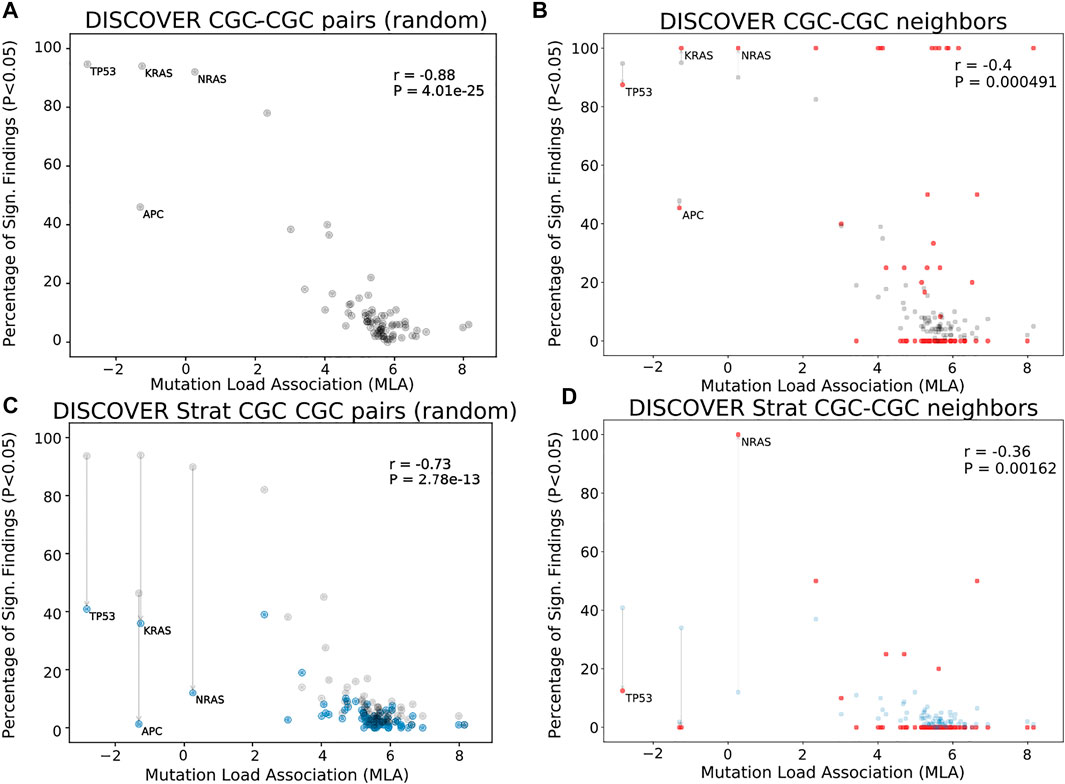
Figures 1A shows the MLA of the reference cancer genes vs the percent significant findings in mutual exclusivity tests performed with DISCOVER for the TCGA COADREAD cohort (498 tumors). We observe a strong negative correlation between MLA values and percent significant findings in mutual exclusivity tests (Pearson correlation -0.88, p-value 4.0e − 25) similar to van de Haar et al., 2019. In Figures 1B, we take into account the PPI information to calculate percent significant findings. Namely, for each CGC gene, we perform mutual exclusivity tests only with its PPI neighbors that are also in CGC. Note that CGC genes which do not have any CGC neighbors are excluded from this analysis. To make a fair comparison between Figures 1A,B, only the CGC genes that have CGC neighbors are shown in Figures 1A. We also ensure that the mutual exclusivity of a gene of interest is checked with same sized group of genes in both Figures 1A,B. To achieve this in Figures 1A, for each gene, we compute mutual exclusivity with a random subsample of the CGC reference set, the same size as the set of CGC neighbors of that gene. We repeat this random sampling 100 times and plot the mean percent significant findings value. For reference, Supplementary Figure S3A, S3D contains versions of Figures 1A,C, where all CGC genes (i.e., with and without CGC neighbors) are plotted and mutual exclusivities are checked between all CGC pairs, as it was done in van de Haar et al., 2019.

FIGURE 1. Comparison of mutual exclusivity results of DISCOVER and DISCOVER Strat on TCGA COADREAD cohort (498 samples) (A) The scatterplot of percentage significance of ME runs (p-value
In Figures 1B, we observe a reduced correlation when network information is included (Pearson correlation -0.4, p-value 4.91e − 4). We also run DISCOVER Strat where stratification is based on CMS subtypes (Guinney et al., 2015). We plot these results in Figures 1C where we again ensure comparability with Figures 1D where both subtype and network information are considered. Comparing Figures 1A and Figures 1C, we verify the findings of van de Haar et al., although with less significance in correlation difference (Pearson correlation −0.73, p-value 2.8e − 13). It should be noted that the subtype stratification inherently causes an overall decrease in percent significant findings, not specific to genes with low MLA. On the contrary the idea of ME corrections through network incorporation, materialized in the comparison of Figures 1A and Figures 1B, inherently leads to an increase in percent significant findings. Most of the decreases occur in genes with small number of CGC neighbors. When we compare Figures 1D to Figures 1B, the decrease in correlation from −0.4 to −0.36 indicates that including subtype information is still useful when used on top of network-based corrections we propose.
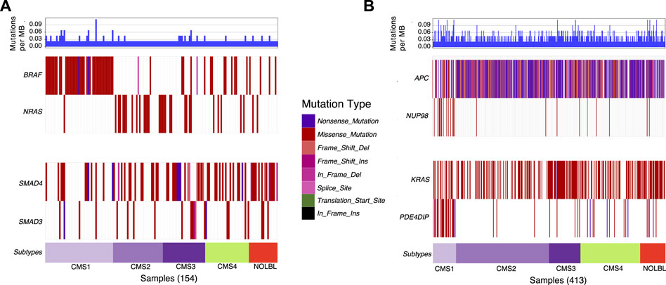
Next, we utilize waterfall plots to compare the outputs of DISCOVER and DISCOVER-Strat to assess how MLA and subtype information can affect mutual exclusivity findings. Figures 2A shows two selected gene pairs that display significant mutual exclusivity based on both DISCOVER and DISCOVER-Strat estimations on TCGA COADREAD dataset. The mutual exclusivity between BRAF and NRAS, two members of the MAPK pathway, is well-known and has been detected in multiple cancer types including melanoma, myeloma and colorectal cancer (Samowitz et al., 2006; Roth et al., 2010; Popovici et al., 2012) BRAF is frequently mutated in patients from CMS1 subtype whereas NRAS shows almost no mutation across these patients. However, since BRAF and NRAS mutations are mutually exclusive across not only CMS1 subtype but also across the other subtypes, DISCOVER-Strat identifies this pair as significantly mutually exclusive. Similarly, SMAD3 and SMAD4 are two members of the TGF-β pathway and the mutual exclusivity between these two transcription factors is previously reported in colorectal cancer (Fleming et al., 2013). Mutations on SMAD3 and SMAD4 are distributed almost uniformly across the subtypes. As such, the mutual exclusivity between the mutations of these two genes is still significant when subtype information is incorporated. Figures 2B similarly shows two selected gene pairs that display significant mutual exclusivity based on DISCOVER but not based on DISCOVER Strat. For the first pair, we observe that NUP98 is mutated almost exclusively in patients from the CMS1 subtype which shows hypermutation due to microsatellite instability. On the other hand, there is a depletion of APC mutations among the patients from the CMS1 subtype which results in a low MLA value. As such, DISCOVER Strat fails to detect a significant ME between these two genes since it explores ME within each subtype separately. A similar observation can also be made for the KRAS-PDE4DIP pair where the former has a low MLA and the latter has a high MLA.

FIGURE 2. Waterfall plots of the distribution of mutations for selected gene pairs. (A) Mutation distribution of two selected gene pairs (BRAF-NRAS and SMAD4-SMAD3) that are found to be significantly mutually exclusive based on both DISCOVER and DISCOVER-Strat estimations. (B) Mutation distribution of two selected gene pairs (APC-NUP98 and KRAS-PDE4DIP) that are found to be significantly mutually exclusive based on DISCOVER but not based on DISCOVER-Strat. Note that the set of samples included in each plot is determined by finding the set of patients that have a mutation in at least one of the listed genes. GenVisR R package is used to generate the waterfall plots (Skidmore et al., 2016). Subtype information is downloaded from (Guinney et al., 2015).
Supplementary Figure S2 compares the MLA of the reference cancer genes with the percent significant findings in mutual exclusivity tests for BRCA. Similar to the results that we obtain for COADREAD data, including network information reduces the correlation between MLA and ME detection rate (Supplementary Figures S2B vs S2C). The magnitude of reduction is even more significant than what we observe for COADREAD data (Pearson correlation −0.93 vs −0.27). Interestingly, including subtype information results in a very slight decrease in correlation coefficient (−0.93 to −0.91) (Supplementary Figures S2B vs S2E) as opposed to what we observe for COADREAD. We observe that including subtype information on top of network information results in no decrease in correlation (Supplementary Figures S2C vs S2F). This difference in the effect of including subtype information for BRCA and COADREAD datasets could be related to the average tumor mutation load of subtypes. BRCA subtypes have comparable average TML values (Her2: 146, LumA:65, LumB: 71, Normal: 55) whereas the CMS1 subtype in COADREAD has a dramatically larger average TML value compared to the other subtypes of COADREAD (CMS1: 1387, CMS2:93, CMS3: 272, CMS4: 212) We repeat the same analysis with the other ME detection methods as well as for other cancer types when t is set to 20 (Supplementary Figures S1-S8). We observe that the percent significant finding values can vary remarkably across the tumor types. Compared to other cancer types, we observe smaller percent significant findings for LUSC (Supplementary Figures S5A,S5D, S5G). Similarly, very few pairs have percent significance value
When we consider the correlation between MLA and percent significant values, we observe that adding network information decreases the correlation coefficient values for all cancer types and for all ME detection methods except for Fisher’s Exact Test. Fisher’s Exact Test results show an increased correlation with the addition of network information for LUSC and SKCM (Supplementary Figures S5-S6 D vs F). Also, the correlation coefficient can not be computed for LUAD and STAD since Fisher’s Exact Test gives a value of 0 for the percent significant findings of all considered genes (Supplementary Figures S4D-S7F). Another interesting observation is the variance in magnitude of decrease in correlation values across different tumor types. In particular, we observe a smaller decrease in correlation values for LUAD compared to other cancer types. The analogous results are also available for t = 5 setting (Supplementary Figures S9-S16). For all the cancer types, the correlation between MLA values and percent significant findings decreases and becomes non-significant for most cases.
We should also note that the majority of CGC genes have only one neighbor within the data setting of the cancer type under consideration. This leads to percentage significant findings of either 0 or 1 in many cases simply because these are the only possible values; for COADREAD see Figures 1B and Figures 1D where 41 out of 74 genes under study have only one CGC neighbor in the COADREAD data settings. To avoid any such possible biases, we repeat the same evaluations after filtering out those CGC genes with only one neighbor. The evaluations still provide significant decreases in correlation coefficient values analogous to the decreases observed in Figures 1B as compared to Figures 1A and Figures 1D as compared to Figures 1C. For detailed results, see Supplementary Figures S17-S24 for t = 20 and Supplementary Figures S25-S32 for t = 5.
Individual genes of interest are those that have increased percent significant findings when network neigborhood information is incorporated while at the same have significant number of CGC neighbors. More specifically, for the former constraint, we identify the CGC genes with at least 0.1 increase in percentage of significant findings value of WeXT, DISCOVER and MEMo when the network information is included as opposed to the scenario when it is not (e.g., for COADREAD, Figures 1A vs Figures 1B). We choose these 3 ME methods since they are top performers based on the defined metrics in Section 3.1. For STAD, SKCM and UCEC, since MEMo results are unavailable, we only consider WeXT and DISCOVER results. For the second constraint, we include the CGC genes with at least 3 CGC neighbors. For COADREAD, this selection procedure results in four genes: EP300, CREBBP, NCOA2 and NCOR2. Among these, EP300 is a well-known tumor suppressor in epithelial cancer types including COADREAD (Gayther et al., 2000). For BRCA, the only identified gene is PIK3R1. PIK3R1 is found to be significantly mutually exclusive with PIK3CA and SPEN based on both WeXT, DISCOVER and MEMo results. PIK3R1 and PIK3CA are members of the PI3K pathway and their mutual exclusivity has been previously established in the literature (Chen et al., 2018). For LUAD, PTPRB is the only identified gene and is found to be mutually exclusive with EGFR, a well-known oncogene in non-small cell lung cancer (Bethune et al., 2010). The set of identified genes for STAD are NCOA2, NCOR2 and CREBBP; all of which are found to be mutually exclusive with TP53. For SKCM, we identify ERBB4, RAC1, EP300 and ITK. ERBB4 is a well-known oncogene in skin cancer and found to be mutually exclusive with ERBB2 (Prickett et al., 2009; Nielsen et al., 2014). ERBB2 and ERBB4 indeed belong to the same family (i.e. ErbB family of receptor tyrosine kinases) and form a heterodimer receptor for Heparin-binding EGF-like growth factor (HB-EGF) (Iwamoto et al., 2017). RAC1 mutation P29S is an established driver in melanoma (Jiang et al., 2018). RAC1 is found to be mutually exclusive with MYH9, a tumor suppressor in melanoma (Singh et al., 2020). Lastly, ITK has been shown to be an oncogene in melanoma (Carson et al., 2015). For UCEC, we identify 33 genes in total. Among these, KIT and PTEN have established roles in UCEC cancer development (Chang et al., 2015; Wang et al., 2020). Moreover, PTEN is found to be strongly mutually exclusive with SPOP, whose mutations are also associated with endometrial cancer (Clark and Burleson, 2020). Lastly, for BLCA and LUSC, no gene satisfies the abovementioned criteria. Overall these results suggest that the CGC genes that show increased ME with network incorporation as well as their mutually exclusive partner genes often have established roles in the development of the particular cancer type.
3.4 Mutual Exclusivity Evaluations Based on Corrections via Tissue-Specific Networks
We first provide our ME evaluations with respect to the metrics defined in Section 3.1 by replacing the non-specific networks with TSNs. We provide two types of comparisons; one where we compare TSN0.5 with the original non-tissue specific Intact network and one where results of TSN0.5 are compared against TSN0. We do the latter to avoid artifacts that may be introduced due to the fact that some genes in the original Intact network might be simply missing from even TSN0 since they may be nonexistent in the GTEX database. For the BLCA dataset, comparing the F1 scores of the ME methods under TSN0 and TSN0.5 settings, we observe that the scores of all methods are higher for the latter network. The largest percent increase of 10% is observed for WeXT when the control group is
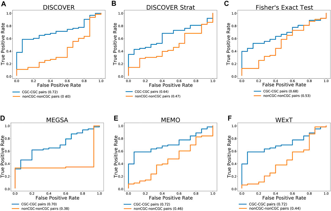
Figure 3 compares the ROC curves of CGC gene pairs and non-CGC gene pairs for COADREAD data where mutual exclusivities are estimated with DISCOVER, DISCOVER Strat, Fisher’s Exact Test, MEGSA, MEMo and WeXT with t = 20. We observe that all the ME methods estimate stronger mutual exclusivities for tissue-specific CGC gene pairs compared to non-tissue-specific CGC gene pairs since AUROCs are greater than 0.5. Additionally, we observe much smaller AUROCs for the control group where we repeat the same analysis with non-CGC gene pairs. Analogous results are available for the other cancer types where both the positive and negative set contains at least 10 number of pairs when t is set to 20. (Supplementary Figures S33–S35). We observe a similar result for SKCM where CGC pairs result in larger AUROCs compared to non-CGC pairs for all ME methods (Supplementary Figure S34). We observe a steep increase in the ROC curves plotted for MEGSA results. This is due to the utilized likelihood ratio test that results in a p-value of 0.5 when the likelihood values are equal to each other. For UCEC, we see a significant difference between the ROC curves of CGC-pairs vs non-CGC pairs for Fisher’s Exact Test and MEGSA; whereas the corresponding difference is negligible for DISCOVER and WeXT.

FIGURE 3. Performance of selected ME tests in terms of discriminating TSN and non-TSN gene pairs based on estimated ME p-values on COADREAD data. Blue curve is plotted with CGC gene pairs and red curve is plotted with non-CGC gene pairs. Mutual exclusivities are estimated with (A) DISCOVER, (B) DISCOVER Strat, (C) Fisher’s Exact Test, (D) MEGSA, (E) MEMo and (G) WeXT respectively.
4 Case Study
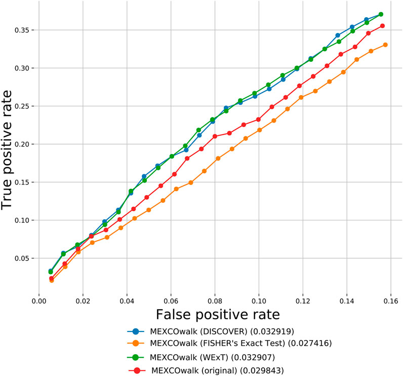
Apart from the defined network-centric ME evaluation framework, we discuss a case study where we assess whether mutual exclusivities estimated by the considered ME methods improve the performance of driver identification methods that utilize mutual exclusivity information. To this end, we compare the original version of MEXCOwalk with its alternatives where mutual exclusivity estimates are provided by the employed ME methods. Assuming that gi and gj genes are mutated in patient sets Si and Sj, respectively; MEXCOWalk simply computes the mutual exclusivity between these two genes with the following formula: |Si ∪ Sj|/(|Si| + |Sj|). MEXCOwalk uses the estimated mutual exclusivity values as part of edge weights. As such, to utilize the p-values output by ME detection methods in MEXCOwalk, we first compute −log (p-value) and then convert the resulting values between 0 and 1. To this end, we replace all −log (p-value)’s larger than 10 with 1. We then find the maximum −log (p-value) less than 10 and divide all other −log (p-value)’s with this value. The reason why we set a threshold for finding the maximum is the large differences across the smallest p-values output by different ME methods. For instance, WeXT outputs a very large range of p-values and if we use the smallest p-value to scale, all other −log (p-value)s will be converted to values that are very close to 0. In the original MEXCOWalk study, a threshold of 0.7 is applied to ME values such that all values ≤0.7 are clamped to 0. This conversion is equivalent to removing those edges from the network since the edge weights include a multiplicative term for ME values. We find that the removal of these edges correspond to a 0.035 percent reduction in graph density. For the current analysis, we determine the threshold value for each ME detection method to achieve the same percent density reduction in the graph. Figure 4 shows the number of recovered CGC genes for fixed output gene sizes from 100 to 2,500 as a ROC curve for original MEXCOwalk as well as for versions of MEXCOwalk where mutual exclusivity values are estimated with DISCOVER, Fisher’s Exact Test and WeXT, respectively. We observe that MEXCOwalk with WeXT’s ME values results in the best AUROC value for COADREAD. Supplementary Figures S36 shows the analogous results for the other cancer types. For, LUSC, STAD and UCEC, MEXCOwalk with DISCOVER gives the best AUROC whereas for BLCA, LUAD and SKCM MEXCOwalk with Fisher’s Exact Test performs the best. An important observation is the worse performance of MEXCOwalk with Fisher’s Exact Test compared to the original MEXCOwalk for COADREAD, STAD and UCEC. As such, using Fisher’s Exact Test in place of MEXCOwalk’s original ME values does have the potential to decrease the performance whereas for the other ME methods we do not observe such a risk. Note that for these analysis we employ t = 5 since t = 20 filtering does not provide enough number of genes to be evaluated.

FIGURE 4. The number of recovered CGC genes for the original MEXCOwalk as well as for its modified versions where mutual exclusivity values are estimated with DISCOVER, Fisher’s Exact Test and WeXT. COADREAD dataset is used with t = 5 setting. The numbers in parentheses indicate the area under the ROC curve for the corresponding curve.
5 Discussion
It is important to investigate whether the employment of an interaction network within our ME evaluation framework causes any ascertainment bias in the findings and to elaborate on how any such potential bias is mediated within the framework. It is established that known cancer genes have larger number of interactions compared to other genes in the network (Hou and Ma, 2014a). This implies a potential bias that needs to be resolved in cancer driver gene identification methods employing interaction network data. Such a bias is less of a problem for the current study, since our aim is not to identify novel cancer driver genes but to utilize the interaction network and known cancer genes to form a ground truth of mutually exclusive interactions for evaluating existing ME methods. On the contrary, the fact that most known cancer genes have well-characterized interactions in the network provides a benefit for our work as it supports the confidence of our true positive examples. Additionally, our framework makes use of not only genes from the reference set
Another important point worth emphasizing is that apart from the aggregate statistics provided in the previous sections as part of the metrics for the network-centric ME evaluations, our proposed framework also provides analogous statistics at the gene-level as well. Such statistics may in fact be of more interest to cancer biologists than the aggregate statistics in certain cases. Several interesting observations can be made through an inspection of these gene-level evaluations, especially for the settings where the conventionally defined F1 score fails in quantifying ME. Genes with low MLA comprise an example setting, where TP53 is a leading member. Consider the case of TP53 in COADREAD evaluations for instance. With respect to the degree-normalized setting, the values of precision, sensitivity, precisionstrict and sensitivitystrict for WeXT are respectively 0.5, 1, 0.25, 0.25 which gives rise to an F1 score of 0.66 and F1strict score of 0.25. On the other hand, MEMo provides the same precision, sensitivity and F1 scores as WeXT whereas its precisionstrict, sensitivitystrict and F1strict scores are all 0. To summarize, although the inspection of the F1 scores does not provide a distinction between the two results, an inspection of the F1strict scores establishes that MEMo is worse than WeXT in this setting. We note that the advantages of inspections based on the strict definitions of the metrics rather than the conventional ones are also apparent in the aggregate analysis as well. In addition to the COADREAD evaluations shown in Table 2, BRCA also contains an example instance where the conventional and the strict versions of the metrics provide different conclusions; see Supplementary Table S7B. In terms of the F1 scores, DISCOVER Strat ranks fourth, whereas comparing F1strict scores it ranks the second. Also, overall we observe that MEMo’s performance gets severely affected when the strict versions of the metrics are employed.
Next, our robustness analysis results reveal some suggestions for potential users of our framework. We recommend using a p-value threshold smaller than 0.1 but larger than 0.05 as lower threshold values are too stringent and lead to too few predicted positives. Regarding robustness_iterations, we tested values both smaller than and higher than the default value of 100 for COADREAD evaluations: 5, 50, 100, 300 and 500. We repeated each experiment 20 times and calculated the standard deviation of the obtained set of F1 and F1strict scores. For the majority of the cases, we observe a large decrease in the standard deviation values when robustness_iterations is increased from 5 to 50. (Supplementary Table S82). This analysis suggests that the robustness_iterations should be set to a at least 50. Lastly, we observe that different PPI networks can lead to large differences in both the F1/F1strict scores and the ranking of the methods. As such, exploring different PPI sources would be beneficial.
To assess whether our findings extend to other datasets other than TCGA, we repeat our evaluations on somatic mutation data of 402 colon cancer patients within the Pancancer Analysis of Whole Genomes (PCAWG) study (Campbell, 2020). Supplementary Tables S83-S86 shows the ME evaluations with respect to the metrics defined in section 4.2. We observe an overall decrease in F1strict scores of the methods. Compared to analogous results in TCGA data, WexT still performs the best in terms of F1strict score whereas the second best performing method is changed from MEMo to DISCOVER. The changes with respect to varying the p-value threshold, robustness_iterations value, input PPI, reference cancer gene set are consistent with the changes that we previously observe for the TCGA COADREAD dataset. When we switch from IntAct to its TSN version, we observe that all the ME methods estimate stronger mutual exclusivities for the tissue-specific CGC gene pairs compared to the non-tissue-specific CGC gene pairs as evident from AUROC values greater than 0.5; see Supplementary Figures S37, S38 shows the results of MLA where we observe slightly smaller correlation values for DISCOVER (−0.84 vs −0.88) as compared to the results obtained from TCGA COADREAD dataset. We observe findings similar to those obtained from the TCGA COADREAD data in that the correlation values drop when the network information is incorporated. To summarize, our conclusions remain the same when we repeat our analyses on an entirely different cohort from the PCAWG study.
The majority of the somatic mutations observed in cancer genomics are passenger mutations. In the evaluations provided in the Results section we employ a simple filtering strategy where we remove silent mutations and mutations on non-coding regions of the genes. Additionally, we also assess the effects of employing a more elaborate mutation filtering procedure. To this end, we download the predictions of the Muiños et al. study on COREAD type (Muinos et al., 2021). This includes the classification of all possible mutations on 12 genes as driver or passenger mutations. Accordingly, we filter out the proposed passenger mutations from our mutation data and repeat all of our relevant analyses. We observe that the ranking of the methods according to the metrics proposed in section 4.2 remain the same where WExT, MEMO, and DISCOVER Strat show reduced F1strict scores, and DISCOVER and Fisher’s Exact Test show higher F1strict scores (Supplementary Tables S87-S90). The TSN results and the MLA analysis results are also similar to our original results (Supplementary Figures S39-S40). Muiños et al. provides classifications of mutations on a subset of genes which have training data larger than a certain size. If such classifications become available for a larger set of genes in the future we can provide a better assessment regarding the filtering procedures employing these classifications.
Mutated genes in cancer prevalently exhibit a long tail phenomenon where few genes are mutated in many patients and large number of genes are mutated in few patients. To check whether assessing the mutual exclusivity of gene pairs with very different mutation frequencies bias the evaluations of the compared ME methods, we repeat our analyses after filtering out the genes with mutation frequencies
We also evaluate a more general ME detection method SELECT, which investigates both types of relationships among pairs, co-occurence and ME simultaneously. SELECT outputs ME associated scores to only a subset of the input gene pairs. Thus one strategy for comparing the results of SELECT against other methods is to focus only on such subsets. The relevant results where we use this strategy are available in Supplementary Tables S94-S97. We report evaluations on two subsets of TCGA COADREAD dataset: 1) the set of CGC-CGC pairs where SELECT results are available, 2) the set of CGC-CGC pairs where SELECT’s version which uses subtype information (i.e., SELECTsubtype) are available. For the former, we observe that SELECT and SELECTsubtype rank the fourth after WExT, MEMO, and DISCOVER. For the latter evaluation, SELECTsubtype performs better than SELECT although both of them still rank the fourth among the other ME methods. Another strategy to fix this problem is to assign the worst ASC score to such pairs without specific ASC scores in the ME direction. We employ this approach as well and observe that it gives no significant difference in the comparisons.
Lastly, it is important to mention certain limitations of the proposed framework. Our framework is based on the presupposition that ME is likely to occur between interacting known cancer genes. Although rare, there may exist two different types of exceptions to this assumption; ME can be observed between non-interacting known drivers and the relationship between an interacting pair of known drivers can be that of co-occurrence rather than that of ME. These constitute respectively the false negative and the false positive events in our framework. An example instance of the former is the mutually exclusive mutations of APC and RNF43 observed in colorectal cancer [Mina et al., 2017] and example instance of the latter is the co-occurrence of CCNE1 and TP53 alterations [Zhang et al., 2014]. Both of these patterns are currently ignored by our framework and incorporation of mechanisms to dissect each such pattern to increase the performance of true ME detection is an important future step. Another limitation of the current framework is that it requires the availability of whole-genome or whole-exome sequencing data.
6 Conclusion
We propose a network-centric framework to evaluate pairwise mutual exclusivity findings reported by different ME algorithms. The first component of our framework consists of useful definitions of statistics employed in the network-centric ME evaluations. We observe that for the majority of the cancer types under study WeXT outperforms the other methods in terms of F1 score measured with respect to appropriately defined control groups. In half of the cancer types DISCOVER and in the other half MEMo perform as the second best methods. When comparing different cancer types we observe that BRCA and COADREAD are among the top two types leading to maximum F1 scores with at least one of the ME methods providing a score greater than 0.5. We note that DISCOVER Strat is only applicable in two cancer types among a total of eight since these are the only cancer types with well-defined subtypes. Furthermore, among these two cancer types, DISCOVER Strat outperforms original DISCOVER algorithm in BRCA, whereas it is the second worst method after Fisher’s Exact Test in COADREAD. This is noteworthy since van de Haar et al. propose subtype stratification as employed by DISCOVER Strat as a way to emphasize true mutual exclusivity by reducing mutation load confounding (van de Haar et al., 2019). We also observe that Fisher’s exact test and MEGSA are more conservative compared to DISCOVER and WeXT, where from the latter group, WeXT outputs notably larger number of significant p-values. The second component of our framework evaluates ME tests by comparing two types of measures obtained with and without network information. First measure is with respect to the percent significant findings of mutually exclusive gene pairs, whereas the second is based on MLC values. In most of the cancer types and for most of the genes we observe an increase with respect to the former whereas a decrease with respect to the latter measure. Finally, we repeat the same analysis by considering TSNs in the network-centric framework. Considerable improvements achieved due to the use of TSNs as opposed tissue nonspecific interaction network are only observed for BLCA and STAD datasets. A more detailed analysis in terms of comparing ROCs of CGC gene pairs and non-CGC gene pairs on cancer types with considerable number of tissue-specific gene pairs indicate the advantages of employing tissue specificity in detecting mutual exclusivity in COADREAD, SKCM, and UCEC. Finally we extend out network-centric evaluation framework to assess whether including network knowledge reduces the mutation load confounding problem.
As noted earlier the proposed framework is intended for the network-centric evaluations of mutual exclusivities of pairs of genes rather than groups of genes. Such a choice stems form the fact that the mutual exclusivities are commonly made use of in driver gene/module identification algorithms which mostly employ pairwise mutual exclusivities. Furthermore the extensive evaluation settings proposed, the number of ME methods under study and their own computational requirements, and the potentially exponential computational complexity inherent in handling groups of genes limits the scope of the current study to evaluations of pairwise ME scorings. Nonetheless most statistical ME methods are capable of providing ME results for groups of genes as well. Regarding the ME tests considered in this study, the main ME test provided by DISCOVER is based on a pairwise test definition but it also extends the definition for possible use in quantifying the ME of a group of genes, although the experiments involving the latter are based only on simulation data. The remaining tests MEGSA, MEMo, and WeXT are all ME tests specifically designed for groups of genes. An important direction for future work is to design a suitable extension of the proposed network-centric framework to evaluate the results of ME tests on groups of genes. Design choices relevant for such an extension would involve an appropriate and computationally efficient definition of the reference groups of genes analogous to a pair of interacting genes from the set
Data Availability Statement
The original contributions presented in the study are included in the article/Supplementary Material, further inquiries can be directed to the corresponding author.
Author Contributions
Authors names are written in alphabetical order. CE and HK conceived the idea and supervised the study. RA implemented the code and performed the initial experiments. CY and AH repeated the experiments for other cancer types. All authors contributed to the preparation of the manuscript. All authors read and approved the manuscript.
Funding
This work was supported by the Scientific and Technological Research Council of Turkey (grant number 117E879 to HK and CE.).
Conflict of Interest
The authors declare that the research was conducted in the absence of any commercial or financial relationships that could be construed as a potential conflict of interest.
Publisher’s Note
All claims expressed in this article are solely those of the authors and do not necessarily represent those of their affiliated organizations, or those of the publisher, the editors and the reviewers. Any product that may be evaluated in this article, or claim that may be made by its manufacturer, is not guaranteed or endorsed by the publisher.
Acknowledgments
We thank Joris van de Haar for sharing us the data used in van de Haar et al., 2019.
Supplementary Material
The Supplementary Material for this article can be found online at: https://www.frontiersin.org/articles/10.3389/fgene.2021.746495/full#supplementary-material
References
Ahmed, R., Baali, I., Erten, C., Hoxha, E., and Kazan, H. (2019). MEXCOwalk: Mutual Exclusion and Coverage Based Random Walk to Identify Cancer Modules. Bioinformatics 36 (3), 872–879. ISSN 1367-4803. doi:10.1093/bioinformatics/btz655
Baali, I., Erten, C., and Kazan, H. (2020). Driveways: A Method for Identifying Possibly Overlapping Driver Pathways in Cancer. Sci. Rep. 10. doi:10.1101/2020.04.01.015388
Babur, Ö., Gönen, M., Aksoy, B. A., Schultz, N., Ciriello, G., Sander, C., et al. Bülent Arman Aksoy (2015). Systematic Identification of Cancer Driving Signaling Pathways Based on Mutual Exclusivity of Genomic Alterations. Genome Biol. 16 (1), 45. doi:10.1186/s13059-015-0612-6
Bailey, M. H., Tokheim, C., Porta-Pardo, E., Sengupta, S., Bertrand, D., Weerasinghe, A., et al. (2018). Comprehensive Characterization of Cancer Driver Genes and Mutations. Cell 173, 371–e18. e18. doi:10.1016/j.cell.2018.02.060
Bethune, G., Bethune, D., Ridgway, N., and Xu, Z. (2010). Epidermal Growth Factor Receptor (Egfr) in Lung Cancer: an Overview and Update. J. Thorac. Dis. 2, 48–51.
Campbell, G. (2020). Pan-cancer Analysis of Whole Genomes. Nature 578, 82–93. doi:10.1038/s41586-020-1969-6
Canisius, S., Martens, J. W. M., Wessels, L. F. A., and Martens, M. (2016). A Novel independence Test for Somatic Alterations in Cancer Shows that Biology Drives Mutual Exclusivity but Chance Explains Most Co-occurrence. Genome Biol. 17 (261), 1–17. ISSN 1474-760X. doi:10.1186/s13059-016-1114-x
Carson, C. C., Moschos, S. J., Edmiston, S. N., Darr, D. B., Nikolaishvili-Feinberg, N., Groben, P. A., et al. (2015). Il2 Inducible T-Cell Kinase, a Novel Therapeutic Target in Melanoma. Clin. Cancer Res. 21 (9), 2167–2176. doi:10.1158/1078-0432.CCR-14-1826
Cerami, E., Gao, J., Dogrusoz, U., Gross, B. E., Sumer, S. O., Aksoy, B. A., et al. (2012). The cBio Cancer Genomics Portal: An Open Platform for Exploring Multidimensional Cancer Genomics Data: Figure 1. Cancer Discov. 2 (5), 401–404. ISSN 2159-8274. doi:10.1158/2159-8290.cd-12-0095
Chang, S.-W., Chao, W.-R., Ruan, A., Wang, P.-H., Lin, J.-C., and Han, C.-P. (2015). A Promising Hypothesis of C-KIT Methylation/Expression Paradox in C-KIT (+) Squamous Cell Carcinoma of Uterine Cervix ----- CTCF Transcriptional Repressor Regulates C-KIT Proto-Oncogene Expression. Diagn. Pathol. 10 (1). doi:10.1186/s13000-015-0438-2
Chen, L., Yang, L., Yao, L., Kuang, X.-Y., Zuo, W.-J., Li, S., et al. (2018). Characterization of Pik3ca and Pik3r1 Somatic Mutations in Chinese Breast Cancer Patients. Nat. Commun. 9 (1). doi:10.1038/s41467-018-03867-9
Cheng, D. T., Mitchell, T. N., Zehir, A., Shah, R. H., Benayed, R., Syed, A., et al. (2015). Memorial Sloan Kettering-Integrated Mutation Profiling of Actionable Cancer Targets (MSK-IMPACT): A Hybridization Capture-Based Next-Generation Sequencing Clinical Assay for Solid Tumor Molecular Oncology. J. Mol. Diagn. 17 (3), 251–264. doi:10.1016/j.jmoldx.2014.12.006
Ciriello, G., Cerami, E., Sander, C., and Schultz, N. (2012). Mutual Exclusivity Analysis Identifies Oncogenic Network Modules. Genome Res. 22 (2), 398–406. doi:10.1101/gr.125567.111
Clark, A., and Burleson, M. (2020). Spop and Cancer: a Systematic Review. Am. J. Cancer Res. 10 (3), 704–726.
Constantinescu, S., Szczurek, E., Mohammadi, P., Rahnenführer, J., and Beerenwinkel, N. (2015). TiMEx: a Waiting Time Model for Mutually Exclusive Cancer Alterations. Bioinformatics 32 (7), 968–975. doi:10.1093/bioinformatics/btv400
Dao, P., Kim, Y.-A., Wojtowicz, D., Madan, S., Sharan, R., and Przytycka, T. M. (2017). BeWith: A Between-Within Method to Discover Relationships between Cancer Modules via Integrated Analysis of Mutual Exclusivity, Co-occurrence and Functional Interactions. Plos Comput. Biol. 13, e1005695. doi:10.1371/journal.pcbi.1005695
Deng, Y., Luo, S., Deng, C., Luo, T., Yin, W., Zhang, H., et al. (2017). Identifying Mutual Exclusivity across Cancer Genomes: Computational Approaches to Discover Genetic Interaction and Reveal Tumor Vulnerability. Brief in Bionform 20, 254–266. doi:10.1093/bib/bbx109
Dimitrakopoulos, C. M., and Beerenwinkel, N. (2017). Computational Approaches for the Identification of Cancer Genes and Pathways. Wires Syst. Biol. Med. 9 (1), e1364. ISSN 1939-5094. doi:10.1002/wsbm.1364
Dinstag, G., and Shamir, R. (2020). PRODIGY: Personalized Prioritization of Driver Genes. Bioinformatics 36 (6), 18311367–18394803. doi:10.1093/bioinformatics/btz815
Fleming, N. I., Jorissen, R. N., Mouradov, D., Christie, M., Sakthianandeswaren, A., Palmieri, M., et al. (2013). SMAD2, SMAD3 and SMAD4 Mutations in Colorectal Cancer. Cancer Res. 73 (2), 7251538–7357445. ISSN 0008-5472. doi:10.1158/0008-5472.CAN-12-2706
Forbes, S. A., Beare, D., Boutselakis, H., Bamford, S., Bindal, N., Tate, J., et al. (2017). Cosmic: Somatic Cancer Genetics at High-Resolution. Nucleic Acids Res. 45, D777–D783. doi:10.1093/nar/gkw1121
Gao, J., Aksoy, B. A., Dogrusoz, U., Dresdner, G., Gross, B., Sumer, S. O., et al. (2013). Integrative Analysis of Complex Cancer Genomics and Clinical Profiles Using the Cbioportal. Sci. Signal. 6 (269), pl1. ISSN 1945-0877. doi:10.1126/scisignal.2004088
Gayther, S. A., Batley, S. J., Linger, L., Bannister, A., Thorpe, K., Chin, S.-F., et al. (2000). Mutations Truncating the Ep300 Acetylase in Human Cancers. Nat. Genet. 24, 300–303. doi:10.1038/73536
GTEX Consortium, (2020). The Gtex Consortium Atlas of Genetic Regulatory Effects across Human Tissues. Science 369 (6509), 1318–1330. ISSN 0036-8075. doi:10.1126/science.aaz1776
Guinney, J., Dienstmann, R., Wang, X., de Reyniès, A., Schlicker, A., Soneson, C., et al. (2015). The Consensus Molecular Subtypes of Colorectal Cancer. Nat. Med. 21, 1350–1356. doi:10.1038/nm.3967
Hou, J. P., and Ma, J. (2014a). Dawnrank: Discovering Personalized Driver Genes in Cancer. Genome Med. 6 (56), 16. doi:10.1186/s13073-014-0056-8
Hou, J. P., and Ma, J. (2014b). DawnRank: Discovering Personalized Driver Genes in Cancer. Genome Med. 6 (7), 56. ISSN 1756-994X. doi:10.1186/s13073-014-0056-8
Hua, X., Hyland, P. L., Huang, J., Song, L., Zhu, B., Caporaso, N. E., et al. (2016). Megsa: A Powerful and Flexible Framework for Analyzing Mutual Exclusivity of Tumor Mutations. Am. J. Hum. Genet. 98, 442–455. doi:10.1016/j.ajhg.2015.12.021
Iwamoto, R., Mine, N., Mizushima, H., and Mekada, E. (2017). Erbb1 and Erbb4 Generate Opposing Signals Regulating Mesenchymal Cell Proliferation during Valvulogenesis. Development 144 (8), 2–e1. doi:10.1242/dev.152710
Jiang, Z. B., Ma, B. Q., Liu, S. G., Li, J., Yang, G. M., Hou, Y. B., et al. (2018). miR‐365 Regulates Liver Cancer Stem Cells via RAC1 Pathway. Mol. Carcinogenesis 58 (1), 55–65. doi:10.1002/mc.22906
Kim, Y.-A., Cho, D.-Y., Dao, P., and Przytycka, T. M. (2015). MEMCover: Integrated Analysis of Mutual Exclusivity and Functional Network Reveals Dysregulated Pathways across Multiple Cancer Types. Bioinformatics 31 (12), i284–i292. doi:10.1093/bioinformatics/btv247
Kim, Y. A., Madan, S., and Przytycka, T. M. (2017). WeSME: Uncovering Mutual Exclusivity of Cancer Drivers and beyond. Bioinformatics 33 (6), 814–821. ISSN 1367-4803. doi:10.1093/bioinformatics/btw242
Leiserson, M. D. M., Blokh, D., Sharan, R., and Raphael, B. J. (2013). Simultaneous Identification of Multiple Driver Pathways in Cancer. Plos Comput. Biol. 9 (5), e1003054. doi:10.1371/journal.pcbi.1003054
Leiserson, M. D. M., Reyna, M. A., and Raphael, B. J. (2016). A Weighted Exact Test for Mutually Exclusive Mutations in Cancer. Bioinformatics 32, i736–i745. doi:10.1093/bioinformatics/btw462
Leiserson, M. D., Wu, H.-T., Vandin, F., and Raphael, B. J. (2015). Comet: A Statistical Approach to Identify Combinations of Mutually Exclusive Alterations in Cancer. Genome Biol. 16, 160, ISSN 1474-7596. doi:10.1186/s13059-015-0700-7
Liu, S., Liu, J., Xie, Y., Zhai, T., Hinderer, E. W., Stromberg, A. J., et al. (2020). MEScan: a Powerful Statistical Framework for Genome-Scale Mutual Exclusivity Analysis of Cancer Mutations. Bioinformatics 11. doi:10.1093/bioinformatics/btaa957
Luck, K., Kim, D. K., Lambourne, L., Spirohn, K., Begg, B. E., Bian, W., et al. (2020). A Reference Map of the Human Binary Protein Interactome. Nature 580, 402–408. doi:10.1038/s41586-020-2188-x
Mina, M., Raynaud, F., Tavernari, D., Battistello, E., Sungalee, S., Saghafinia, S., et al. (2017). Conditional Selection of Genomic Alterations Dictates Cancer Evolution and Oncogenic Dependencies. Cancer Cell 32 (2), 155–168. e6ISSN 1535-6108. doi:10.1016/j.ccell.2017.06.010
Muiños, F., Martínez-Jiménez, F., Pich, O., Gonzalez-Perez, A., and Lopez-Bigas, N. (2021). In Silico saturation Mutagenesis of Cancer Genes. Nature 596, 428–432. doi:10.1038/s41586-021-03771-1
Nielsen, T. O., Poulsen, S. S., Journe, F., Ghanem, G., and Sorensen, B. S. (2014). Her4 and its Cytoplasmic Isoforms Are Associated with Progression-free Survival of Malignant Melanoma. Melanoma Res. 24 (1), 88–91. doi:10.1097/cmr.0000000000000040
Orchard, S., Ammari, M., Aranda, B., Breuza, L., Briganti, L., Broackes-Carter, F., et al. (2014). The MIntAct Project-IntAct as a Common Curation Platform for 11 Molecular Interaction Databases. Nucl. Acids Res. 42 (D), D358–D363. ISSN 0305-1048. doi:10.1093/nar/gkt1115
Popovici, V., Budinska, E., Tejpar, S., Weinrich, S., Estrella, H., Hodgson, G., et al. (2012). Identification of a Poor-Prognosis BRAF-mutant-like Population of Patients with colon Cancer. Jco 30 (12), 1288–1295. ISSN 1527-7755. doi:10.1200/JCO.2011.39.5814
Prickett, T. D., NeenaAgrawal, S., Agrawal, N. S., Wei, X., Yates, K. E., Lin, J. C., et al. (2009). Analysis of the Tyrosine Kinome in Melanoma Reveals Recurrent Mutations in Erbb4. Nat. Genet. 41 (10), 1127–1132. doi:10.1038/ng.438
Roth, A. D., Tejpar, S., Delorenzi, M., Yan, P., Fiocca, R., Klingbiel, D., et al. (2010). Prognostic Role of KRAS and BRAF in Stage II and III Resected Colon Cancer: Results of the Translational Study on the PETACC-3, EORTC 40993, SAKK 60-00 Trial. Jco 28 (3), 466–474. ISSN 1527-7755. doi:10.1200/JCO.2009.23.3452
Samowitz, W. S., Albertsen, H., Sweeney, C., Herrick, J., Caan, B. J., Anderson, K. E., et al. (2006). Association of smoking, CpG island methylator phenotype, and v600e BRAF mutations in colon cancer. JNCI J. Natl. Cancer Inst. 98 (23), 1731–1738. doi:10.1093/jnci/djj468
Sarto Basso, R., Hochbaum, D. S., and Vandin, F. (2019). Efficient Algorithms to Discover Alterations with Complementary Functional Association in Cancer. PLOS Comput. Biol. 15, e1006802. doi:10.1371/journal.pcbi.1006802
Singh, S. K., Sinha, S., Padhan, J., Jangde, N., Ray, R., and Rai, V. (2020). Myh9 Suppresses Melanoma Tumorigenesis, Metastasis and Regulates Tumor Microenvironment. Med. Oncol. 37 (10), 2020. doi:10.1007/s12032-020-01413-6
Skidmore, Z. L., Wagner, A. H., Lesurf, R., Campbell, K. M., Kunisaki, J., Griffith, O. L., et al. (2016). GenVisR: Genomic Visualizations in R. Bioinformatics 32 (19), 3012–3014. doi:10.1093/bioinformatics/btw325
Sondka, Z., Bamford, S., Cole, C. G., Ward, S. A., Dunham, I., and Forbes, S. A. (2018). The Cosmic Cancer Gene Census: Describing Genetic Dysfunction across All Human Cancers. Nat. Rev. Cancer 18 (11), 696–705. doi:10.1038/s41568-018-0060-1
Song, J., Peng, W., and Wang, F. (2020). An Entropy-Based Method for Identifying Mutual Exclusive Driver Genes in Cancer. Ieee/acm Trans. Comput. Biol. Bioinf. 17 (3), 758–768. doi:10.1109/tcbb.2019.2897931
Szczurek, E., and Beerenwinkel, N. (2014). Modeling Mutual Exclusivity of Cancer Mutations. Plos Comput. Biol. 10 (3), e1003503–12. doi:10.1371/journal.pcbi.1003503
Thomas, R. K., Baker, A. C., Debiasi, R. M., Winckler, W., Laframboise, T., Lin, W. M., et al. (2007). High-throughput Oncogene Mutation Profiling in Human Cancer. Nat. Genet. 39, 347–351. doi:10.1038/ng1975
Tokheim, C. J., Papadopoulos, N., Kinzler, K. W., Vogelstein, B., and Karchin, R. (2016). Evaluating the Evaluation of Cancer Driver Genes. Proc. Natl. Acad. Sci. USA 113, 14330–14335. doi:10.1073/pnas.1616440113
van de Haar, J., Canisius, S., Yu, M. K., Voest, E. E., Wessels, L. F. A., and Ideker, T. (2019). Identifying Epistasis in Cancer Genomes: A Delicate Affair. Cell 177, 1375–1383. doi:10.1016/j.cell.2019.05.005
Vandin, F., Upfal, E., and Raphael, B. J. (2012). De Novo discovery of Mutated Driver Pathways in Cancer. Genome Res. 22 (2), 375–385. doi:10.1101/gr.120477.111
Vogelstein, B., Papadopoulos, N., Velculescu, V. E., Zhou, S., Diaz, L. A., and Kinzler, K. W. (2013). Cancer Genome Landscapes. Science 339 (6127), 1546–1558. doi:10.1126/science.1235122
Wang, T., Ruan, S., Zhao, X., Shi, X., Teng, H., Zhong, J., et al. (2020). Oncovar: an Integrated Database and Analysis Platform for Oncogenic Driver Variants in Cancers. NAR 49 (D1), D1289–D1301. doi:10.1093/nar/gkaa1033
Yeang, C.-H., McCormick, F., and Levine, A. (2008). Combinatorial Patterns of Somatic Gene Mutations in Cancer. FASEB j. 22, 2605–2622. doi:10.1096/fj.08-108985
Zhang, J., Wu, L.-Y., Zhang, X.-S., and Zhang, S. (2014). Discovery of Co-occurring Driver Pathways in Cancer. BMC bioinformatics 15, 271. doi:10.1186/1471-2105-15-271
Zhang, J., and Zhang, S. (2018). The Discovery of Mutated Driver Pathways in Cancer: Models and Algorithms. Ieee/acm Trans. Comput. Biol. Bioinf. 15 (3), 988–998. doi:10.1109/TCBB.2016.264096310.1109/tcbb.2016.2640963
Keywords: mutual exclusivity, network-centric mutual exclusivity evaluation, cancer drivers, cancer genomics, tumor mutation load
Citation: Ahmed R, Erten C, Houdjedj A, Kazan H and Yalcin C (2021) A Network-Centric Framework for the Evaluation of Mutual Exclusivity Tests on Cancer Drivers. Front. Genet. 12:746495. doi: 10.3389/fgene.2021.746495
Received: 23 July 2021; Accepted: 27 October 2021;
Published: 26 November 2021.
Edited by:
Marco Pellegrini, Italian National Research Council, ItalyReviewed by:
Marco Mina, HAYA Therapeutics, SwitzerlandVsevolod Jurievich Makeev, Russian Academy of Sciences, Russia
Copyright © 2021 Ahmed, Erten, Houdjedj, Kazan and Yalcin. This is an open-access article distributed under the terms of the Creative Commons Attribution License (CC BY). The use, distribution or reproduction in other forums is permitted, provided the original author(s) and the copyright owner(s) are credited and that the original publication in this journal is cited, in accordance with accepted academic practice. No use, distribution or reproduction is permitted which does not comply with these terms.
*Correspondence: Hilal Kazan , aGlsYWwua2F6YW5AYW50YWx5YS5lZHUudHI=
 Rafsan Ahmed
 Cesim Erten
 Aissa Houdjedj
 Hilal Kazan
 Cansu Yalcin2