
94% of researchers rate our articles as excellent or good
Learn more about the work of our research integrity team to safeguard the quality of each article we publish.
Find out more
ORIGINAL RESEARCH article
Front. Genet. , 11 August 2021
Sec. Genetics of Common and Rare Diseases
Volume 12 - 2021 | https://doi.org/10.3389/fgene.2021.688984
This article is part of the Research Topic Genetic Studies on Spondyloarthritis: from Disease Predictors to Therapeutic Targets View all 10 articles
 Isabel Pimenta1,2
 Hugo Mateus1,3
 Santiago Rodrigues-Manica1,4Rita Pinheiro-Torres1,4Agna Neto1,4Lúcia Domingues1,5
 Carolina Lage Crespo1
 Atlas Sardoo1,6
 Pedro Machado7
 Jaime C. Branco1,4
 Susana N. Silva8*Fernando M. Pimentel-Santos1,4*
Background: Spondyloarthritis (SpA) are the most common group of chronic inflammatory rheumatic diseases affecting about 1.5% of the adult Caucasian population. Low back pain is the most common symptom. The aetiopathogenesis of SpA is multifactorial, with well-known genetic and environmental contributions. Furthermore, muscle properties might also be involved in the pathophysiological process and these could be modulated by the genetic background. Alpha-actinin-3 (ACTN3) and Vitamin D receptor (VDR) genes are well-known genes related with muscle performance. Our aim was to analyze four SNPs of these genes and to evaluate their influence in axial SpA (axSpA) susceptibility, phenotype and muscle properties.
Methods: We performed a pilot study based on case-control approach involving 56 participants: 28 axSpA patients and 28 healthy controls matched by age, gender and levels of physical activity. Clinical, epidemiological and muscle characterization data—muscle physical properties (stiffness, tone, and elasticity), strength, mass, and performance, were collected. Two different muscles were considered for analysis, the Multifidus and Gastrocnemius. Four SNPs of ACTN3 (rs1815739) and VDR (rs2228570, rs731236, and rs7975232), were selected, analyzed and correlated with clinical, epidemiological and muscle characterization data.
Results: In total, 51 individuals (27 axSpA patients and 24 matched controls) were eligible for further genetic analysis, 66.7% being male and with a mean age of 36 years. Muscle physical properties, muscle strength and muscle mass were similar in both groups; however, axSpA patients showed a decrease in muscle performance. None of the studied SNPs were associated with disease susceptibility/phenotype, muscle physical properties, muscle strength or muscle mass. However, ACTN3 rs1815739 and VDR rs2228570 were shown to be associated with muscle performance.
Conclusion: Our results suggest an association between ACTN3 and VDR polymorphisms and muscle performance in axSpA.
Spondyloarthritis (SpA) is one of the most common groups of chronic inflammatory rheumatic diseases, affecting about 1.5% of the Caucasian adult population (Apostolakos et al., 2014; Costantino et al., 2018). SpA is typically characterized by inflammation of the spine and sacroiliac joints accompanied by pain, stiffness and in late stages, by reduced mobility. The disease may also affect the peripheral joints, periarticular structures (enthesitis, dactylitis), and extra-articular systems (acute anterior uveitis, psoriasis, and inflammatory bowel diseases) (Pimentel-Santos et al., 2012; Stolwijk et al., 2016; Costantino et al., 2018).
Entheses play a critical role in SpA ethiopathogeny and in the normal function of the musculoskeletal system. This structure not only allows the transmission of muscle contractile forces into the skeletal attachment site, but also participates in the dissipation of force from tendon into bone (Apostolakos et al., 2014).
The entheses have a unique immune microenvironment that can be stimulated through the combination of several factors (such as genetic predisposition, mechanical stress in the joints, and microbiota immune activation), leading to prostaglandin E2 release and IL-23-IL17 axis activation (Schett et al., 2017). This phenomenon leads to an influx of innate immune cells, promoting chronic inflammation in the entheses, followed by mesenchymal tissues responses and osteogenesis (Benjamin and Mcgonagle, 2001; Schett et al., 2017). In spite of all this information the underlying pathophysiological mechanisms for axial spondyloarthritis (axSpA) susceptibility remain unknown (Asquith et al., 2014; Gill et al., 2015; Van Mechelen and Lories, 2016).
The strong genetic component associated with the presence of HLA-B27 has been validated in different populations (Rosenbaum and Davey, 2011; Rudwaleit et al., 2011; Pimentel-Santos et al., 2013; Costantino et al., 2018). On the other hand, several loci and haplotypes relevant to disease susceptibility were identified through “Genome Wide Association Studies” (GWAS) (Sieper et al., 2009; International Genetics of Ankylosing Spondylitis et al., 2013; Osgood and Knight, 2018). Finally, expression studies also allowed the identification of genes related to inflammation, cartilage, bone and muscle metabolism (Pimentel-Santos et al., 2011). However, these studies can only explain a small portion of disease genetic predisposition and phenotype (Costantino et al., 2018).
In recent years, a link between biomechanical stress and axSpA susceptibility and severity has been raised. In axSpA patients and in animal models of the disease, the occurrence of microtrauma related to physical activities seems to induce inflammation and osteoproliferation within the spine (Mcgonagle et al., 2001; Jacques et al., 2014; Ramiro et al., 2015). Another study demonstrated an increased stiffness of the axial muscles in patients with axSpA (Andonian et al., 2015). Conceptually, microtrauma induced by daily activities (or by the muscle itself), may play an essential role in disease susceptibility/severity. Moreover, many studies support the notion that the gut microbiota plays an important role in axSpA through alterations of intestinal permeability, stimulation of immune responses, and molecular mimicry (Asquith et al., 2014; Gill et al., 2015). Thus, it is reasonable to speculate that general axSpA susceptibility and progression may result from a combination of host genetics, microbiota and micro-trauma. Still, how these factors interact with each other remains largely unknown (Benjamin and Mcgonagle, 2001; Simone et al., 2018). Further insights into genetic factors influencing muscle properties in axSpA patients will help unveil these complex interactions.
Human Alpha- actinin 3 (ACTN3) and Vitamin D receptor (VDR) genes are associated with physical fitness and/or performance and muscular efficiency (Ceglia, 2009; Rejnmark, 2011; Pickering and Kiely, 2017). ACTN3 protein is a fast-twitch-specific isoform uniquely expressed in type-II muscle fibers, having an important role in the generation of contractile forces at high speeds (Pickering and Kiely, 2017). The VDR gene exhibits different PCR-RFLP single-nucleotide polymorphisms (SNPs), being BsmI, FokI, ApaI, and TaqI the most studied, which are thought to be associated with higher VDR activity (Ceglia, 2009; Hamilton, 2010; Rejnmark, 2011) or with protein function, also changing cellular responses to therapies (Hunt et al., 2009).
Thus, the aim of the present study is to characterize the association between ACTN3 and VDR SNPs with axSpA susceptibility or disease characteristics, namely disease activity, functional, and metrological assessments, and in particular, the association with muscle physical properties, muscle strength, mass, and performance.
This pilot study enrolled 56 Caucasian individuals, of which 28 unrelated patients. All patients were previously diagnosed with axSpA and fulfill the Assessment of Spondyloarthritis international Society (ASAS) axSpA classification criteria (Rudwaleit et al., 2009) and the 28 healthy controls were matched by gender, age and level of physical activity. Cases were recruited from a Rheumatology outpatient clinic.
Eligible axSpA participants were recruited according to the following inclusion criteria: (1) axSpA, meeting the ASAS classification criteria, and symptom duration under 10 years; (2) Age between 18 and 50 years old; (3) Non-steroidal anti-inflammatory drugs (NSAIDs) and/or corticosteroids (equivalent to ≤ 10 mg of prednisone), in stable doses ≥ 4 weeks before screening was allowed; (4) Ability to provide informed consent. The exclusion criteria were: (1) Body mass index (BMI) ≥ 35 kg/m2, (2) Previous exposure to synthetic disease-modifying anti-rheumatic drugs (DMARDs) or biological disease-modifying anti-rheumatic drugs (bDMARDs); (3) Current pregnancy or breastfeeding; (4) Infection that required hospital stay, intravenous antibiotic treatment in the previous 30 days or oral antibiotic treatment within 14 days before screening; (5) Neoplastic disease (except for successfully treated squamous or basal cell carcinoma); (6) Any non-treated conditions (e.g., diabetes mellitus, ischemic heart disease); (7) Intra or peri-articular and tendon sheaths injections within 28 days prior to screening; (8) Rachis ankylosis (with syndesmophytes in all levels from D12 to S1, on lateral spine radiograph).
The current study was submitted and approved by both ethical committees of NOVA Medical School-Faculdade de Ciências Médicas, Universidade NOVA de Lisboa, Portugal and Centro Hospitalar Lisboa Ocidental, Hospital de Egas Moniz, EPE, Lisboa, Portugal. The study was conducted in accordance with International Conference on Harmonization good clinical practices and the Declaration of Helsinki. Voluntary written informed consent was obtained from all subjects before starting study procedures.
All 56 participants were submitted to a standardized protocol for extensive epidemiologic and muscle characterization. Physical activity was assessed according to the International Physical Activity Questionnaire (IPAQ) (Craig et al., 2003). The axSpA patients were also clinically evaluated (including Bath Ankylosing Spondylitis Disease Activity Index (BASDAI), Bath Ankylosing Spondylitis Functional Index (BASFI), disease duration, therapy). Additionally, Bath Ankylosing Spondylitis Metrology Index (BASMI) and myofascial characterization were performed by a single investigator (FPS), using different approaches:
(1) Muscle physical properties (stiffness, tone, and elasticity), assessed by a non-invasive device, the MyotonPro®, focusing on axial/torso (multifidus and longissimus dorsi) and on peripheral/lower limbs (gastrocnemius) muscles. The participants were in a prone position and measurements were taken after a 10 min rest.
(2) Muscle strength, measured using the Lafayette Manual Muscle Testing System for torso extension and lower limb extension; global strength evaluated through 5 times Sit-to-Stand test (STS5) (Cruz-Jentoft et al., 2019).
(3) Muscle Mass, assessed by bioimpedance, using direct segmental 8-point multifrequency bioelectric impedance analysis (InBody770, InBody Co., Ltd., Seoul, South Korea).
(4) Muscle performance, assessed by 60 s Sit-to-Stand test (ST60) and Gait speed (Cruz-Jentoft et al., 2019), using a 3D full-body kinematic model (Kinetikos® Coimbra, Portugal).
Due to the absence of already established reference values for some variables related to physical muscular characteristics (stiffness, decrement/inverse of elasticity, tone), we have defined categories (low, intermediate and high) for the whole population group (for details see Supplementary Material), without taking into account the differences between genders because of the reduced sample size. Clinical variables were categorized according to the following cut offs in “not active/active disease” (BASDAI < 4, BASDAI ≥ 4), “Low/high functional ability” (BASFI < 4, BASFI ≥ 4) and low/high reduction in spinal range of motion (BASMI < 3, BASMI ≥ 3).
Genomic DNA from all participants was isolated from peripheral blood samples using PureLinkTM Genomic DNA Mini Kit (Invitrogen) according to the manufacturer’s protocol instructions for blood lysates.
All samples were screened and genotyped for ACTN3 and VDR genes polymorphisms: R557X (rs1815739) and ApaI (rs7975232), FokI (rs2228570), TaqI (rs731236), respectively, that have previously been associated with muscle performance in both men and women.
All SNPs studied (Table 1) were previously selected considering the Minor Allele Frequency (MAF) above or equal to 5% for European Caucasian population (HapMap CEU). SNPs under study belong to several parts of the gene: regulatory region, coding region or non-coding region.
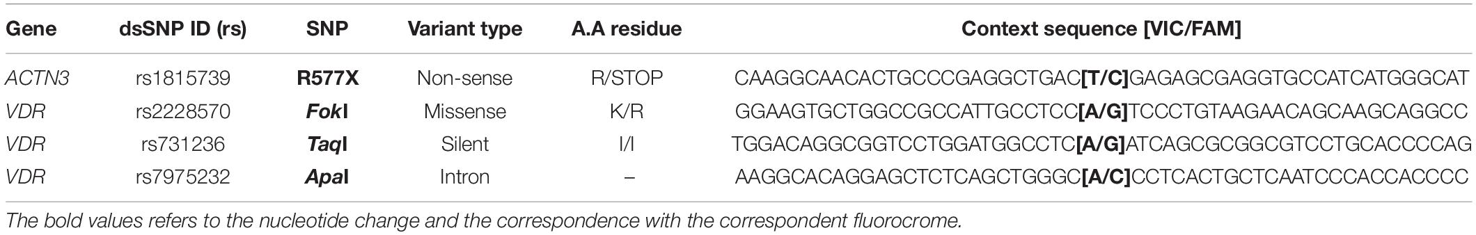
Table 1. Identification of all genetic variants included in the study.
The genotyping analysis was performed by quantitative polymerase chain reaction (qPCR) carried out on a 96-well QS5 Real-Time PCR (RT-PCR) System (Applied Biosystems; Thermo Fisher Scientific, Inc., Waltham, MA, United States), following the manufacturer’s instructions with the use of the commercially available TaqMan® SNP Genotyping Assays (Applied Biosystems) detailed in Table 1. To confirm genotyping and ensure accurate results, inconclusive samples were reanalyzed, and genotyping was repeated in 10–15% of randomly chosen samples, with 100% concordance.
Data analysis was performed using the Statistical Package for the Social Sciences for Windows 22.0 version (SPSS, Inc.). All genotypes were coded accordingly in order to proceed with the statistical analysis. The analysis of Hardy-Weinberg frequencies for all alleles present in patients’ populations was carried out using exact probability tests available using the SNPStat software1 (Sole et al., 2006).
Participants’ demographic, clinical, and biomechanical characteristics were described and compared between healthy individual and patients with axSpA, using Chi-Square (χ2) for discrete data and Wilcoxon-Mann-Whitney-test for non-parametric continuous data.
Since this is not a conclusive final study, but an exploratory one on the role of selected polymorphisms in the ACTN3 and VDR genes, and the data to be obtained should be looked at as proof of concept, the Bonferroni adjustment was deemed as not necessary as it is too conservative.
Logistic regression was used to estimate the risk of each muscle property modification when associated with each genotype: risk estimates were calculated under the codominant model and expressed as crude odds ratios (OR) and corresponding 95% confidence intervals (CI). Association between SNPs and the quantitative variables BASDAI, BASFI, BASMI, stiffness, decrement/inverse of elasticity, tone and strength were tested by linear regression. Results were considered significant when the corresponding two-tailed p-values were < 0.05. The most common homozygous genotype was considered the reference classes for such calculations.
Even though the study started with 56 enrolled participants, in equal number of controls and patients, due to missing data in some physical measures and in individuals’ polymorphisms identification, the total sample population ended up comprising 51 individuals (27 axSpA patients and 24 controls). The axSpA patients, 66.7% male with a mean age of 36 ± 7 years old and a mean of 7.0 ± 0.9 years of disease duration. From the total of patients, 80.8% were HLA-B27 positive, 18.5% had active disease (BASDAI ≥ 4) and 14.8% presented high functional impairment (BASFI > 4). The majority of patients did not exhibit reduction in mobility (88.9% had BASMI < 3). There was no difference between patients and controls in terms of age, gender, level of physical activity and body mass.
Baseline characteristics are shown in detail, in Table 2, where all data comparison between patients and controls can be found.
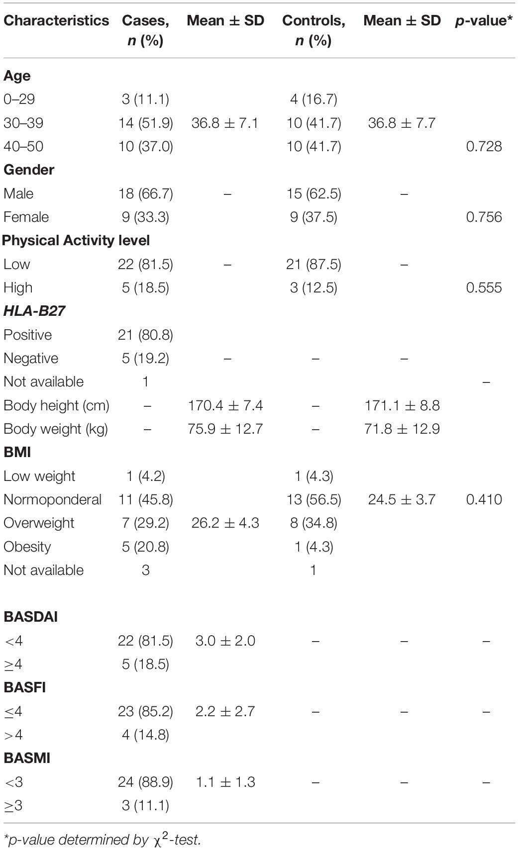
Table 2. Study population characterization. Cases group (n = 27) and Healthy control group (n = 24).
We recorded and analyzed the results obtained from lumbar paravertebral muscle (Multifidus muscle) physical properties, namely: Muscle tonus (M.F); Muscle decrement, i.e., the inverse of elasticity (M.D) and Muscle stiffness (M.S). The regression analysis was performed individually for each characteristic (crude analysis) (Table 3).
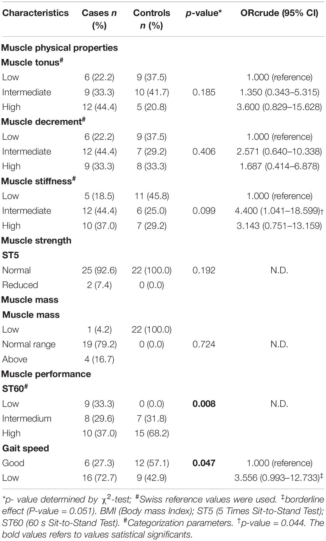
Table 3. Characterization of multifidus muscle physical properties, strength and mass.
No significant differences in muscle physical properties, muscle strength (ST5) and muscle mass were identified between patients and controls. However, it seems that patients tend to express higher levels of stiffness in multifidus muscle (trunk) (Table 3). Muscle performance, measured by ST60 and gait speed, was significantly reduced in patients, compared to controls (p < 0.05).
After genotyping analysis, the SNPs distribution was performed and their genetic contribution to disease susceptibility was evaluated (Table 4). To perform the correlations, we deemed it important to verify if the four SNPs were in Hardy-Weinberg Equilibrium (HWE). All the populations followed the Hardy-Weinberg Equilibrium (HWE), except for TaqI SNP (p-value = 0.021).
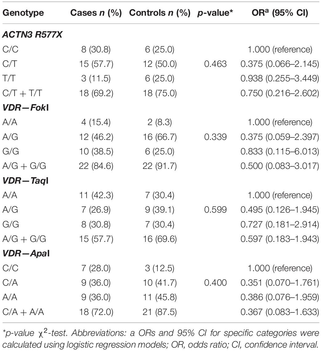
Table 4. Genotype distribution between axSpA patients and controls, for ACTN3 and VDR polymorphisms: rs1815739 (R577X); rs2228570 (FokI), rs731236 (TaqI), and rs7975232 (ApaI).
Considering that three SNPs from the same gene (VDR) were analyzed, we also evaluated the possibility of establishing an haplotype. However, the allele combination obtained for our populations did not reveal a statistically relevant combination to be correlated with disease susceptibility. After a multiple-SNP analysis, the results showed the existence of Linkage Disequilibrium between ApaI and TaqI SNPs from VDR (D’ = 0.9382, p-value ≤ 0.001).
Another question we wanted to address was related to the potential role of ACTN3 and VDR genes polymorphisms in the etiology of the disease evaluating the risk magnitude. The genotypic frequencies were determined for both groups and for all SNPs under study.
According to the results obtained, the studied SNPs did not seem to be associated with an increased risk of axSpA susceptibility.
In addition, axSpA clinical parameters (BASDAI, BASFI, and BASMI) were also measured in the patient population and the association with the allelic distribution for each SNP was investigated (Table 5). ACTN3 and VDR polymorphisms did not markedly influence axSpA disease activity, physical function or severity, as measured by BASDAI, BASFI, and BASMI.
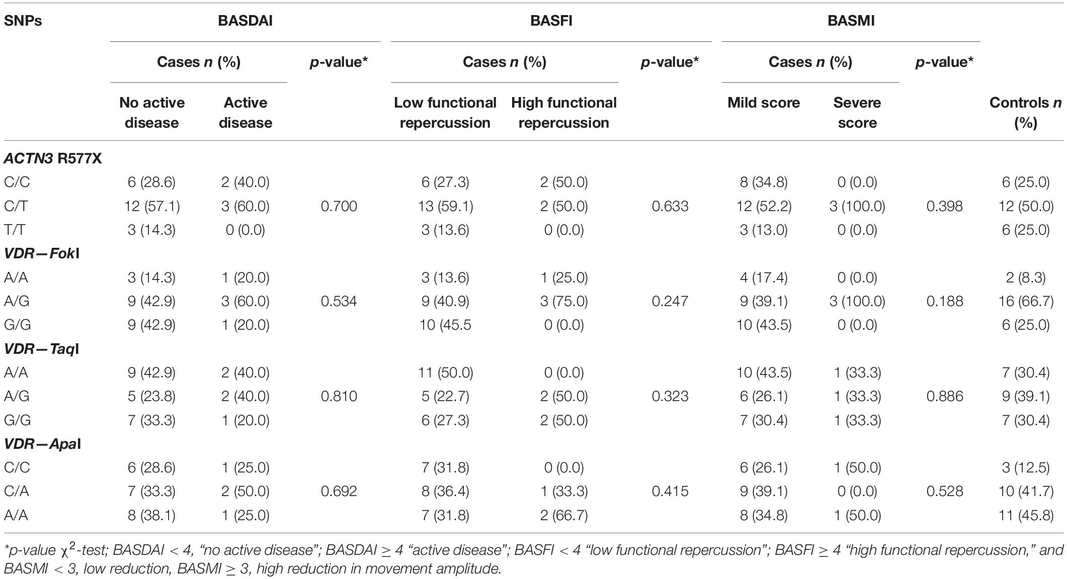
Table 5. Clinical characteristics across ACTN3 and VDR genes polymorphisms.
We hypothesize how relevant the genetic background might be to explain muscle properties (physical-stiffness, tone, elasticity; strength; mass) and in particular physical performance (ST60 and Gait Speed), where statistically significant differences between patients and controls were registered. To understand the hypothetical effect of single SNP in these parameters, we applied the logistic regression model adjusted to the presence of the genetic component; the results are presented in the table below (Table 6).
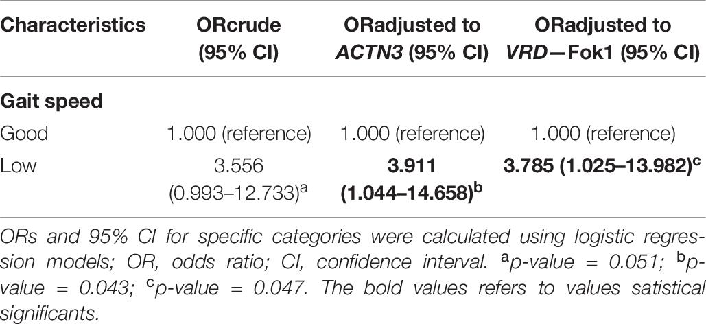
Table 6. Association of ACTN3 and VDR FokI polymorphisms and gait speed parameters.
Data shown refers only to ACTN3 and VDR Fok1 polymorphisms, since the logistic regression of the other two SNPs did not show statistically significant results.
This pilot study, involving 27 young axSpA patients with short disease duration, has not shown any difference in muscle physical properties, global muscle strength or mass compared to controls. However, a reduction in muscle performance assessed by two different approaches, ST60 and Gait speed, was found. Even using a small participant’s sample, the overall results allowed us to generate relevant data on disease susceptibility evidencing low physical performance in spondiloarthritis patients. Thus, we considered interesting to examine if genes associated with muscle performance, such as ACTN3 and VDR, might contribute to explain such differences.
As expected, due to our selection criteria, both groups (SpA patients and controls) are largely similar, regarding age, gender, levels of physical activity. When analyzing disease characteristics, the majority of patients exhibit low disease activity, low functional and low metrological repercussion. Most of our patients are HLA-B27 positive (80.8%), which is in line with the percentages already found for the Portuguese population (Pimentel-Santos et al., 2012). Due to the small size of our study population and low number of female individuals, we did not take into account possible gender differences in any of our analysis.
In this study, we selected several SNPs of well-known genes—ACTN3 (Ma et al., 2013; Pratt et al., 2019) and VDR (Pratt et al., 2019)—related with muscle performance to evaluate their influence in axSpA susceptibility, axSpA phenotype and in muscle properties. In this context, we were interested in studying these genetic variants in the axSpA context looking for the variants related with low muscle performance and simultaneously evaluate any association with muscle physical properties, strength, and lean mass. This would represent an additional method to identify patients that might benefit from a program of physical exercise and to the identification of the best modalities to be used in clinical practice.
In our study, we demonstrate that ACTN3 and VDR are not significantly associated with either susceptibility to axSpA or measures of its activity, disability or severity, as measured by BASDAI, BASFI, and BASMI, respectively. Indeed, no association has been identified in GWAS between these genes and SpA (Australo-Anglo-American Spondyloarthritis et al., 2010; International Genetics of Ankylosing Spondylitis et al., 2013). Nevertheless, some VDR polymorphisms have been linked to some musculoskeletal diseases, such as idiopathic scoliosis susceptibility or curve severity, herniation and spinal tissues degeneration and rheumatoid arthritis (Saad et al., 2015; Di Spigna et al., 2016; Vieira et al., 2018; Li et al., 2019).
No association was established between the studied SNPs and muscle physical properties, namely stiffness, tone or elasticity. To the best of our knowledge, this association was never studied. Again, no association for overall strength (ST5) was registered in our cohort. However, in several previous studies, ACTN3 577R allele and VDR were associated with higher levels of strength. The rs540874 polymorphism of ACTN3 gene was associated with the muscle function of lower limb (women with the G allele were likely to have higher strength compared with the ones with A allele) but not with the higher limb, in postmenopausal women. Interestingly, in the same study, the improvement of muscle strength after an intervention (exercise and Vitamin D supplementation) was possibly correlated with rs540874, rs618838, and rs2229456 polymorphisms (Xue et al., 2018). A significant association between VDR genotype and quadriceps (23% difference) and grip (7% difference) strength was observed in non-obese elderly women (Geusens et al., 1997).
The analysis of these genetic markers regarding lean muscle mass did not show, again, any difference between both groups. It is well-known that genetic factors account for approximately 50-80% of inter-individual variation in lean body mass, with impacts detected on both “training-naïve” muscle mass and its growth response (Puthucheary et al., 2011). Indeed, these genes have been found to contribute to variation in lean body mass and bone mass density, contributing to understanding the molecular bases of sarcopenia and osteoporosis (Tan et al., 2012; Gonzalez-Mercado et al., 2013; Cho et al., 2017; Scimeca et al., 2018). However, in one study involving older Caucasian men, whole body and thigh non-skeletal lean mass were independent of ACTN3 R/X polymorphisms (Mccauley et al., 2010). In contrast, VDR expression decreases with age and VDR genotype seems to be associated with fat-free mass in elderly men and women (Puthucheary et al., 2011). Differences in populations’ characteristics, study methodologies and reduced number of participants (underpowered studies) to detect genes with small effects, potentially lead to discrepancies between results.
This study has shown a clear reduction in axSpA muscle performance without changes in muscle physical properties, strength, and mass. This observation allows us to speculate about a possible muscle dysfunction. As genetics has a strong influence in overall axSpA susceptibility, it is of main interest to investigate a possible genetic base to explain this impairment in muscle performance. Our results indicate that ACTN3 R577X and VDR Fok1 SNPs might influence Gait Speed [OR, 3.911; 95% CI (1.044–14.658) and OR, 3.785; 95% CI (1.025–13.982)]. Several studies have shown an association of these variants on muscle performance. In 2003, Yang and colleagues (Yang et al., 2003) demonstrated a significant association between ACTN3 genotype and athletic performance. They found that both male and female elite sprint athletes have significantly higher frequencies of the 577R allele compared to controls. Later on, several papers consistently reported a strong association between RR genotype and elite power performance (Paparini et al., 2007; Papadimitriou et al., 2008; Eynon et al., 2009; Chiu et al., 2011; Ma et al., 2013). Similar evidence was documented for VDR polymorphisms (Micheli et al., 2011; Puthucheary et al., 2011). In elite Italian soccer players, an interaction of two polymorphisms (ACE and ACTN3) predicted explosive leg-muscle strength, however, the contribution of genetic factors was only 23.92% (Massidda et al., 2012).
The genotype distribution of all SNPs was in HWE, except for the TaqI polymorphism (p = 0.021), suggesting the influence of genetic drift due to the small population size. Furthermore, we also evaluated the possibility to establish a specific haplotype for VDR gene polymorphisms, however, no haplotype was identified as relevant. Our results allowed us to identify Linkage Disequilibrium between ApaI and TaqI SNPs, indicating that both are segregated together. This corroborates some studies (Hamilton, 2010) and means that both SNPs are always transmitted in block (Cieslinska et al., 2018) (data not shown), even though it was not possible to identify a risk haplotype for our population. Despite the literature revealing that VDR SNPs constitute a haplotype, due to their proximity, we could not confirm those reports (Hunt et al., 2009).
To our knowledge, this pilot study is the very first including a genetic susceptibility analysis for muscle properties in the axSpA context. Overall, our results suggest an association between ACTN3 and VDR polymorphisms and muscle performance in the axSpA context. This opens the door to a better stratification of patients regarding exercise programs, but also to identify other candidate genes that might help to characterize the genetic background of the disease.
The raw data supporting the conclusions of this article will be made available by the authors, without undue reservation.
The studies involving human participants were reviewed and approved by the NOVA Medical School | Faculdade de Ciências Médicas, Universidade NOVA de Lisboa, Portugal Centro Hospitalar Lisboa Ocidental, Hospital de Egas Moniz, EPE, Lisboa, Portugal. The patients/participants provided their written informed consent to participate in this study.
FP-S, JB, and SS mainly developed the conceptualization. IP, HM, FP-S, and SS performed the methodology. SS and FP-S proceeded validation and prepared the visualization. SS and IP did formal analysis. FP-S, SR-M, RP-T, AN, LD, CL, IP, HM, and SS mainly performed the investigation. FP-S collaborated resources acquired in restrict. AN, FP-S, and SS performed the data curation. FP-S, SS, and IP contributed to the writing – original draft preparation. FP-S, SS, SR-M, RP-T, AN, LD, CL, AS, PM, and JB contributed to the writing – review and editing. FP-S, JB, and SS supervised this project. LD contributed to the project administration. FP-S and JB contributed to the funding acquisition. All authors contributed to the article and approved the submitted version.
This study was supported by the funding through project MyoSpA, from iNOVA4 health. PM was supported by the National Institute for Health Research (NIHR) University College London Hospitals (UCLH) Biomedical Research Centre (BRC).
The authors declare that the research was conducted in the absence of any commercial or financial relationships that could be construed as a potential conflict of interest.
All claims expressed in this article are solely those of the authors and do not necessarily represent those of their affiliated organizations, or those of the publisher, the editors and the reviewers. Any product that may be evaluated in this article, or claim that may be made by its manufacturer, is not guaranteed or endorsed by the publisher.
We gratefully acknowledge all patients and controls who generously participated in this study. The acknowledgments are also extended to Manuela Correia for the logistic control.
The Supplementary Material for this article can be found online at: https://www.frontiersin.org/articles/10.3389/fgene.2021.688984/full#supplementary-material
Andonian, B. J., Masi, A. T., Aldag, J. C., Barry, A. J., Coates, B. A., Emrich, K., et al. (2015). Greater Resting Lumbar Extensor Myofascial Stiffness in Younger Ankylosing Spondylitis Patients Than Age-Comparable Healthy Volunteers Quantified by Myotonometry. Arch. Phys. Med. Rehabil. 96, 2041–2047. doi: 10.1016/j.apmr.2015.07.014
Apostolakos, J., Durant, T. J., Dwyer, C. R., Russell, R. P., Weinreb, J. H., and Alaee, F. (2014). The enthesis: a review of the tendon-to-bone insertion. Muscles Ligaments Tendons J. 4, 333–342. doi: 10.32098/mltj.03.2014.12
Asquith, M., Elewaut, D., Lin, P., and Rosenbaum, J. T. (2014). The role of the gut and microbes in the pathogenesis of spondyloarthritis. Best Pract. Res. Clin. Rheumatol. 28, 687–702. doi: 10.1016/j.berh.2014.10.018
Australo-Anglo-American Spondyloarthritis, C., Reveille, J. D., Sims, A. M., Danoy, P., Evans, D. M., Leo, P., et al. (2010). Genome-wide association study of ankylosing spondylitis identifies non-MHC susceptibility loci. Nat. Genet. 42, 123–127. doi: 10.1038/ng.513
Benjamin, M., and Mcgonagle, D. (2001). The anatomical basis for disease localisation in seronegative spondyloarthropathy at entheses and related sites. J. Anat. 199, 503–526. doi: 10.1046/j.1469-7580.2001.19950503.x
Ceglia, L. (2009). Vitamin D and its role in skeletal muscle. Curr. Opin. Clin. Nutr. Metab. Care 12, 628–633. doi: 10.1097/mco.0b013e328331c707
Chiu, L. L., Wu, Y. F., Tang, M. T., Yu, H. C., Hsieh, L. L., and Hsieh, S. S. (2011). ACTN3 genotype and swimming performance in Taiwan. Int J Sports Med 32, 476–480. doi: 10.1055/s-0030-1263115
Cho, J., Lee, I., and Kang, H. (2017). ACTN3 Gene and Susceptibility to Sarcopenia and Osteoporotic Status in Older Korean Adults. Biomed. Res. Int. 2017:4239648.
Cieslinska, A., Kostyra, E., Fiedorowicz, E., Snarska, J., Kordulewska, N., Kiper, K., et al. (2018). Single Nucleotide Polymorphisms in the Vitamin D Receptor Gene (VDR) May Have an Impact on Acute Pancreatitis (AP) Development: A Prospective Study in Populations of AP Patients and Alcohol-Abuse Controls. Int. J. Mol. Sci. 2018:19.
Costantino, F., Breban, M., and Garchon, H. J. (2018). Genetics and Functional Genomics of Spondyloarthritis. Front. Immunol. 9:2933. doi: 10.3389/fimmu.2018.02933
Craig, C. L., Marshall, A. L., Sjostrom, M., Bauman, A. E., Booth, M. L., Ainsworth, B. E., et al. (2003). International physical activity questionnaire: 12-country reliability and validity. Med. Sci. Sports Exerc. 35, 1381–1395.
Cruz-Jentoft, A. J., Bahat, G., Bauer, J., Boirie, Y., Bruyere, O., Cederholm, T., et al. (2019). Sarcopenia: revised European consensus on definition and diagnosis. Age Ageing 48:601. doi: 10.1093/ageing/afz046
Di Spigna, G., Del Puente, A., Covelli, B., Abete, E., Varriale, E., Salzano, S., et al. (2016). Vitamin D receptor polymorphisms as tool for early screening of severe bone loss in women patients with rheumatoid arthritis. Eur. Rev. Med. Pharmacol. Sci. 20, 4664–4669.
Eynon, N., Duarte, J. A., Oliveira, J., Sagiv, M., Yamin, C., Meckel, Y., et al. (2009). ACTN3 R577X polymorphism and Israeli top-level athletes. Int. J. Sports Med. 30, 695–698.
Geusens, P., Vandevyver, C., Vanhoof, J., Cassiman, J. J., Boonen, S., and Raus, J. (1997). Quadriceps and grip strength are related to vitamin D receptor genotype in elderly nonobese women. J. Bone Miner Res. 12, 2082–2088. doi: 10.1359/jbmr.1997.12.12.2082
Gill, T., Asquith, M., Rosenbaum, J. T., and Colbert, R. A. (2015). The intestinal microbiome in spondyloarthritis. Curr. Opin. Rheumatol. 27, 319–325. doi: 10.1097/bor.0000000000000187
Gonzalez-Mercado, A., Sanchez-Lopez, J. Y., Regla-Nava, J. A., Gamez-Nava, J. I., Gonzalez-Lopez, L., Duran-Gonzalez, J., et al. (2013). Association analysis of vitamin D receptor gene polymorphisms and bone mineral density in postmenopausal Mexican-Mestizo women. Genet. Mol. Res. 12, 2755–2763. doi: 10.4238/2013.july.30.13
Hunt, R., Sauna, Z. E., Ambudkar, S. V., Gottesman, M. M., and Kimchi-Sarfaty, C. (2009). Silent (synonymous) SNPs: should we care about them? Methods Mol. Biol. 578, 23–39. doi: 10.1007/978-1-60327-411-1_2
International Genetics of Ankylosing Spondylitis, C., Cortes, A., Hadler, J., Pointon, J. P., Robinson, P. C., Karaderi, T., et al. (2013). Identification of multiple risk variants for ankylosing spondylitis through high-density genotyping of immune-related loci. Nat. Genet. 45, 730–738. doi: 10.1038/ng.2667
Jacques, P., Lambrecht, S., Verheugen, E., Pauwels, E., Kollias, G., Armaka, M., et al. (2014). Proof of concept: enthesitis and new bone formation in spondyloarthritis are driven by mechanical strain and stromal cells. Ann. Rheum Dis. 73, 437–445. doi: 10.1136/annrheumdis-2013-203643
Li, J., Chen, S. Y., Liu, H. H., Yin, X. D., Cao, L. T., Xu, J. H., et al. (2019). Associations of Vitamin D Receptor Single Nucleotide Polymorphisms with Susceptibility to Systemic Sclerosis. Arch. Med. Res. 50, 368–376. doi: 10.1016/j.arcmed.2019.09.006
Ma, F., Yang, Y., Li, X., Zhou, F., Gao, C., Li, M., et al. (2013). The association of sport performance with ACE and ACTN3 genetic polymorphisms: a systematic review and meta-analysis. PLoS One 8:e54685. doi: 10.1371/journal.pone.0054685
Massidda, M., Corrias, L., Ibba, G., Scorcu, M., Vona, G., and Calo, C. M. (2012). Genetic markers and explosive leg-muscle strength in elite Italian soccer players. J. Sports Med. Phys. Fitness 52, 328–334.
Mccauley, T., Mastana, S. S., and Folland, J. P. (2010). ACE I/D and ACTN3 R/X polymorphisms and muscle function and muscularity of older Caucasian men. Eur. J. Appl. Physiol. 109, 269–277. doi: 10.1007/s00421-009-1340-y
Mcgonagle, D., Stockwin, L., Isaacs, J., and Emery, P. (2001). An enthesitis based model for the pathogenesis of spondyloarthropathy. additive effects of microbial adjuvant and biomechanical factors at disease sites. J. Rheumatol. 28, 2155–2159.
Micheli, M. L., Gulisano, M., Morucci, G., Punzi, T., Ruggiero, M., Ceroti, M., et al. (2011). Angiotensin-converting enzyme/vitamin D receptor gene polymorphisms and bioelectrical impedance analysis in predicting athletic performances of Italian young soccer players. J. Strength Cond. Res. 25, 2084–2091. doi: 10.1519/jsc.0b013e31820238aa
Osgood, J. A., and Knight, J. C. (2018). Translating GWAS in rheumatic disease: approaches to establishing mechanism and function for genetic associations with ankylosing spondylitis. Brief Funct. Genomics 17, 308–318.
Papadimitriou, I. D., Papadopoulos, C., Kouvatsi, A., and Triantaphyllidis, C. (2008). The ACTN3 gene in elite Greek track and field athletes. Int. J. Sports Med. 29, 352–355. doi: 10.1055/s-2007-965339
Paparini, A., Ripani, M., Giordano, G. D., Santoni, D., Pigozzi, F., and Romano-Spica, V. (2007). ACTN3 genotyping by real-time PCR in the Italian population and athletes. Med. Sci. Sports Exerc. 39, 810–815. doi: 10.1097/mss.0b013e3180317491
Pickering, C., and Kiely, J. (2017). ACTN3: More than Just a Gene for Speed. Front. Physiol. 8:1080. doi: 10.3389/fphys.2017.01080
Pimentel-Santos, F. M., Ligeiro, D., Matos, M., Mourao, A. F., Costa, J., Santos, H., et al. (2011). Whole blood transcriptional profiling in ankylosing spondylitis identifies novel candidate genes that might contribute to the inflammatory and tissue-destructive disease aspects. Arthritis Res. Ther. 13:R57.
Pimentel-Santos, F. M., Matos, M., Ligeiro, D., Mourao, A. F., Ribeiro, C., Costa, J., et al. (2013). HLA alleles and HLA-B27 haplotypes associated with susceptibility and severity of ankylosing spondylitis in a Portuguese population. Tissue Antigens 82, 374–379. doi: 10.1111/tan.12238
Pimentel-Santos, F. M., Mourao, A. F., Ribeiro, C., Costa, J., Santos, H., Barcelos, A., et al. (2012). Spectrum of ankylosing spondylitis in Portugal. Development of BASDAI, BASFI, BASMI and mSASSS reference centile charts. Clin. Rheumatol. 31, 447–454. doi: 10.1007/s10067-011-1854-7
Pratt, J., Boreham, C., Ennis, S., Ryan, A. W., and De Vito, G. (2019). Genetic Associations with Aging Muscle: A Systematic Review. Cells 2019:9.
Puthucheary, Z., Skipworth, J. R., Rawal, J., Loosemore, M., Van Someren, K., and Montgomery, H. E. (2011). Genetic influences in sport and physical performance. Sports Med. 41, 845–859. doi: 10.2165/11593200-000000000-00000
Ramiro, S., Landewé, R., Van Tubergen, A., Boonen, A., Stolwijk, C., Dougados, M., et al. (2015). Lifestyle factors may modify the effect of disease activity on radiographic progression in patients with ankylosing spondylitis: a longitudinal analysis. RMD Open 1:e000153. doi: 10.1136/rmdopen-2015-000153
Rejnmark, L. (2011). Effects of vitamin d on muscle function and performance: a review of evidence from randomized controlled trials. Ther. Adv. Chronic. Dis. 2, 25–37. doi: 10.1177/2040622310381934
Rosenbaum, J. T., and Davey, M. P. (2011). Time for a gut check: evidence for the hypothesis that HLA-B27 predisposes to ankylosing spondylitis by altering the microbiome. Arthritis Rheum. 63, 3195–3198. doi: 10.1002/art.30558
Rudwaleit, M., Van Der Heijde, D., Landewe, R., Akkoc, N., Brandt, J., Chou, C. T., et al. (2011). The Assessment of SpondyloArthritis International Society classification criteria for peripheral spondyloarthritis and for spondyloarthritis in general. Ann. Rheum. Dis. 70, 25–31. doi: 10.1136/ard.2010.133645
Rudwaleit, M., Van Der Heijde, D., Landewe, R., Listing, J., Akkoc, N., Brandt, J., et al. (2009). The development of Assessment of SpondyloArthritis international Society classification criteria for axial spondyloarthritis (part II): validation and final selection. Ann. Rheum. Dis. 68, 777–783. doi: 10.1136/ard.2009.108233
Saad, M. N., Mabrouk, M. S., Eldeib, A. M., and Shaker, O. G. (2015). Genetic Case-Control Study for Eight Polymorphisms Associated with Rheumatoid Arthritis. PLoS One 10:e0131960. doi: 10.1371/journal.pone.0131960
Schett, G., Lories, R. J., D’agostino, M. A., Elewaut, D., Kirkham, B., Soriano, E. R., et al. (2017). Enthesitis: from pathophysiology to treatment. Nat. Rev. Rheumatol. 13, 731–741. doi: 10.1038/nrrheum.2017.188
Scimeca, M., Centofanti, F., Celi, M., Gasbarra, E., Novelli, G., Botta, A., et al. (2018). Vitamin D Receptor in Muscle Atrophy of Elderly Patients: A Key Element of Osteoporosis-Sarcopenia Connection. Aging Dis. 9, 952–964. doi: 10.14336/ad.2018.0215
Sieper, J., Rudwaleit, M., Baraliakos, X., Brandt, J., Braun, J., and Burgos-Vargas, R. (2009). The Assessment of SpondyloArthritis international Society (ASAS) handbook: a guide to assess spondyloarthritis. Ann. Rheum. Dis. 68(Suppl. 2), ii1–ii44.
Simone, D., Al Mossawi, M. H., and Bowness, P. (2018). Progress in our understanding of the pathogenesis of ankylosing spondylitis. Rheumatology 57, vi4–vi9.
Sole, X., Guino, E., Valls, J., Iniesta, R., and Moreno, V. (2006). SNPStats: a web tool for the analysis of association studies. Bioinformatics 22, 1928–1929. doi: 10.1093/bioinformatics/btl268
Stolwijk, C., Van Onna, M., Boonen, A., and Van Tubergen, A. (2016). Global Prevalence of Spondyloarthritis: A Systematic Review and Meta-Regression Analysis. Arthritis Care Res. 68, 1320–1331. doi: 10.1002/acr.22831
Tan, L. J., Liu, S. L., Lei, S. F., Papasian, C. J., and Deng, H. W. (2012). Molecular genetic studies of gene identification for sarcopenia. Hum. Genet. 131, 1–31. doi: 10.1007/s00439-011-1040-7
Van Mechelen, M., and Lories, R. J. (2016). Microtrauma: no longer to be ignored in spondyloarthritis? Curr. Opin. Rheumatol. 28, 176–180. doi: 10.1097/bor.0000000000000254
Vieira, L. A., Dos Santos, A. A., Peluso, C., Barbosa, C. P., Bianco, B., and Rodrigues, L. M. R. (2018). Influence of lifestyle characteristics and VDR polymorphisms as risk factors for intervertebral disc degeneration: a case-control study. Eur. J. Med. Res. 23:11.
Xue, Y., Nie, M., Wang, O., Wang, C. Y., Han, G. Y., Shen, Q., et al. (2018). [Association of alpha-actinin-3 gene polymorphism and muscle strength of postmenopausal women]. Zhonghua Yi Xue Za Zhi 98, 1408–1413.
Keywords: spondyloarthropathies, muscle, muscle performance, ACTN3, VDR
Citation: Pimenta I, Mateus H, Rodrigues-Manica S, Pinheiro-Torres R, Neto A, Domingues L, Lage Crespo C, Sardoo A, Machado P, Branco JC, Silva SN and Pimentel-Santos FM (2021) The Effect of ACTN3 and VDR Polymorphisms on Skeletal Muscle Performance in Axial Spondyloarthropathies. Front. Genet. 12:688984. doi: 10.3389/fgene.2021.688984
Received: 12 April 2021; Accepted: 12 July 2021;
Published: 11 August 2021.
Edited by:
Roberta Ramonda, University of Padua, ItalyReviewed by:
Paola Galozzi, University of Padua, ItalyCopyright © 2021 Pimenta, Mateus, Rodrigues-Manica, Pinheiro-Torres, Neto, Domingues, Lage Crespo, Sardoo, Machado, Branco, Silva and Pimentel-Santos. This is an open-access article distributed under the terms of the Creative Commons Attribution License (CC BY). The use, distribution or reproduction in other forums is permitted, provided the original author(s) and the copyright owner(s) are credited and that the original publication in this journal is cited, in accordance with accepted academic practice. No use, distribution or reproduction is permitted which does not comply with these terms.
*Correspondence: Susana N. Silva, c25zaWx2YUBubXMudW5sLnB0; Fernando M. Pimentel-Santos, cGltZW50ZWwuc2FudG9zQG5tcy51bmwucHQ=
Disclaimer: All claims expressed in this article are solely those of the authors and do not necessarily represent those of their affiliated organizations, or those of the publisher, the editors and the reviewers. Any product that may be evaluated in this article or claim that may be made by its manufacturer is not guaranteed or endorsed by the publisher.
Research integrity at Frontiers
Learn more about the work of our research integrity team to safeguard the quality of each article we publish.