- 1Division of Gastroenterology, Department of Internal Medicine, Taitung Mackay Memorial Hospital, Taitung, Taiwan
- 2Department of Medicine, Mackay Medical College, New Taipei, Taiwan
- 3Department of Pathology, Taitung Mackay Memorial Hospital, Taitung, Taiwan
Background and Aims: Chronic hepatitis C virus (HCV) infection is associated with dysregulation of glucose homeostasis, including insulin resistance (IR) and type 2 diabetes. However, independent risk factors associated with IR in chronic HCV-infected patients have not been detailly elucidated. Previous data regarding the impact of HCV elimination by direct-acting antiviral agents (DAAs) on glucose homeostasis is insufficient and controversial. This study aimed to analyze the independent factors associated with IR and to evaluate the changes in glucose homeostasis in chronic HCV-infected patients treated with DAAs therapies.
Methods: We screened 704 patients with chronic HCV infection who underwent treatment with interferon-free DAAs. Patients’ baseline characteristics, biochemical and virological data were collected. The outcome measurements were their IR and β-cell function assessed by the homeostasis model assessment (HOMA) method at baseline and 12-weeks post-treatment.
Results: High IR (HOMA-IR ≥ 2.5) was observed in 35.1% of the patients. Multivariable logistic regression analysis revealed that body mass index (BMI) >25 kg/m2, treatment experience, elevated baseline levels of alanine aminotransferase (ALT) and triglyceride, as well as Fibrosis-4 score >3.25 were independently associated with high IR. In patients who achieved sustained virological response (SVR), no significant change in mean HOMA-IR was observed from baseline to 12-weeks post-treatment (2.74 ± 2.78 to 2.54 ± 2.20, p = 0.128). We observed a significant improvement in β-cell secretion stress from 121.0 ± 110.1 to 107.6 ± 93.0 (p = 0.015). Subgroup analysis revealed that SVR was associated with a significant reduction in mean HOMA-IR in patients with baseline HOMA-IR ≥ 2.5 (5.31 ± 3.39 to 3.68 ± 2.57, p < 0.001), HCV genotype 1 (3.05 ± 3.11 to 2.62 ± 2.05, p = 0.027), and treatment experience (4.00 ± 3.37 to 3.01 ± 2.49, p = 0.039).
Conclusions: There were several independent factors associated with IR in patients with chronic HCV infection, including obesity, treatment experience, high serum ALT and triglyceride levels, as well as advanced hepatic fibrosis. After viral elimination by DAAs, we observed a significant reduction in mean HOMA-IR in patients with baseline high IR, HCV genotype 1, and treatment experience.
Introduction
Chronic hepatitis C virus (HCV) infection is a major cause of chronic hepatitis, cirrhosis, and hepatocellular carcinoma. It is also associated with multiple extrahepatic complications that influence the clinical outcomes of patients. An important extrahepatic manifestation is abnormal glucose metabolism including insulin resistance (IR), β-cell dysfunction, and diabetes mellitus (DM) (1–3). A prospective study showed that patients with chronic HCV infection were more than 11 times as likely as those without HCV infection to develop diabetes (4). A meta-analysis revealed a 1.8-fold higher risk of type 2 DM in HCV-infected patients than in hepatitis B virus (HBV)-positive/HCV-negative patients (5). Additionally, glucose abnormalities can worsen hepatic outcomes in HCV-infected patients (6). IR promotes the progression of hepatic fibrosis. This may be due to the direct effect of insulin on the proliferation of hepatic stellate cells and secretion of extracellular matrix (7, 8). Further, high glucose levels and hyperinsulinemia can lead to upregulation of connective growth factor that is involved in the pathogenesis of liver fibrosis (9). In HCV-related chronic liver disease, type 2 diabetes and IR are independently associated with rapid progression of liver fibrosis and increased risk of hepatocellular carcinoma (10, 11), as well as the elevated rate of hepatic morbidity and mortality (12).
The mechanisms underlying HCV-induced IR and DM are complex and poorly explained. The HCV genome is composed of structural (core, E1, and E2) and nonstructural (NS2-NS5B) genes. The complex effects of HCV core and nonstructural 5A (NS5A) proteins have been observed to play important roles in glucose metabolism (13). In normal circumstances, when insulin attaches to the hepatocyte receptor, the insulin receptor substrate (IRS) is phosphorylated and then causes activation of Akt. The activated Akt causes the translocation of glucose transporter-4 to the surface of the hepatocyte and facilitates glucose entry into the hepatocyte. Akt also induces the synthesis of glycogen and inhibition of hepatic gluconeogenesis (14). The HCV core protein impairs insulin signaling via several post-receptor mechanisms, including the activation of suppressor of cytokine signaling (SOCS) family members and consequent decrease in IRS-1 (15). The core protein also increases phosphorylation of IRS-1 at serine rather than tyrosine in hepatocytes, again preventing the downstream activation of Akt (14). HCV NS5A upregulates protein phosphatase 2A and inactivates Akt, which reduces the expression of glucose transporters GLUT1 and GLUT2, leading to a reduction in glucose uptake in the hepatocytes, and thus, induces IR (16). Additionally, HCV protein increases oxidative stress and mitochondrial dysfunction and leads to overexpression of inflammatory cytokines, such as tumor necrosis factor alpha, interleukin (IL)-6, and IL-8, resulting in systemic effect including hyperinsulinemia, hypertriglycemia, and down-regulation of adiponectin (17). Epidemiological data suggest that all the major HCV genotypes lead to IR. Some viral-specific factors that influence glucose metabolism have also been discussed. Molecular evidence suggests that HCV core proteins in genotypes 3a and 1b promote IRS-1 degradation via different mechanisms (18). IR was clinically found to be associated with HCV genotype 1 and a high viral load, although the results were inconsistent (4, 19, 20).
Elimination of HCV by interferon (IFN)-based regimen may perturb glucose homeostasis. Previous observational studies have shown that successful eradication of HCV improves IR in patients receiving IFN-based therapy (20, 21). The β-cell hyperfunction is also ameliorated after antiviral treatment (22). Therefore, elimination or suppression of HCV can reduce the incidence of type 2 diabetes (23, 24). However, a few studies revealed no change in fasting glucose, insulin, and homeostatic model assessment-IR (HOMA-IR) after antiviral treatment (25, 26). The results of these studies are inconsistent, but they indicate that the benefits of glucose metabolism may also be HCV genotype specific.
Treatment has dramatically improved in the era of direct-acting antiviral agents (DAAs). These modern drugs have extremely high treatment efficacy, safety, and less adverse events. These new medications have a higher rate of viral clearance than IFN-based therapy but lack the direct effect of IFN on glucose metabolism. Moreover, the impact of HCV clearance by DAAs on glucose abnormalities remains to be evaluated.
Given the presumption that chronic HCV infection involves the development of IR, we hypothesized that HCV eradication by DAAs treatment could improve IR. The aim of the present study was to explore potential factors associated with IR in chronic HCV-infected patients and to assess the impact of HCV elimination on glucose homeostasis in patients who received DAAs therapies.
Methods
Study Design
This was a single-center, observational study. Chronic HCV-infected patients with DAAs treatment were enrolled. Laboratory data including glucose parameters at baseline and 12-weeks post-treatment were measured. Several factors associated with baseline high IR were analyzed. Further, we calculated the dynamic changes in IR and β-cell secretion function in patients whose viruses were successfully eradicated. This study was conducted in accordance with the principles of the Declaration of Helsinki, and the study design was approved by the Institutional Review Board of the Mackay Memorial Hospital. Informed consent was obtained from the study participants.
Patients
We enrolled patients with chronic HCV infection who underwent treatment with IFN-free DAAs therapy between January 2017 and July 2020. The inclusion criteria were patients (1) aged ≥ 20 years; (2) with detectable HCV viral load in the blood prior to treatment; (3) who agreed to receive laboratory testing at baseline, during antiviral treatment, and at 12 weeks after treatment; and (4) who provided informed consent. Patients were excluded if they: (1) missed regular clinical follow-ups after treatment; (2) had a medical history of DM or used glucose-lowering drugs; (3) died from other etiology during the study period; and (4) had concurrent conditions, including human immunodeficiency virus or HBV coinfection, Wilson’s disease, primary biliary cirrhosis, hemochromatosis, and autoimmune hepatitis. Basic patient information, body mass index (BMI), HCV viral loads and genotype, alcohol consumption, and medical history were collected. Patients received different DAAs regimens, including asunaprevir + daclatasvir (2.5%), paritaprevir + ritonavir + ombitasvir + dasabuvir (7.8%), elbasvir + grazoprevir (10.7%), glecaprevir + pibrentasvir (16.6%), and sofosbuvir-based regimens (62.4%). Each patient received one of these DAAs regimens based on the HCV genotype, previous treatment status, presence of cirrhosis, current medications, and comorbidities.
Laboratory Examinations
Infection with chronic HCV was defined as persistent viremia for at least six months before treatment. Serum HCV ribonucleic acid (RNA) levels were quantified using a Roche Amplicor polymerase chain reaction (PCR) assay, in which the lowest level of detection was 15 IU/mL. HCV genotyping was performed using primer-specific PCR and direct PCR deep sequencing with an ABI 3730 sequencer. The sustained virological response (SVR) was defined as undetectable serum HCV RNA levels 12 weeks following treatment cessation. Information on the levels of fasting glucose, insulin, aspartate transaminase (AST), alanine aminotransferase (ALT), bilirubin, albumin, cholesterol, triglyceride, alpha-fetoprotein, white blood cells (WBC), as well as hemoglobin and platelets at baseline and 12-weeks post-treatment were collected. We used Fibrosis-4 (FIB-4) index as the non-invasive measurement for hepatic fibrosis. The FIB-4 index was calculated using the following formula: (AST [IU/L] × age [years])/(platelet count [109/L] × ALT [IU/L]1/2).
The glucose homeostasis was assessed by the HOMA method. The formulas for the HOMA model are as follows: HOMA-IR = fasting glucose (mg/dL) × fasting insulin level (μU/mL)/405; HOMA-β = fasting insulin level (μU/mL) × 360/(fasting glucose [mg/dL]–63). We dichotomized patients at a cutoff point of HOMA-IR 2.5 and analyzed factors associated with IR.
Statistical Analysis
Categorical data are presented as numbers (percentages) and continuous variables are presented as mean ± standard deviation. The significance of the difference between categorical variables was determined using the Chi-square test or Fisher’s exact test depending on the sample size; continuous variables were compared using Student’s t-test. For continuous variables with wide distribution, we presented data as median (interquartile range) and applied the Mann-Whitney test to evaluate the differences. After univariate analysis, we performed multivariable logistic regression modelling to identify factors that were independently associated with baseline high IR. To compare quantitative glucose parameters between baseline and post-treatment, we performed Paired t-test. All statistical analyses were defined as two-sided hypotheses with a significance level defined at p < 0.05. Statistical analyses were performed using SPSS 22.0 (SPSS Inc., Chicago, IL, USA).
Results
Baseline Characteristics
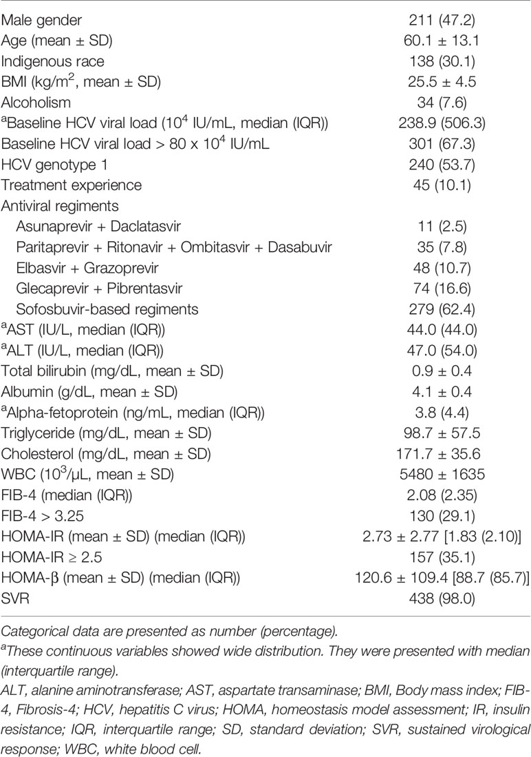
During the study period, 704 patients were treated with IFN-free DAAs. We excluded 192 patients with medical history of DM or who used glucose-lowering drugs. Additionally, 65 patients were excluded from the study due to failure of checking insulin levels at either baseline or follow-up (n=34), missing regular follow-up (n=24), and death during treatment (n=7). Finally, we enrolled 447 patients in the study. The baseline characteristics, laboratory results, glucose parameters, DAAs regimens, and treatment responses are shown in Table 1. The patients were predominantly women (52.8%), with a mean BMI of 25.5 ± 4.5 kg/m2, which was in the upper range of normal weight. The most frequent HCV genotype was genotype 1 (53.7%), followed by genotype 2 (35.1%). The prevalence of high viral loads (> 80 × 104 IU/mL) was 67.3%. Sofosbuvir-based regimens were most commonly used, accounting for 62.4%.
Variables Associated With Baseline IR
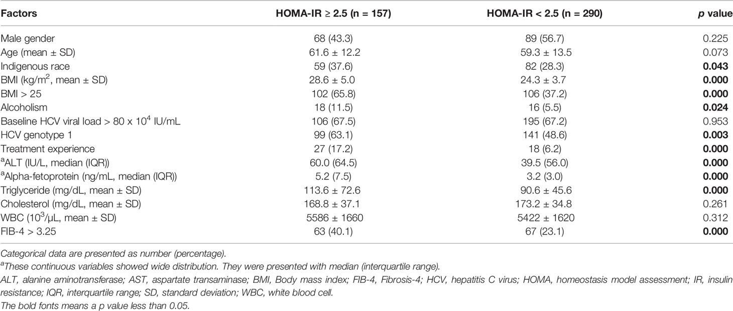
At baseline, 157 patients (35.1%) presented with high IR (HOMA-IR ≥ 2.5). Univariate analysis (Table 2) showed that several factors were significantly associated with high IR, including indigenous race, high BMI (> 25 kg/m2), alcoholism, baseline elevated levels of ALT, alpha-fetoprotein, and triglyceride, as well as hepatic fibrosis (defined as FIB-4 > 3.25). The association between IR and older age was not significant (p=0.073). For virus-specific parameters, HCV genotype 1 and HCV treatment experience were significantly associated with high IR (both p < 0.01). The baseline HCV viral load did not influence IR in our patients (p=0.953). Baseline bilirubin, creatinine, uric acid, WBC counts, hemoglobin, and low-density lipoprotein (LDL) were not significantly associated with baseline high IR.
Further, we performed multivariable logistic regression analysis (Table 3), which revealed that BMI > 25 kg/m2, treatment experience, baseline elevated ALT and triglyceride levels, as well as FIB-4 > 3.25 were independently associated with high IR (all p < 0.05). After adjustment of other relevant variables, some factors showed insignificant associations, including race, HCV genotype, alcoholism, and serum alpha-fetoprotein levels.
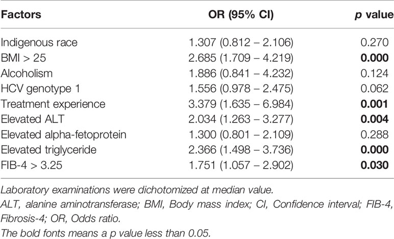
Table 3 Multivariable logistic regression analysis for factors associated with high insulin resistance.
Outcome of Viral Elimination on Glucose Parameters
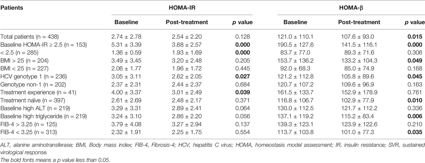
Of 447 patients, 438 (98.0%) achieved SVR. To analyze the relationship between viral elimination and changes in glucose parameters, HOMA-IR and HOMA-β were considered as continuous variables (Table 4). HOMA-IR did not change significantly from baseline to 12-weeks after treatment (2.74 ± 2.78 to 2.54 ± 2.20, p = 0.128). There was a significant improvement in β-cell secretion stress (121.0 ± 110.1 to 107.6 ± 93.0, p = 0.015). In patients with baseline HOMA-IR ≥ 2.5, we observed a significant improvement in mean IR (5.31 ± 3.39 to 3.68 ± 2.57, p < 0.001). Additionally, the subgroup analysis revealed that a statistically significant reduction in mean IR after DAAs therapies was observed in patients with HCV genotype 1 (3.05 ± 3.11 to 2.62 ± 2.05, p = 0.027) and treatment experience (4.00 ± 3.37 to 3.01 ± 2.49, p = 0.039) (Figure 1). However, in patients with baseline HOMA-IR < 2.5, there was a significant increase in mean IR after HCV eradication. We also analyzed pre- and post-treatment HOMA-IR in patients with different races, BMI, HCV viral loads, baseline laboratory results, and FIB-4 score. There was no significant difference in HOMA-IR between values at baseline and 12-weeks post-treatment.
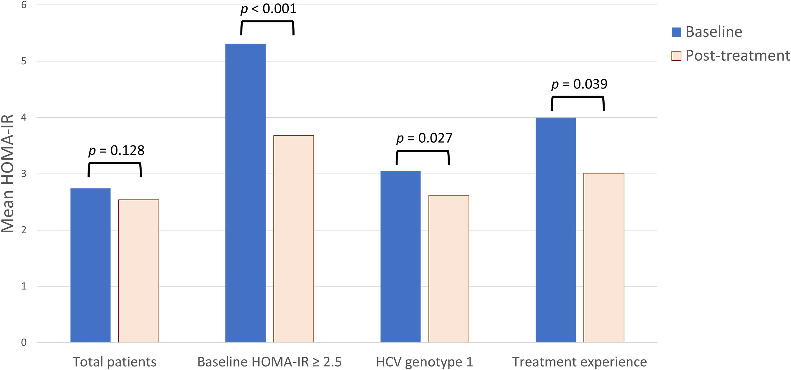
Figure 1 The change of insulin resistance (IR) in hepatitis C virus (HCV)-infected patients treated with direct-acting antiviral agents. Homeostasis model assessment (HOMA)-IR at baseline and 12-weeks after antiviral treatment were measured. In general, there was no significant change in mean HOMA-IR. In subgroup analysis, IR significantly reduced after antiviral treatment in patients with baseline HOMA-IR ≥ 2.5, HCV genotype 1, and treatment experience.
Discussion
We presented an observational analysis of glucose homeostasis in patients with chronic HCV infection and the outcomes of HCV elimination by DAAs therapy. We identified several factors contributing to IR in non-diabetic patients with chronic HCV infection. Obesity, elevated ALT and triglyceride levels, treatment experience, and advanced fibrosis were independently associated with IR. Obesity and metabolic syndrome were generally validated as risk factors for IR. In our result, serum levels of triglyceride, a well-known predictor of type 2 diabetes, were also independently related to IR. HCV NS5A and core proteins are known to directly inhibit microsomal triglyceride transfer protein activity. The accumulation of hepatic triglycerides reduces insulin-stimulated glycogen synthesis and enhances hepatic gluconeogenesis, consequently leading to hepatic and peripheral IR. We did not observe any virus-specific effects of IR. Neither HCV genotype nor viral loads were observed to be independent factors for HOMA-IR ≥ 2.5. At the molecular level, the core protein of genotype 3a promotes degradation of IRS-1 via downregulation of peroxisome proliferator-activated receptor gamma (PPAR-γ) and upregulation of SOCS-7; and the core protein of genotype 1b interferes with the insulin signaling by activating the mammalian target of rapamycin (18). Previous studies have investigated the association of HCV genotype 1 with IR, but the results were inconsistent (4, 20, 27, 28). In the present study, we also observed a higher prevalence of IR in patients with HCV genotype 1. However, the statistical power was attenuated in multivariable logistic regression analysis. This could be because that the genotype-specific mechanisms interfering with the insulin signaling pathway only partially influenced systemic IR. The established risk factors for diabetes, such as obesity and hyperlipidemia, remain the major contributors of IR in HCV-infected patients.
The multivariable logistic regression analysis in the present study demonstrated that treatment experience and advanced hepatic fibrosis were independently associated with IR. These findings were less discussed in previous articles. As explained earlier, the HCV core protein and NS5A have complex effects on the insulin signaling pathway, influence hepatic and systemic glucose homeostasis, and thus, induce IR. We hypothesized that prolonged inflammation resulting from chronic HCV infection is associated with dysregulation of glucose homeostasis. A previous history of treatment with IFN-based therapy may imply a longer duration of infection with HCV. Additionally, hepatic fibrosis indirectly supports the probability of a longer inflammation by HCV infection. In HCV-infected liver, inflammation is a key process to drive the fibrogenic response. The immune system secretes proinflammatory cytokines and profibrogenic factors, leading to hepatocellular injury, production of extracellular matrix, proliferation of myofibroblasts, and stimulation of fibrosis (29, 30). Our finding revealed an association between hepatic fibrosis and IR. It is noteworthy that our result only supported a correlation, not a causal relationship. Advanced liver fibrosis may impair insulin clearance, resulting in elevated serum insulin levels. On the contrary, IR also contributes to the progression of liver fibrosis. The mechanism could be a direct stimulation of liver stellate cells by hyperinsulinemia, resulting in increased production of the connective tissue growth factor and subsequent accumulation of extracellular matrix (9, 31).
We analyzed the outcome of glucose homeostasis in chronic HCV patients receiving DAAs. There was a significant improvement in β-cell stress. As mentioned earlier, levels of hepatic IRS1 and IRS2 decrease in HCV-infected patients. Clearance of HCV is accompanied by normalization of hepatic expression of IRS and improved β-cell function (21), which then influences glucose homeostasis. The IR remained statistically unchanged after DAAs therapy. This result is consistent with the results of previous studies that observed no difference in HOMA-IR between baseline and post-treatment phases in patients with HCV who underwent treatment (26, 32). However, this contradicts the hypothesis that IR may be improved after HCV eradication, especially as HCV infection is a risk factor for the development of diabetes. There may be several explanations for the lack of an association between SVR and improvement of IR. The development of IR in HCV-infected patients could be a combination of both host- and virus-mediated pathways. The complex hepatic and non-hepatic mechanisms affecting glucose homeostasis may lead to heterogeneous post-SVR outcomes regarding IR. We performed subgroup analysis for further elucidation of the possible causal relationship. We observed a significant improvement in IR in patients with baseline HOMA-IR ≥ 2.5, which indirectly supports that HCV is an important contributor to IR, especially among patients with high-IR status. Additionally, this high-IR status could be reversed after HCV elimination by DAAs. Our subgroup analysis suggested that elimination of HCV genotype 1 was also associated with reduced IR. This relationship was not observed in patients with other genotypes. The result is consistent with previous research, which showed that successful viral clearance was found to be associated with reduced HOMA-IR in patients with HCV genotype 1 (32). The genotype-specific outcome of IR suggested the direct viral effect as an important contributor to glucose abnormalities. In our result, advanced hepatic fibrosis was independently associated with IR. However, we did not observe a significant improvement in IR after HCV eradication in patients with advanced fibrosis. Liver fibrosis is a chronic process. Patients who received DAA therapy could have a rapid decrease in inflammation rather than regression of fibrosis. The duration of our study seemed too short for significant hepatocyte remodeling or IR improvement.
As discussed above, patients with chronic HCV infection show an increased risk of IR and progression to type 2 DM. There are no definite virus-specific factors associated with the increasing prevalence of IR. Thus, we suggest that all patients with HCV infection be screened for glucose abnormalities. It will help physicians to identify patients who have a higher risk of developing type 2 diabetes and may require active surveillance, as diabetes can also increase the risk of cirrhosis and hepatocellular carcinoma (33, 34). With the advent of DAAs, we may be able to eradicate HCV with greater certainty and a lower risk of adverse effects. An SVR is generally associated with normalization of liver enzymes, improvement in liver function, as well as regression of liver necroinflammation and fibrosis (35). Compared with untreated patients, patients whose HCV was eliminated by DAAs have a significantly lower risk of developing hepatocellular carcinoma (36). Furthermore, after successful virus elimination by treatment with DAAs, improvement in IR can be an additional benefit and may reduce diabetes-related morbidities. A prospective study found that HCV eradication by DAAs reduces the incidence of type 2 DM (37). However, a general worsening of the lipid profile after DAAs was observed (38, 39). The impact of the balance between improved glucose metabolism and negative changes in lipid profile on the outcome of cardiovascular and cardiometabolic risk must be determined. A significant reduction in major adverse cardiovascular events has recently been observed following HCV clearance by DAAs (40, 41). Further research is required to fully understand the underlying mechanisms.
This study has some limitations. First, this was an observational, open-label study and had the potential for selection bias. Second, a majority of the patients (98.0%) achieved SVR; therefore, it was impossible to compare glucose outcomes between patients with successful eradication and those with treatment failure. Third, the short-term study failed to demonstrate an improvement in IR; thus, the long-term outcome should be investigated to understand the effect of virus elimination on glucose homeostasis. Finally, we could not clarify the impact of HCV infection duration on IR, and could only demonstrate that treatment experience and advanced hepatic fibrosis were associated with IR.
In conclusion, we showed that obesity, treatment experience, baseline elevated levels of ALT and triglyceride, as well as advanced hepatic fibrosis were independently associated with high IR in chronic HCV-infected patients. The prevalence of IR was not influenced by HCV genotypes and viral load. This study provided evidence that HCV-related IR could be partially reversed by HCV eradication, especially in patients with high baseline IR, HCV genotype 1, and treatment experience. Further research for long-term outcomes of glucose homeostasis and diabetes-related morbidities in chronic HCV patients receiving DAAs is required.
Data Availability Statement
The raw data supporting the conclusions of this article will be made available by the authors, without undue reservation.
Ethics Statement
The studies involving human participants were reviewed and approved by Institutional Review Board of Mackay Memorial Hospital. The patients/participants provided their written informed consent to participate in this study.
Author Contributions
C-HC: Design of the work, Writing. C-YC: Investigation, Supervision. H-LC: Interpretation of data, Review & Editing. I-TL: Analysis of data. C-HW: Methodology, Analysis of data. Y-KL: Design of the work, Review & Editing. M-JB: Resources, Supervision. All authors have seen the contents of the manuscript and agree to be accountable for all aspects of the work in ensuring that questions related to the accuracy or integrity of any part of the work are appropriately investigated and resolved. All authors contributed to the article and approved the submitted version.
Conflict of Interest
The authors declare that the research was conducted in the absence of any commercial or financial relationships that could be construed as a potential conflict of interest.
Publisher’s Note
All claims expressed in this article are solely those of the authors and do not necessarily represent those of their affiliated organizations, or those of the publisher, the editors and the reviewers. Any product that may be evaluated in this article, or claim that may be made by its manufacturer, is not guaranteed or endorsed by the publisher.
References
1. Lecube A, Hernández C, Genescà J, Simó R. Proinflammatory Cytokines, Insulin Resistance, and Insulin Secretion in Chronic Hepatitis C Patients: A Case-Control Study. Diabetes Care (2006) 29(5):1096–101. doi: 10.2337/diacare.2951096
2. Shintani Y, Fujie H, Miyoshi H, Tsutsumi T, Tsukamoto K, Kimura S, et al. Hepatitis C Virus Infection and Diabetes: Direct Involvement of the Virus in the Development of Insulin Resistance. Gastroenterology (2004) 126(3):840–8. doi: 10.1053/j.gastro.2003.11.056
3. Alexander GJ. An Association Between Hepatitis C Virus Infection and Type 2 Diabetes Mellitus: What Is the Connection? Ann Internal Med (2000) 133(8):650–2. doi: 10.7326/0003-4819-133-8-200010170-00018
4. Mehta SH, Brancati FL, Strathdee SA, Pankow JS, Netski D, Coresh J, et al. Hepatitis C Virus Infection and Incident Type 2 Diabetes. Hepatology (Baltimore Md) (2003) 38(1):50–6. doi: 10.1053/jhep.2003.50291
5. White DL, Ratziu V, El-Serag HB. Hepatitis C Infection and Risk of Diabetes: A Systematic Review and Meta-Analysis. J Hepatol (2008) 49(5):831–44. doi: 10.1016/j.jhep.2008.08.006
6. Desbois AC, Cacoub P. Diabetes Mellitus, Insulin Resistance and Hepatitis C Virus Infection: A Contemporary Review. World J Gastroenterol (2017) 23(9):1697–711. doi: 10.3748/wjg.v23.i9.1697
7. Hull MW, Rollet K, Moodie EE, Walmsley S, Cox J, Potter M, et al. Insulin Resistance Is Associated With Progression to Hepatic Fibrosis in a Cohort of HIV/hepatitis C Virus-Coinfected Patients. AIDS (London England) (2012) 26(14):1789–94. doi: 10.1097/QAD.0b013e32835612ce
8. Svegliati-Baroni G, Ridolfi F, Di Sario A, Casini A, Marucci L, Gaggiotti G, et al. Insulin and Insulin-Like Growth Factor-1 Stimulate Proliferation and Type I Collagen Accumulation by Human Hepatic Stellate Cells: Differential Effects on Signal Transduction Pathways. Hepatology (Baltimore Md) (1999) 29(6):1743–51. doi: 10.1002/hep.510290632
9. Paradis V, Perlemuter G, Bonvoust F, Dargere D, Parfait B, Vidaud M, et al. High Glucose and Hyperinsulinemia Stimulate Connective Tissue Growth Factor Expression: A Potential Mechanism Involved in Progression to Fibrosis in Nonalcoholic Steatohepatitis. Hepatology (Baltimore Md) (2001) 34(4 Pt 1):738–44. doi: 10.1053/jhep.2001.28055
10. Foxton MR, Quaglia A, Muiesan P, Heneghan MA, Portmann B, Norris S, et al. The Impact of Diabetes Mellitus on Fibrosis Progression in Patients Transplanted for Hepatitis C. Am J Transplant (2006) 6(8):1922–9. doi: 10.1111/j.1600-6143.2006.01408.x
11. Arase Y, Kobayashi M, Suzuki F, Suzuki Y, Kawamura Y, Akuta N, et al. Effect of Type 2 Diabetes on Risk for Malignancies Includes Hepatocellular Carcinoma in Chronic Hepatitis C. Hepatology (Baltimore Md) (2013) 57(3):964–73. doi: 10.1002/hep.26087
12. Calzadilla-Bertot L, Vilar-Gomez E, Torres-Gonzalez A, Socias-Lopez M, Diago M, Adams LA, et al. Impaired Glucose Metabolism Increases Risk of Hepatic Decompensation and Death in Patients With Compensated Hepatitis C Virus-Related Cirrhosis. Dig Liver Dis (2016) 48(3):283–90. doi: 10.1016/j.dld.2015.12.002
13. Kawaguchi Y, Mizuta T. Interaction Between Hepatitis C Virus and Metabolic Factors. World J Gastroenterol (2014) 20(11):2888–901. doi: 10.3748/wjg.v20.i11.2888
14. Banerjee S, Saito K, Ait-Goughoulte M, Meyer K, Ray RB, Ray R. Hepatitis C Virus Core Protein Upregulates Serine Phosphorylation of Insulin Receptor Substrate-1 and Impairs the Downstream Akt/Protein Kinase B Signaling Pathway for Insulin Resistance. J Virol (2008) 82(6):2606–12. doi: 10.1128/jvi.01672-07
15. Pascarella S, Clément S, Guilloux K, Conzelmann S, Penin F, Negro F. Effects of Hepatitis C Virus on Suppressor of Cytokine Signaling mRNA Levels: Comparison Between Different Genotypes and Core Protein Sequence Analysis. J Med Virol (2011) 83(6):1005–15. doi: 10.1002/jmv.22072
16. Georgopoulou U, Tsitoura P, Kalamvoki M, Mavromara P. The Protein Phosphatase 2A Represents a Novel Cellular Target for Hepatitis C Virus NS5A Protein. Biochimie (2006) 88(6):651–62. doi: 10.1016/j.biochi.2005.12.003
17. Maqbool MA, Imache MR, Higgs MR, Carmouse S, Pawlotsky JM, Lerat H. Regulation of Hepatitis C Virus Replication by Nuclear Translocation of Nonstructural 5A Protein and Transcriptional Activation of Host Genes. J Virol (2013) 87(10):5523–39. doi: 10.1128/jvi.00585-12
18. Pazienza V, Clément S, Pugnale P, Conzelman S, Foti M, Mangia A, et al. The Hepatitis C Virus Core Protein of Genotypes 3a and 1b Downregulates Insulin Receptor Substrate 1 Through Genotype-Specific Mechanisms. Hepatology (Baltimore Md) (2007) 45(5):1164–71. doi: 10.1002/hep.21634
19. Hsu CS, Liu CJ, Liu CH, Wang CC, Chen CL, Lai MY, et al. High Hepatitis C Viral Load Is Associated With Insulin Resistance in Patients With Chronic Hepatitis C. Liver Int (2008) 28(2):271–7. doi: 10.1111/j.1478-3231.2007.01626.x
20. Tsochatzis E, Manolakopoulos S, Papatheodoridis GV, Hadziyannis E, Triantos C, Zisimopoulos K, et al. Serum HCV RNA Levels and HCV Genotype do Not Affect Insulin Resistance in Nondiabetic Patients With Chronic Hepatitis C: A Multicentre Study. Aliment Pharmacol Ther (2009) 30(9):947–54. doi: 10.1111/j.1365-2036.2009.04094.x
21. Kawaguchi T, Ide T, Taniguchi E, Hirano E, Itou M, Sumie S, et al. Clearance of HCV Improves Insulin Resistance, Beta-Cell Function, and Hepatic Expression of Insulin Receptor Substrate 1 and 2. Am J Gastroenterol (2007) 102(3):570–6. doi: 10.1111/j.1572-0241.2006.01038.x
22. Delgado-Borrego A, Jordan SH, Negre B, Healey D, Lin W, Kamegaya Y, et al. Reduction of Insulin Resistance With Effective Clearance of Hepatitis C Infection: Results From the HALT-C Trial. Clin Gastroenterol Hepatol (2010) 8(5):458–62. doi: 10.1016/j.cgh.2010.01.022
23. Huang JF, Dai CY, Yu ML, Huang CF, Huang CI, Yeh ML, et al. Pegylated Interferon Plus Ribavirin Therapy Improves Pancreatic β-Cell Function in Chronic Hepatitis C Patients. Liver Int (2011) 31(8):1155–62. doi: 10.1111/j.1478-3231.2011.02545.x
24. Romero-Gómez M, Fernández-Rodríguez CM, Andrade RJ, Diago M, Alonso S, Planas R, et al. Effect of Sustained Virological Response to Treatment on the Incidence of Abnormal Glucose Values in Chronic Hepatitis C. J Hepatol (2008) 48(5):721–7. doi: 10.1016/j.jhep.2007.11.022
25. Arase Y, Suzuki F, Suzuki Y, Akuta N, Kobayashi M, Kawamura Y, et al. Sustained Virological Response Reduces Incidence of Onset of Type 2 Diabetes in Chronic Hepatitis C. Hepatology (Baltimore Md) (2009) 49(3):739–44. doi: 10.1002/hep.22703
26. Doyle MA, Galanakis C, Mulvihill E, Crawley A, Cooper CL. Hepatitis C Direct Acting Antivirals and Ribavirin Modify Lipid But Not Glucose Parameters. Cells (2019) 8(3):252. doi: 10.3390/cells8030252
27. Knobler H, Schihmanter R, Zifroni A, Fenakel G, Schattner A. Increased Risk of Type 2 Diabetes in Noncirrhotic Patients With Chronic Hepatitis C Virus Infection. Mayo Clin Proc (2000) 75(4):355–9. doi: 10.4065/75.4.355
28. Moucari R, Asselah T, Cazals-Hatem D, Voitot H, Boyer N, Ripault MP, et al. Insulin Resistance in Chronic Hepatitis C: Association With Genotypes 1 and 4, Serum HCV RNA Level, and Liver Fibrosis. Gastroenterology (2008) 134(2):416–23. doi: 10.1053/j.gastro.2007.11.010
29. Khatun M, Ray RB. Mechanisms Underlying Hepatitis C Virus-Associated Hepatic Fibrosis. Cells (2019) 8(10):1249. doi: 10.3390/cells8101249
30. Rockey DC, Bell PD, Hill JA. Fibrosis–a Common Pathway to Organ Injury and Failure. N Engl J Med (2015) 372(12):1138–49. doi: 10.1056/NEJMra1300575
31. Ratziu V, Munteanu M, Charlotte F, Bonyhay L, Poynard T. Fibrogenic Impact of High Serum Glucose in Chronic Hepatitis C. J Hepatol (2003) 39(6):1049–55. doi: 10.1016/s0168-8278(03)00456-2
32. Thompson AJ, Patel K, Chuang WL, Lawitz EJ, Rodriguez-Torres M, Rustgi VK, et al. Viral Clearance Is Associated With Improved Insulin Resistance in Genotype 1 Chronic Hepatitis C But Not Genotype 2/3. Gut (2012) 61(1):128–34. doi: 10.1136/gut.2010.236158
33. Veldt BJ, Chen W, Heathcote EJ, Wedemeyer H, Reichen J, Hofmann WP, et al. Increased Risk of Hepatocellular Carcinoma Among Patients With Hepatitis C Cirrhosis and Diabetes Mellitus. Hepatology (Baltimore Md) (2008) 47(6):1856–62. doi: 10.1002/hep.22251
34. Chen CL, Yang HI, Yang WS, Liu CJ, Chen PJ, You SL, et al. Metabolic Factors and Risk of Hepatocellular Carcinoma by Chronic Hepatitis B/C Infection: A Follow-Up Study in Taiwan. Gastroenterology (2008) 135(1):111–21. doi: 10.1053/j.gastro.2008.03.073
35. Carrat F, Fontaine H, Dorival C, Simony M, Diallo A, Hezode C, et al. Clinical Outcomes in Patients With Chronic Hepatitis C After Direct-Acting Antiviral Treatment: A Prospective Cohort Study. Lancet (London England) (2019) 393(10179):1453–64. doi: 10.1016/s0140-6736(18)32111-1
36. Rinaldi L, Perrella A, Guarino M, De Luca M, Piai G, Coppola N, et al. Incidence and Risk Factors of Early HCC Occurrence in HCV Patients Treated With Direct Acting Antivirals: A Prospective Multicentre Study. J Trans Med (2019) 17(1):292. doi: 10.1186/s12967-019-2033-x
37. Adinolfi LE, Petta S, Fracanzani AL, Nevola R, Coppola C, Narciso V, et al. Reduced Incidence of Type 2 Diabetes in Patients With Chronic Hepatitis C Virus Infection Cleared by Direct-Acting Antiviral Therapy: A Prospective Study. Diabetes Obes Metab (2020) 22(12):2408–16. doi: 10.1111/dom.14168
38. Hashimoto S, Yatsuhashi H, Abiru S, Yamasaki K, Komori A, Nagaoka S, et al. Rapid Increase in Serum Low-Density Lipoprotein Cholesterol Concentration During Hepatitis C Interferon-Free Treatment. PloS One (2016) 11(9):e0163644. doi: 10.1371/journal.pone.0163644
39. Gitto S, Cicero AFG, Loggi E, Giovannini M, Conti F, Grandini E, et al. Worsening of Serum Lipid Profile After Direct Acting Antiviral Treatment. Ann Hepatol (2018) 17(1):64–75. doi: 10.5604/01.3001.0010.7536
40. Adinolfi LE, Petta S, Fracanzani AL, Coppola C, Narciso V, Nevola R, et al. Impact of Hepatitis C Virus Clearance by Direct-Acting Antiviral Treatment on the Incidence of Major Cardiovascular Events: A Prospective Multicentre Study. Atherosclerosis (2020) 296:40–7. doi: 10.1016/j.atherosclerosis.2020.01.010
Keywords: direct-acting antiviral agents, genotype, hepatitis C, homeostasis model assessment, insulin resistance, type 2 dabetes
Citation: Cheng C-H, Chu C-Y, Chen H-L, Lin I-T, Wu C-H, Lee Y-K and Bair M-J (2022) Virus Elimination by Direct-Acting Antiviral Agents Impacts Glucose Homeostasis in Chronic Hepatitis C Patients. Front. Endocrinol. 12:799382. doi: 10.3389/fendo.2021.799382
Received: 21 October 2021; Accepted: 20 December 2021;
Published: 13 January 2022.
Edited by:
Lixin Li, Central Michigan University, United StatesReviewed by:
Ferdinando Carlo Sasso, Università della Campania Luigi Vanvitelli, ItalyAixia Sun, Michigan State University, United States
Copyright © 2022 Cheng, Chu, Chen, Lin, Wu, Lee and Bair. This is an open-access article distributed under the terms of the Creative Commons Attribution License (CC BY). The use, distribution or reproduction in other forums is permitted, provided the original author(s) and the copyright owner(s) are credited and that the original publication in this journal is cited, in accordance with accepted academic practice. No use, distribution or reproduction is permitted which does not comply with these terms.
*Correspondence: Ming-Jong Bair, YTU5NjNAbW1oLm9yZy50dw==
 Chun-Han Cheng
 Chia-Ying Chu3
 Huan-Lin Chen
 Ming-Jong Bair