95% of researchers rate our articles as excellent or good
Learn more about the work of our research integrity team to safeguard the quality of each article we publish.
Find out more
EDITORIAL article
Front. Earth Sci. , 01 August 2022
Sec. Paleontology
Volume 10 - 2022 | https://doi.org/10.3389/feart.2022.973459
This article is part of the Research Topic Technological Frontiers in Dinosaur Science Mark a New Age of Opportunity for Early Career Researchers View all 9 articles
Editorial on the Research Topic
Technological Frontiers in Dinosaur Science Mark a New Age of Opportunity for Early Career Researchers
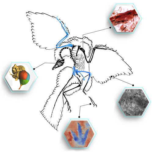
Implementation of innovative techniques and methodologies is currently fundamental to better understand and improve our knowledge of extinct faunas, like those of the dinosaurs. Today, dinosaur science is experiencing a revolution thanks to these innovative techniques that stem from multiple scientific disciplines (Figure 1). In fact, young researchers are taking the lead in research with new scientific approaches (e.g. Falkingham et al., 2018; Lefebvre et al., 2020; Manafzadeh and Gatesy, 2020; Bailleul et al., 2021; Nieto et al., 2021; Serrano-Martínez et al., 2021; Van Bijlert et al., 2021; Ma et al., 2022; Wiemann et al., 2022). Young scientists are the key to bringing new perspectives into science and pushing technological and methodological developments. By publishing this collection we wanted to showcase this, and present high-impact scientific projects mainly developed by worldwide young researchers in vertebrate palaeontology.

FIGURE 1. The famous Berlin specimen of the early bird Archaeopteryx summarizing some innovative outcomes that can be developed thanks to new techniques and methods like digitization, 3D reconstruction of soft tissues, track morphology and neuroanatomy or the retrieval of fossil cells and molecules (images modified from Field et al., 2013 and Castanera et al., 2018, and courtesy of D. Vidal and J. Wiemann).
The innovative approaches presented in this volume will, not only advance palaeontological knowledge but also provide new tools to all vertebrate palaeontologists, since working protocols and tasks can be easier and faster to carry on with these techniques. One impressive example is the implementation of deep learning algorithms when automating image processing, like in the labelling and segmentation of computed tomography images or when osteohistological features need to be identified. Yu et al. analyze how deep neural networks can efficiently segment protoceratopsian dinosaur fossils, which can save significant time from current manual segmentation. In the case of the implementation of deep neural network-based methods, Quin et al. have tested this technique for getting segmented maps with different osteon regions on a histological dataset from various taxa within alvarezsaurian theropods. These works set an important base for analyzing larger sets of images and samples, but also developing more complex deep learning algorithms.
It is a fact that the use of 3D models in descriptive studies and for soft tissue reconstructions is growing exponentially year after year. One really nice example is the partial ankylosaur skull from the mid-Cretaceous of Queensland (Australia), which Frauenfelder et al. have digitized by synchrotron radiation X-ray tomography and then reconstructed in 3D to facilitate the virtual preparation of the separate cranial bones. Although encased in a limestone concretion, thanks to this technique the palatal anatomy of Australian ankylosaurs has been elucidated, being indeed an important element in resolving ankylosaur phylogenetic relationships. It is essential to study well-preserved fossils when assessing anatomical features for descriptions, comparisons and phylogenetic analyses. However, as most of us—palaeontologists—know, this is not the case most of the time. As Demuth et al. state in the first sentence of their abstract “taphonomic and diagenetic processes inevitably distort the original skeletal morphology of fossil vertebrate remains”. These authors propose a novel reconstruction workflow combining retopology and retrodeformation, allowing the original morphology of both symmetrically and asymmetrically damaged areas of fossils to be reconstructed. They test this workflow in 3D reconstructions of the sternum of the crownward stem-bird Ichthyornis and some cervical vertebrae of the sauropod Galeamopus in the modelling software MAYA. DeVries et al. also propose a retrodeformation and reconstruction workflow for digital restoration of fossils using the armatures of the open-source software Blender, with the specific goal of recording the specific changes and decisions made for retrodeformation. The authors test this technique with fossil bones of an unnamed basal thyreophoran dinosaur from the Jurassic of Niger.
For a few decades we are able to analyze how extinct animals moved (see, e.g., Sellers et al., 2017), and even to 3D reconstruct more realistically their musculoskeletal system (see, e.g., Díez Díaz et al., 2020; Vidal et al., 2020). But not always the important role of the articular cartilage is kept in mind. Voegele et al. present a new method for modeling joints that allows testing hypotheses about articular cartilage morphology in extinct taxa. They examine the left elbow joint of the sauropod dinosaur Dreadnoughtus using articular cartilage reconstructions constrained by extant phylogenetic bracketing, and by following a different approach focused on joint contact surfaces. Also related to the study of cartilage in extinct animals, little seems to be known about chondrocytic fossilization. To further understand the spectrum of cellular preservation in this tissue, Bailleul and Zhou analyze the morphology and the chemistry of some intralacunar content seen in avian cartilage from the Early Cretaceous Jehol biota (more specifically Yanornis and Confuciusornis). They combine standard paleohistology with Scanning Electron Microscopy and Energy Dispersive Spectroscopy, but also paired with actuotaphonomy experiments. A field of study that also provides very important data for the study of the biomechanics of extinct animals is palaeoichnology. Sciscio et al. test traditional and more novel landmark-based geometric morphometric (GM) analysis to describe the sauropod Jurassic trackway of the Courtedoux-Tchâfouè (TCH), in NW Switzerland. This method helps to assess if there is variation in morphology within the sample, which is greatly useful for ichnotaxonomic studies.
As a whole, this Research Topic explores innovative techniques and approaches, crossing technological frontiers, that have recently led to a rapid advancement of our understanding of the biology of the life of the past. Traditional methodologies, such as cladistic or anatomical comparison, which has been followed in dinosaur palaeontology for more than a century, are still fundamental to the understanding of evolution. But we can not just stagnate here and, hence, it is also essential that these “more classic” methods be linked with these new approaches to break new grounds towards a more inclusive, technological and revolutionary palaeontology. Thanks to these methods we can automate repetitive tasks, make data more accessible, digitally restore fossils and even better comprehend behavioural patterns of extinct animals.
Despite studying specimens millions of years old, palaeontology is an extremely innovative field that can gain expertise from other disciplines and apply it to its purposes. The contributions to this volume are clear examples of how the next generation of palaeontologists will continue contributing to such a unique multi-disciplinary research approach.
VD drafted the first version of the manuscript. VD, EC, DV, and MB developed it and provided comments and editions. DV created Figure 1.
The authors declare that the research was conducted in the absence of any commercial or financial relationships that could be construed as a potential conflict of interest.
All claims expressed in this article are solely those of the authors and do not necessarily represent those of their affiliated organizations, or those of the publisher, the editors and the reviewers. Any product that may be evaluated in this article, or claim that may be made by its manufacturer, is not guaranteed or endorsed by the publisher.
We would like to thank all reviewers for their contributions, and all authors of the submitted manuscripts. We also would like to thank Ursula Raba, Kanzis Mattu and the Frontiers Team for their support and suggestions.
Bailleul, A. M., Lu, J., and Li, Z. (2021). DiceCT Applied to Fossilized Hard Tissues: A Preliminary Case Study Using a Miocene Bird. J. Exp. Zool. Mol. Dev. Evol. 336 (4), 364–375. doi:10.1002/jez.b.23037
Castanera, D., Belvedere, M., Marty, D., Paratte, G., Lapaire-Cattin, M., Lovis, C., et al. (2018). A Walk in the Maze: Variation in Late Jurassic Tridactyl Dinosaur Tracks from the Swiss Jura Mountains (NW Switzerland) PeerJ 6, e4579.
Díez Díaz, V., Demuth, O. E., Schwarz, D., and Mallison, H. (2020). The tail of the late jurassic sauropod Giraffatitan Brancai: Digital reconstruction of its epaxial and hypaxial musculature, and implications for tail biomechanics. Front. Earth Sci. 8, 160. doi:10.3389/feart.2020.00160
Falkingham, P. L., Bates, K. T., Avanzini, M., Bennett, M., Bordy, E. M., Breithaupt, B. H., Castanera, D., et al. (2018). A Standard Protocol for Documenting Modern and Fossil Ichnological Data. Palaeontology 61 (4), 469–480. doi:10.1111/pala.12373
Field, D. J., D’Alba, L., Vinther, J., Webb, S. M., Gearty, W., Shawkey, M. D., et al. (2013). Melanin Concentration Gradients in Modern and Fossil Feathers. PLoS One 8 (3), e59451.
Lefebvre, R., Allain, R., Houssaye, A., and Cornette, R. (2020). Disentangling Biological Variability and Taphonomy: Shape Analysis of the Limb Long Bones of the Sauropodomorph Dinosaur plateosaurus. PeerJ 8, e9359. doi:10.7717/peerj.9359
Ma, W., Pittman, M., Butler, R. J., and Lautenschlager, S. (2022). Macroevolutionary Trends in Theropod Dinosaur Feeding Mechanics. Curr. Biol. 32 (3), 677–686. doi:10.1016/j.cub.2021.11.060
Manafzadeh, A. R., and Gatesy, S. M. (2020). A Coordinate-system-independent Method for Comparing Joint Rotational Mobilities. J. Exp. Biol. 223 (18), jeb227108. doi:10.1242/jeb.227108
Nieto, M. N., Degrange, F. J., Sellers, K. C., Pol, D., and Holliday, C. M. (2021). Biomechanical Performance of the Cranio‐mandibular Complex of the Small Notosuchian Araripesuchus Gomesii (Notosuchia, Uruguaysuchidae). The Anatomical Record, 1–13.
Sellers, W. I., Pond, S. B., Brassey, C. A., Manning, P. L., and Bates, K. T. (2017). Investigating the Running Abilities ofTyrannosaurus Rexusing Stress-Constrained Multibody Dynamic Analysis. PeerJ 5, e3420. doi:10.7717/peerj.3420
Serrano-Martínez, A., Knoll, F., Narváez, I., Lautenschlager, S., and Ortega, F. (2021). Neuroanatomical and Neurosensorial Analysis of the Late Cretaceous Basal Eusuchian Agaresuchus Fontisensis (Cuenca, Spain). Pap. Palaeontol. 7 (1), 641–656. doi:10.1002/spp2.1296
Van Bijlert, P. A., van Soest, A. J. K., and Schulp, A. S. (2021). Natural frequency method: Estimating the Preferred Walking Speed of tyrannosaurus Rex Based on Tail Natural Frequency. R. Soc. open Sci. 8 (4), 201441. doi:10.1098/rsos.201441
Vidal, D., Mocho, P., Aberasturi, A., Sanz, J. L., and Ortega, F. (2020). High Browsing Skeletal Adaptations in Spinophorosaurus Reveal an Evolutionary Innovation in Sauropod Dinosaurs. Sci. Rep. 10 (1), 6638. doi:10.1038/s41598-020-63439-0
Keywords: dinosauria, digitization, 3D modelling, interdisciplinarity, early research career
Citation: Díez Díaz V, Cuesta E, Vidal D and Belvedere M (2022) Editorial: Technological Frontiers in Dinosaur Science Mark a New Age of Opportunity for Early Career Researchers. Front. Earth Sci. 10:973459. doi: 10.3389/feart.2022.973459
Received: 20 June 2022; Accepted: 24 June 2022;
Published: 01 August 2022.
Edited and reviewed by:
Bruce S Lieberman, University of Kansas, United StatesCopyright © 2022 Díez Díaz, Cuesta, Vidal and Belvedere. This is an open-access article distributed under the terms of the Creative Commons Attribution License (CC BY). The use, distribution or reproduction in other forums is permitted, provided the original author(s) and the copyright owner(s) are credited and that the original publication in this journal is cited, in accordance with accepted academic practice. No use, distribution or reproduction is permitted which does not comply with these terms.
*Correspondence: Verónica Díez Díaz, ZGllemRpYXoudmVyb25pY2FAZ21haWwuY29t
Disclaimer: All claims expressed in this article are solely those of the authors and do not necessarily represent those of their affiliated organizations, or those of the publisher, the editors and the reviewers. Any product that may be evaluated in this article or claim that may be made by its manufacturer is not guaranteed or endorsed by the publisher.
Research integrity at Frontiers

Learn more about the work of our research integrity team to safeguard the quality of each article we publish.