95% of researchers rate our articles as excellent or good
Learn more about the work of our research integrity team to safeguard the quality of each article we publish.
Find out more
ORIGINAL RESEARCH article
Front. Cell. Infect. Microbiol. , 20 June 2024
Sec. Extra-intestinal Microbiome
Volume 14 - 2024 | https://doi.org/10.3389/fcimb.2024.1398446
This article is part of the Research Topic Investigating the Role of Periodontal Microbiota in Health and Disease View all 5 articles
 Behnoush Selahbarzin1,2
 Hossein Mahmoudvand1,3
 Amal Khudair Khalaf4
 Fahimeh Kooshki5
 Fatemeh Farhadi6
 Parastoo Baharvand7*
Introduction: Children with intellectual disability (ID) often face challenges in maintaining proper oral hygiene due to their motor, sensory, and intellectual impairments, which can lead to compromised oral health; therefore, there is a need to enhance the oral health status of these populations and establish an effective system for administering preventive interventions. Here, we aimed to evaluate the prevalence of Entamoeba gingivalis and Trichomonas tenax among children with ID in Lorestan province, in Western Iran through parasitological and molecular methods.
Methods: The current descriptive investigation involved 215 in children with ID and 215 healthy children (non-ID) who were referred to health facilities in Lorestan province, Iran between October 2022 and March 2024. The prevalence of protozoa in the oral cavity was found through the utilization of both microscopic analysis and conventional polymerase chain reaction (PCR) techniques.
Results: The total prevalence of the E. gingivalis and T. tenax in children with ID was found to be 87 (40.5%) and 92 (42.8%) through microscopic and PCR methods, respectively. Among the positive samples, 57 (61.9%) and 35 (38.1%) children tested positive for E. gingivalis and T. tenax, respectively. In contrast, among the 215 non-ID children in the control group, 39 (18.1%) and 42 (19.5%) tested positive by microscopic and PCR methods, respectively. Among positive samples in non-ID children, 23 (54.7%) and 19 (45.3%) children were positive for E. gingivalis and T. tenax, respectively. Multiple logistic regression analysis indicated that residing in urban areas, parental education, monthly family income, and tooth brushing p<0.001) were identified as independent risk factors for oral cavity parasites.
Conclusion: This study identified a notable prevalence of oral cavity parasites in children with ID in Lorestan province, Western Iran. It is imperative to recognize the primary risk factors associated with these parasites, particularly inadequate teeth brushing, in order to enhance public and oral health strategies for children with ID. Therefore, pediatric dental professionals should remain vigilant regarding these risk factors to effectively recognize and address oral health issues in this population, thereby mitigating the occurrence of oral diseases and infections.
Oral and dental hygiene are crucial factors in maintaining overall health and well-being. Neglecting basic oral care practices not only harms the teeth and gums but also significantly elevates the risk of heart disease, cancer, and diabetes (Dörfer et al., 2017). The oral cavity, along with the teeth, serves as the primary digestive organ in the human body. From a scientific perspective, the initial breakdown of food commences in the mouth, facilitated by oral enzymes and tooth movement (Gift and Atchison, 1995). Given these insights, any malfunctions in the oral cavity and teeth can lead to severe digestive and functional complications (Wu et al., 2016). Thus, the maintenance of oral and dental hygiene is crucial and can potentially lead to significant systemic issues (Wu et al., 2016). Data from the World Health Organization reveals that despite the emphasis on oral health, a significant portion of the global population, especially children, struggle with dental and gum diseases, a predicament that is widespread among underprivileged segments of society (Sadida et al., 2021).
Maintaining optimal oral health is a crucial component of overall well-being, particularly for children, and holds even greater significance for those with special health requirements (Kilian et al., 2016). Despite the heightened prevalence of dental issues among individuals with disabilities or illnesses, they often receive inadequate oral care compared to the general population (Mishu et al., 2022). Researches indicated that dental treatment stands as the most neglected health necessity among disabled individuals. Therefore, the principal objective of dental care provision for this demographic should prioritize preventive measures to address dental diseases, necessitating meticulous planning and effective service delivery (Peres et al., 2019). Intellectual disability (ID) is a genetic condition characterized by substantially below-average intellectual functioning and deficiencies in adaptive behavior (Chelly et al., 2006). This condition typically emerges in childhood and is distinguished by diminished intelligence and adaptive skills (Chelly et al., 2006).
Entamoeba gingivalis and Trichomonas tenax are anaerobic parasites found in the human oral cavity (Bonner et al., 2014; Marty et al., 2017). These parasites can be transmitted through saliva, contaminated food containers, drinking water, and other means (Bonner et al., 2014; Marty et al., 2017). They typically inhabit the upper regions of the digestive system, including the mouth, teeth, gum margins, and spaces between teeth, as well as the respiratory tract (Arpağ and Kaya, 2020; Eslahi et al., 2021). While traditionally considered benign microorganisms by many dentists, recent studies have linked these parasites to oral conditions like periodontal disease, gingivitis, and osteomyelitis (Bao et al., 2020; Yaseen et al., 2021; Azadbakht et al., 2022). Children with ID often face challenges in maintaining proper oral hygiene due to their motor, sensory, and intellectual impairments, which can lead to compromised oral health; therefore, there is a need to enhance the oral health status of these populations and establish an effective system for administering preventive interventions. Here, we aimed to evaluate the prevalence of E. gingivalis and T. tenax among children with ID in Lorestan province, in Western Iran through parasitological and molecular methods.
The Lorestan province, located in the western region of Iran, shares borders with the provinces of Isfahan, Kermanshah, Markazi, Khuzestan, Hamedan, and Ilam. Encompassing an area of approximately 28,000 square kilometers, the province is home to a population of nearly 2 million individuals (Figure 1).
The current descriptive investigation involved 215 ID children who were referred to health facilities in Lorestan province, Iran between October 2022 and March 2024. Additionally, a control group consisting of 215 healthy children without ID (non- ID) who were referred to health centers during the same study period was included. Exclusion criteria encompassed refusal to participate in the study, recent use of systemic antibiotics within the past three months, and immunocompromised status.
Information and consent documents were created and distributed to the parents of participants in both experimental groups. Subsequently, a questionnaire was administered to gather demographic data such as age, gender, place of residence, parental education level, head of household’s occupation, monthly family income, toothbrushing habits, and mouthwash usage. The questionnaire was completed with the assistance of the participants prior to data collection.
Specimens were collected from each child for microscopic evaluation by obtaining two samples using sterile swabs from saliva and dental plaques. Additionally, a third sample was placed in tubes with sterile normal saline for molecular testing purposes (Azadbakht et al., 2022).
Following the preparation of smears on glass slides, the slides underwent staining procedures using trichrome and Giemsa stains, and were subsequently examined using a light microscope (Alam-Eldin and Abdulaziz, 2015).
DNA purification of the saliva/dental plaque samples was carried out using commercial kit (Qiagen, Germany) according to the producer’s instruction.
The extracted DNAs were used for conventional PCR analysis for identification of oral parasites. Two distinct sets of PCR primers were employed to amplify and for the detection of E. gingivalis and T. tenax as previously described by Azadbakht et al. (2022) including: SrRNA gene for E. gingivalis using the primers of Forward: 5′- GCGCATTTCGAACAGGAATGTAGA -3′ and Reveres: 5′-CAAAGCCTTTTCAATAGTATCTTCATTCA-3′, as well as 18S ribosomal RNA gene for T. tenax using the primers of forward (5′-ATGACCAGTTCCATCGATGCCATTC-3′) and reverse (5′-CTCCAAAGATTCTGCCACTAACAAG -3′). The size of the PCR bands for the E. gingivalis and T. tenax was 454 and 496 bp, respectively. The PCR thermal procedure involved an initial denaturation step lasting 6 minutes at 94°C, followed by 35 cycles of denaturation at 94°C for 30s, primer annealing at 60°C for 1 minute, and elongation at 72°C for 1 minute. A final elongation step was conducted for 10 minutes at 72°C (Azadbakht et al., 2022). Appropriate positive and negative controls were employed to validate the quality of DNA extraction and PCR outcomes and to eliminate the possibility of contamination. Positive controls included patient samples containing motile E. gingivalis and T. tenax that produced the anticipated amplicon sizes, while nuclease-free water served as the negative control. The amplicons were visualized by agarose gel (1%) electrophoresis.
Following the acquisition of necessary data, descriptive statistics were employed to elucidate the dataset. The relationship between the variables of interest and the prevalence of oral cavity protozoa was assessed through Chi-square and Fisher exact tests. Additionally, logistic regression was used to calculate the odds ratio along with its 95% confidence intervals. These statistical analyses were conducted using SPSS version 25 software, with statistical significance set at a threshold of P < 0.05.
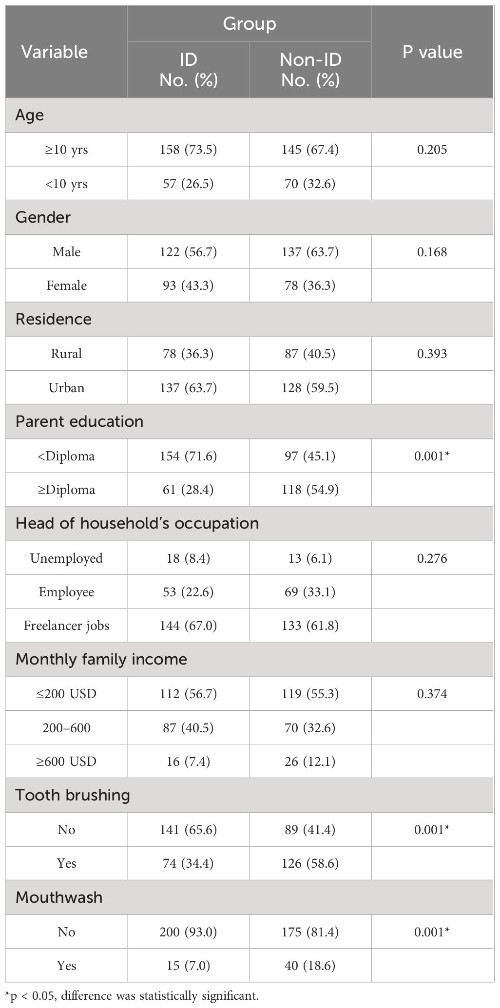
In the present case-control investigation, totally, 430 participants including 215 children with ID and 215 non-ID children referred to health facilities of Lorestan Province, Iran, were studied to evaluate the prevalence of oral cavity parasites (Table 1). The mean age of the participants in the ID and non-ID groups was 10.6 ± 2.21 and 11.1 ± 3.41 years, respectively. The majority of participants were male in the ID (122, 56.7%) and non-ID (137, 63.7%) groups. In terms of residence, 137 (63.7%) and 128 (59.5%) participants in the ID and non-ID groups lived in urban areas, respectively, and the rest lived in rural parts. Among the participants in the ID and non-ID groups, 154 (71.6%) and 97 (45.1%) children had parents with degrees lower than diploma, respectively. This difference was statistically significant between the groups (p < 0.001). Moreover, in 53 (22.6%) of participants in the ID group and 69 (33.1%) in the non-ID group, the head of the household was an employee. Between the children in the ID and non-ID groups, 74 (34.4%) and 126 (58.6%) brushed their teeth daily, respectively. Considering the use of mouthwash, 17 (7.0%) and 39 (18.2%) children in the ID and non-ID groups used the mouthwash, respectively. This difference was statistically significant between the groups (p<0.001) (Table 1).

Table 1 Demographic, socio-economic, and risk factors of participants in children with intellectual disability (ID) and non- intellectual disability children (non-ID) groups.
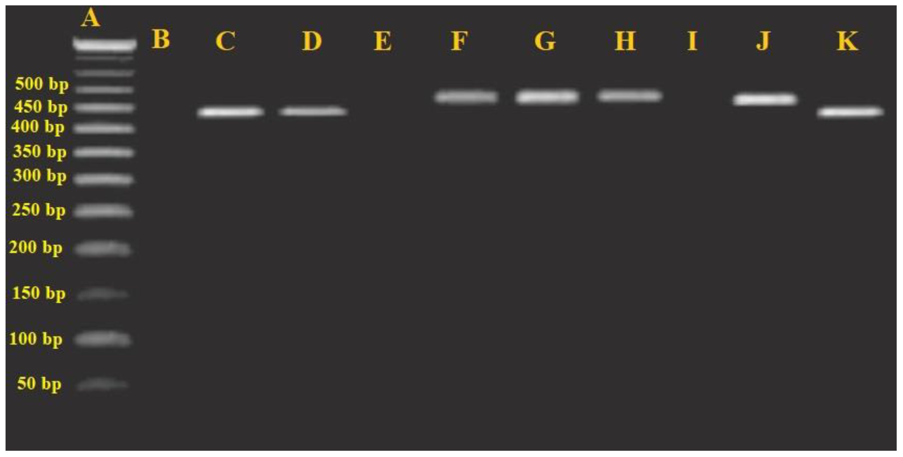
The total prevalence of the E. gingivalis and T. tenax in children with ID was found to be 87 (40.5%) and 92 (42.8%) through microscopic and PCR methods, respectively (Figure 2), respectively. Among the positive samples, 57 (61.9%) and 35 (38.1%) children tested positive for E. gingivalis and T. tenax, respectively. In contrast, among the 215 non-ID children in the control group, 39 (18.1%) and 42 (19.5%) tested positive by microscopic and PCR methods, respectively. Among positive samples in non-ID children, 23 (54.7%) and 19 (45.3%) children were positive for E. gingivalis and T. tenax, respectively (Table 2). The findings indicate that the likelihood of being exposed to oral cavity parasites in non-ID children was significantly lower compared to the case group (p<0.001, OR = 0.325; CI= 0.211–0.500).

Figure 2 Agarose gel electrophoresis analysis of the PCR products. (A) Ladder (50 bp); (B) negative control; (E, I) negative samples; (C) Entamoeba gingivalis positive control, 454 bp; (D, K) E. gingivalis positive sample; (F) T. tenax positive control, 496 bp; (G, H, J) and (J) T. tenax positive sample.

Table 2 Comparison the prevalence of oral cavity parasites in children with intellectual disability (ID) and non- intellectual disability children (non-ID) groups.
In the investigation of age-related subgroups, no statistically significant correlation was found between the occurrence of both oral cavity parasites and the age of participants in both the ID (p=0.172) and non-ID groups (p=0.918). Similarly, no significant association was observed between gender and the prevalence of both oral cavity parasites among participants in the ID (p=0.437) and non-ID groups (p=0.279). However, a notable relationship was identified between the participants’ place of residence and the prevalence of both oral cavity parasites in both the ID (p=0.008) and non-ID groups (p<0.001).
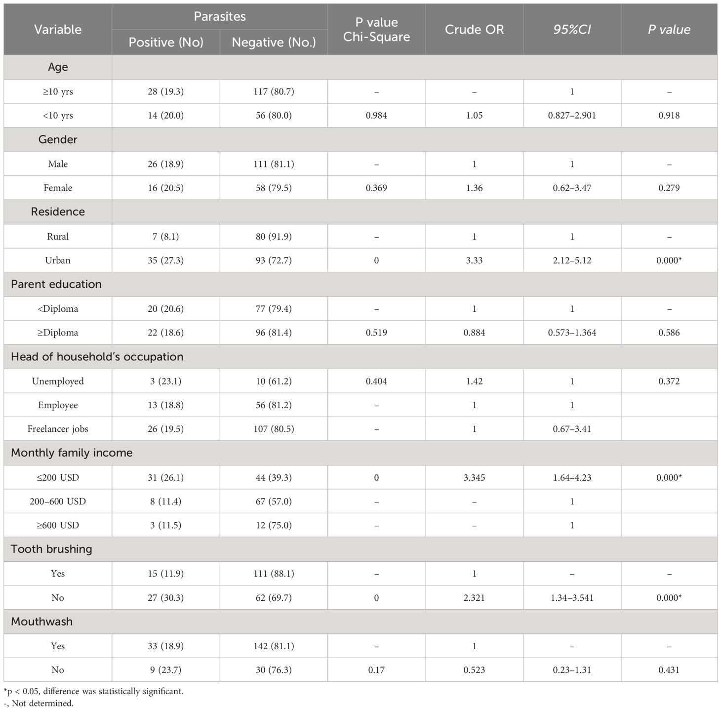
In terms of socio-economic factors, a significant correlation was identified between monthly family income and the prevalence of oral cavity parasites in both the ID (p<0.001) and non-ID groups (p<0.001). Conversely, there was no notable association between parental education level and the prevalence of both E. gingivalis and T. tenax in the ID group (p=0.975) or the non-ID group (p=0.586). Similarly, no significant correlation was found between the head of the household’s occupation and the prevalence of both E. gingivalis and T. tenax in both the ID (p=0.612) and non-ID groups (p=0.312). Analysis of tooth brushing habits revealed a significant association between tooth brushing and the prevalence of both E. gingivalis and T. tenax in both the ID (p<0.001) and non-ID groups (p<0.001). However, there was no significant relationship between the use of mouthwash and the prevalence of both E. gingivalis and T. tenax in either the ID (p = 0.821) or non-ID groups (p = 0.431) (Tables 3, 4). Multiple logistic regression analysis indicated that residing in urban areas (crude OR = 2.63, 95% CI: 1.08–6.81, p = 0.029), parental education (crude OR = 3.37, 95% CI: 2.154–5.170, p<0.001), monthly family income (crude OR = 2.194, 95% CI: 1.096–4.390, P = 0.026), and tooth brushing (crude OR = 2.482, 95% CI: 1.617–3.810, p<0.001) were identified as independent risk factors for oral cavity parasites. Additionally, the analysis revealed that residence (crude OR = 0.421, 95% CI: 0.226–0.783, p = 0.006), monthly family income (crude OR = 2.194, 95% CI: 1.096–4.390, p = 0.026), and tooth brushing (crude OR = 2.482, 95% CI: 1.617–3.810, p<0.001) were also independent risk factors for oral cavity parasites among the examined risk factors and socio-economic parameters.

Table 3 Frequency of oral cavity parasites in children with intellectual disability based on the demographic, socio-economic, and risk factors.

Table 4 Frequency of oral cavity parasites in healthy children (non- intellectual disability) based on the demographic, socio-economic, and risk factors.
Today, it has been proven that the maintaining optimal oral health is a critical aspect of overall well-being, especially for children, and is of particular importance for individuals with special health needs (Gift and Atchison, 1995). Since, children with intellectual disability often face challenges in maintaining proper oral hygiene due to their motor, sensory, and intellectual impairments, which can lead to compromised oral health; therefore, there is a need to enhance the oral health status of these populations and establish an effective system for administering preventive interventions. Here, we aimed to evaluate the prevalence of Entamoeba gingivalis and Trichomonas tenax among children with ID in Lorestan province, in Western Iran through parasitological and molecular methods.
The present study showed that the total prevalence of the E. gingivalis and T. tenax was 40.5% and 42.8% by microscopic and PCR methods, respectively. Among positive samples, 61.9% and 38.1% children were positive for E. gingivalis and T. tenax, respectively. In a study conducted by Sharifi et al. (2020) on 315 adolescents in Kerman province, Iran, it was found that 9.2% and 11.4% tested positive for E. gingivalis and T. tenax through culture and PCR methods, respectively (Sharifi et al., 2020). Additionally, Mehr et al. (2015) demonstrated that the prevalence of T. tenax among individuals with Down syndrome and periodontitis in Tabriz, Iran was 18.8% as determined by PCR analysis (Mehr et al., 2015). Azadbakht et al. (2023) reported that among hemodialysis patients in Lorestan province, Western Iran, the frequencies of E. gingivalis and T. tenax were 17.1% and 14.5%, respectively (Azadbakht et al., 2023). Furthermore, Kooshki et al. (2023) observed that in children with cancer from Lorestan, Iran, the prevalence of E. gingivalis and T. tenax was 25.5% and 31.1% based on microscopic and PCR methods, respectively (Kooshki et al., 2023). The difference in the prevalence of these oral cavity parasites are likely influenced by factors such as the study population, sample size, and methodology employed.
Based on the statistical analysis, there was no significant relationship among age, gender, and prevalence of E. gingivalis and T. tenax parasites in children without ID. It has been proven that women display more favorable attitudes towards dental appointments, possess higher levels of oral health knowledge, and exhibit superior oral health practices compared to males (Lipsky et al., 2021). However, our results showed that there was no significant relationship among gender and prevalence of E. gingivalis and T. tenax parasites in non-ID children. Studies also demonstrated that children under 12 years of age are more susceptible to oral and dental diseases due to less knowledge and education, lifestyle habits and more contact with infectious agents (Hazavehei et al., 2015). In the present study, although the prevalence of oral cavity parasites was higher in children less than 10 years old, this difference was not significant. Similarly, in the study conducted by Kooshki et al. (2023) showed that there was no considerable correlation between the gender and the prevalence of E. gingivalis and T. tenax parasites in children with cancer from Lorestan, Iran (Kooshki et al., 2023).
Although, previous investigations have indicated a positive correlation between the level of education attained by parents and the improved oral health outcomes observed in their children. This relationship is believed to stem from the fact that parents with higher educational levels tend to possess greater awareness, financial means, and healthcare opportunities essential for fostering optimal health in their offspring (Minervini et al., 2023). Our study revealed that there was a higher prevalence of oral cavity parasites in children whose parents had lower levels of education; however, no statistically significant difference was noted.
Previous studies showed higher prevalence of oral and dental diseases in lower socioeconomic status may be due to lack of prevention and treatment services as well as poor diet high in sugar (Steele et al., 2015). On the other hand, children from higher income households have more chances to access dental care, including a more specific diagnostic assessment and have one or more filled teeth, explains some of the differences in oral and dental health due to socioeconomic status (Steele et al., 2015). In the present study although no correlation was observed among between head of household’s occupation and frequency of oral cavity parasites in children with ID; however, there a significant association was reported among the monthly family income and frequency of oral cavity parasites in children with ID.
Our results indicated that residing in urban areas and tooth brushing were identified as independent risk factors for oral cavity parasites in children with ID. In line with our results, Azadbakht et al. (2023) reported a significant association between brushing teeth and prevalence of oral protozoa in hemodialysis participants (Azadbakht et al., 2023). Another study conducted by Azadbakht et al. (2022) showed that there was a significant abscission among brushing teeth and the prevalence of oral cavity parasites in pregnant women in Lorestan province, western Iran; whereas no significant correlation was found by age, education, and use of mouthwash (Kooshki et al., 2023). Conversely, Kooshki et al. (2023) have reported that the living in urban regions were considerably linked with the prevalence of oral cavity parasites in children with cancer in Lorestan province, Iran (Kooshki et al., 2023).
Although this study has limitations such as a small sample size, it is noteworthy as it is the first study in the world to demonstrate the significant prevalence of oral cavity parasites in children with mental retardation. Hence, it is imperative for dental professionals and healthcare providers involved in monitoring health to possess knowledge of oral cavity parasites and their risk factors. This knowledge is essential for effectively identifying and addressing oral health concerns in children with intellectual disabilities. This proactive approach is essential for preventing or reducing potential periodontal and other oral complications that may arise in children with intellectual disabilities.
The study identified a notable prevalence of oral cavity parasites in children with intellectual disability in Lorestan province, Western Iran. It is imperative to recognize the primary risk factors and related socio-economic factors associated with these parasites, particularly inadequate teeth brushing, in order to enhance public and oral health strategies for children with intellectual disability. Therefore, pediatric dental professionals should remain vigilant regarding these risk factors to effectively recognize and address oral health issues in this population, thereby mitigating the occurrence of oral diseases and infections.
All data generated or analyzed during this study are included in this published article.
The studies involving humans were approved by The Ethical Committee of Lorestan University of Medical Sciences approved the study protocol under the No. of IR.LUMS.REC.1401.064. The studies were conducted in accordance with the local legislation and institutional requirements. Written informed consent for participation in this study was provided by the participants’ legal guardians/next of kin.
BS: Conceptualization, Writing – review & editing. HM: Investigation, Methodology, Writing – review & editing. AK: Validation, Visualization, Writing – review & editing. FF: Investigation, Methodology, Writing – review & editing. FK: Supervision, Validation, Writing – original draft. PB: Data curation, Supervision, Formal analysis, Project administration, Software, Writing – review & editing.
The author(s) declare financial support was received for the research, authorship, and/or publication of this article.
The authors thank all the patients and medical staff who contributed to this study.
The authors declare that the research was conducted in the absence of any commercial or financial relationships that could be construed as a potential conflict of interest.
All claims expressed in this article are solely those of the authors and do not necessarily represent those of their affiliated organizations, or those of the publisher, the editors and the reviewers. Any product that may be evaluated in this article, or claim that may be made by its manufacturer, is not guaranteed or endorsed by the publisher.
Alam-Eldin, Y. H., Abdulaziz, A. M. (2015). Identification criteria of the rare multi-flagellate Lophomonas blattarum: comparison of different staining techniques. Parasitol. Res. 114, 3309–3314. doi: 10.1007/s00436-015-4554-4
Arpağ, O. F., Kaya, ÖM. (2020). Presence of Trichomonas tenax and Entamoeba gingivalis in peri-implantitis lesions. Quintessence Int. 51, 212–218. doi: 10.3290/j.qi.a43948
Azadbakht, K., Baharvand, P., Al-Abodi, H. R., Yari, Y., Hadian, B., Fani, M., et al. (2023). Molecular epidemiology and associated risk factors of oral cavity parasites in hemodialysis patients in western Iran. J. Parasitic Dis. 47 (1), 146–151. doi: 10.1007/s12639-022-01551-w
Azadbakht, K., Baharvand, P., Artemes, P., Niazi, M., Mahmoudvand, H. (2022). Prevalence and risk factors of oral cavity parasites in pregnant women in Western Iran. Parasite Epidemiol. Control. 19, e00275. doi: 10.1016/j.parepi.2022.e00275
Bao, X., Wiehe, R., Dommisch, H., Schaefer, A. S. (2020). Entamoeba gingivalis causes oral inflammation and tissue destruction. J. Dent. Res. 99, 561–567. doi: 10.1177/0022034520901738
Bonner, M., Amard, V., Bar-Pinatel, C., Charpentier, F., Chatard, J. M., Desmuyck, Y., et al. (2014). Detection of the amoeba entamoeba gingivalis in periodontal pockets. Parasite. 21, 30. doi: 10.1051/parasite/2014029
Chelly, J., Khelfaoui, M., Francis, F., Chérif, B., Bienvenu, T. (2006). Genetics and pathophysiology of intellectual disability. Eur. J. Hum. Genet. 14, 701–713. doi: 10.1038/sj.ejhg.5201595
Dörfer, C., Benz, C., Aida, J., Campard, G. (2017). The relationship of oral health with general health and NCDs: a brief review. Int. Dental J. 67, 14–18. doi: 10.1111/idj.12360
Eslahi, A. V., Olfatifar, M., Abdoli, A., Houshmand, E., Johkool, M. G., Zarabadipour, M., et al. (2021). The neglected role of trichomonas tenax in oral diseases: A systematic review and meta-analysis. Acta Parasitol. 66 (3), 715–732. doi: 10.1007/s11686-021-00340-4
Gift, H. C., Atchison, K. A. (1995). Oral health, health, and health-related quality of life. Med. Care 33 (11 Suppl), NS57–NS77. doi: 10.1097/00005650-199511001-00008
Hazavehei, S. M., Shirahmadi, S., Taheri, M., Noghan, N., Rezaei, N. (2015). Promoting oral health in 6–12 year-old students: A systematic review. J. Educ. Community Health 1, 66–84. doi: 10.20286/jech-010466
Kilian, M., Chapple, I. L., Hannig, M., Marsh, P. D., Meuric, V., Pedersen, A. M., et al. (2016). The oral microbiome–an update for oral healthcare professionals. Br. Dental J. 221, 657–666. doi: 10.1038/sj.bdj.2016.865
Kooshki, F., Khalaf, A. K., Mahmoudvand, H., Baharvand, P., Gandomi Rouzbahani, F., Selahbarzin, B. (2023). Molecular epidemiology and associated risk factors of parasites in oral cavity of children with Malignancies in Western Iran. Iran J. Parasitol. 18, 324–330. doi: 10.18502/ijpa.v18i3.13755
Lipsky, M. S., Su, S., Crespo, C. J., Hung, M. (2021). Men and oral health: a review of sex and gender differences. Am. J. men's Health 15, 15579883211016361. doi: 10.1177/15579883211016361
Marty, M., Lemaitre, M., Kemoun, P., Morrier, J. J., Monsarrat, P. (2017). Trichomonas tenax and periodontal diseases: A concise review. Parasitology. 144, 1417–1425. doi: 10.1017/S0031182017000701
Mehr, A. K., Zarandi, A., Anush, K. (2015). Prevalence of oral trichomonas tenax in periodontal lesions of down syndrome in Tabriz, Iran. J. Clin. Diagn. Res. 9, ZC88–ZC90. doi: 10.7860/JCDR/2015/14725.6238
Minervini, G., Franco, R., Marrapodi, M. M., Di Blasio, M., Ronsivalle, V., Cicciù, M. (2023). Children oral health and parents education status: a cross sectional study. BMC Oral. Health 23, 787. doi: 10.1186/s12903-023-03424-x
Mishu, M. P., Faisal, I. D., Macnamara, A., Sabbah, W., Peckham, E., Newbronner, L., et al. (2022). A qualitative study exploring the barriers and facilitators for maintaining oral health and using dental service in people with severe mental illness: perspectives from service users and service providers. Int. J. Environ. Res. Public Health 19, 4344. doi: 10.3390/ijerph19074344
Peres, M. A., Macpherson, L. M., Weyant, R. J., Daly, B., Venturelli, R., Mathur, I. D., et al. (2019). Oral diseases: a global public health challenge. Lancet 394, 249–260. doi: 10.1016/S0140-6736(19)31146-8
Sadida, Z. J., Indriyanti, R., Setiawan, A. S. (2021). Does growth stunting correlate with oral health in children?: a systematic review. Eur. J. Dentistry. 16, 32–40. doi: 10.1055/s-0041–1731887
Sharifi, M., Jahanimoghadam, F., Babaei, Z., Mohammadi, M. A., Sharifi, F., Hatami, N., et al. (2020). Prevalence and Associated-Factors for Entamoeba gingivalis in adolescents in Southeastern Iran by culture and PCR, 2017. Iran J. Public Health 49 (2), 351–359. doi: 10.18502/ijph.v49i2.3104
Steele, J., Shen, J., Tsakos, G., Fuller, E., Morris, S., Watt, R., et al. (2015). The interplay between socioeconomic inequalities and clinical oral health. J. Dental Res. 94, 19–26. doi: 10.1177/0022034514553978
Wu, B., Fillenbaum, G. G., Plassman, B. L., Guo, L. (2016). Association between oral health and cognitive status: a systematic review. J. Am. Geriatrics Society. 64, 739–751. doi: 10.1111/jgs.14036
Keywords: parasites, children, prevalence, Iran, oral cavity
Citation: Selahbarzin B, Mahmoudvand H, Khalaf AK, Kooshki F, Farhadi F and Baharvand P (2024) Prevalence, socio-economic, and associated risk factors of oral cavity parasites in children with intellectual disability from Lorestan province, Iran. Front. Cell. Infect. Microbiol. 14:1398446. doi: 10.3389/fcimb.2024.1398446
Received: 09 March 2024; Accepted: 21 May 2024;
Published: 20 June 2024.
Edited by:
Thuy Do, University of Leeds, United KingdomReviewed by:
Mümtaz Güran, Eastern Mediterranean University, TürkiyeCopyright © 2024 Selahbarzin, Mahmoudvand, Khalaf, Kooshki, Farhadi and Baharvand. This is an open-access article distributed under the terms of the Creative Commons Attribution License (CC BY). The use, distribution or reproduction in other forums is permitted, provided the original author(s) and the copyright owner(s) are credited and that the original publication in this journal is cited, in accordance with accepted academic practice. No use, distribution or reproduction is permitted which does not comply with these terms.
*Correspondence: Parastoo Baharvand, RHIuYmFoYXJ2bmRAZ21haWwuY29t
Disclaimer: All claims expressed in this article are solely those of the authors and do not necessarily represent those of their affiliated organizations, or those of the publisher, the editors and the reviewers. Any product that may be evaluated in this article or claim that may be made by its manufacturer is not guaranteed or endorsed by the publisher.
Research integrity at Frontiers

Learn more about the work of our research integrity team to safeguard the quality of each article we publish.