94% of researchers rate our articles as excellent or good
Learn more about the work of our research integrity team to safeguard the quality of each article we publish.
Find out more
ERRATUM article
Front. Cell. Infect. Microbiol. , 06 March 2024
Sec. Clinical Microbiology
Volume 14 - 2024 | https://doi.org/10.3389/fcimb.2024.1391783
This article is an erratum on:
Resistance against two lytic phage variants attenuates virulence and antibiotic resistance in Pseudomonas aeruginosa
An Erratum on
Resistance against two lytic phage variants attenuates virulence and antibiotic resistance in Pseudomonas aeruginosa
By García-Cruz JC, Rebollar-Juarez X, Limones-Martinez A, Santos-Lopez CS, Toya S, Maeda T, Ceapă CD, Blasco L, Tomás M, Díaz-Velásquez CE, Vaca-Paniagua F, Díaz-Guerrero M, Cazares D, Cazares A, Hernández-Durán M, López-Jácome LE, Franco-Cendejas R, Husain FM, Khan A, Arshad M, Morales-Espinosa R, Fernández-Presas AM, Cadet F, Wood TK, and García-Contreras R (2024) 13:1280265. doi: 10.3389/fcimb.2023.1280265
Due to a production error, the captions for Figures 1–4 were mismatched. The corrected Figures and their respective captions appear below.

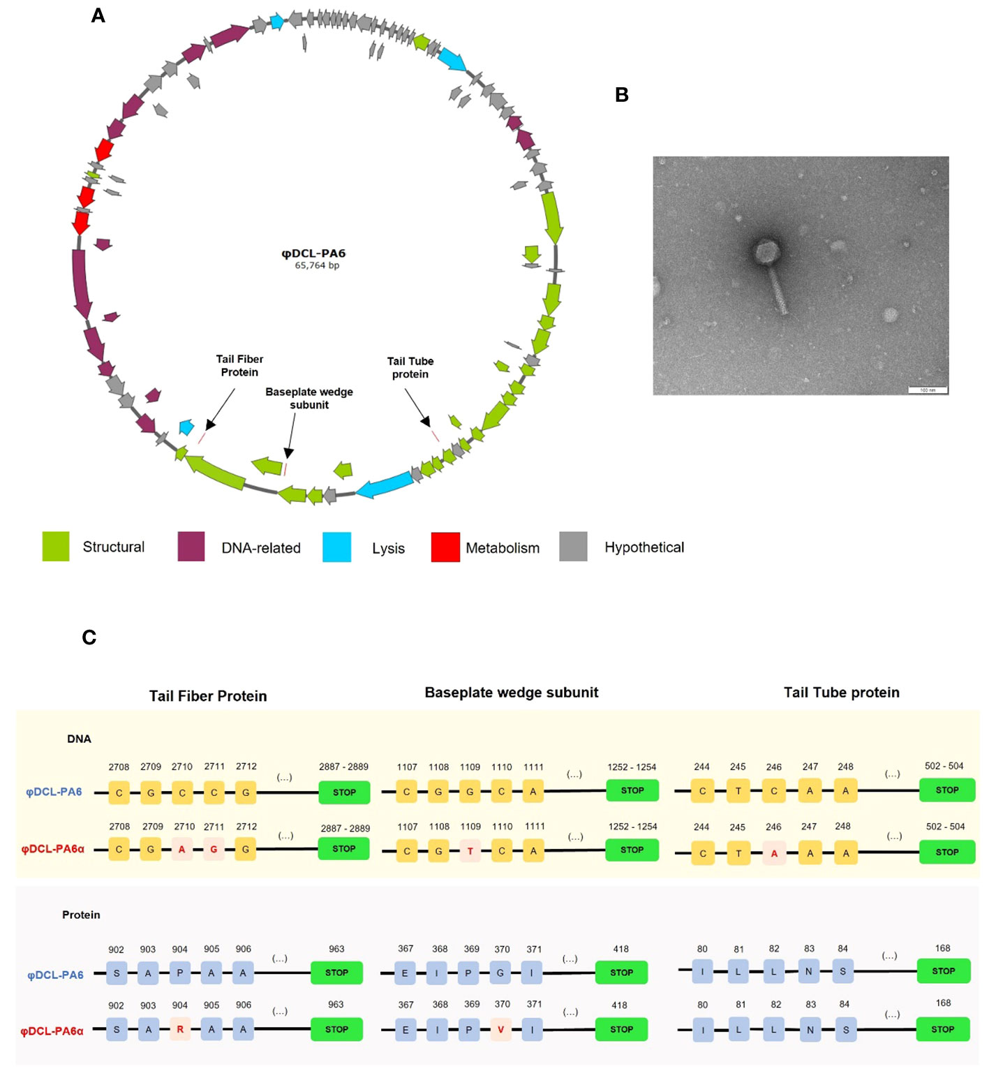
Figure 1 (A) Graphical representation of the genome of phage φDCL-PA6 constructed with SnapGene v6.2.2. The black arrows point to the mutations of the phage variant φDCL-PA6α located in three structural genes corresponding to the tail fiber protein, the baseplate wedge protein, and the tail tube protein. (B) Transmission electron microscopy (TEM) image of the phage variant φDCL-PA6α that belongs to the order Caudovirales. The TEM scale bar represents 100 nm. (C) Mutations identified on the phage variant φDCL-PA6α. Four punctual mutations lead to changes in only two amino acids on the tail fiber protein and the baseplate wedge subunit. Each yellow square represents a nucleotide, while each blue square represents an amino acid, with their respective position on the top. Red squares represent changes in the nucleotide or amino acid sequence. Green boxes represent the stop codon.

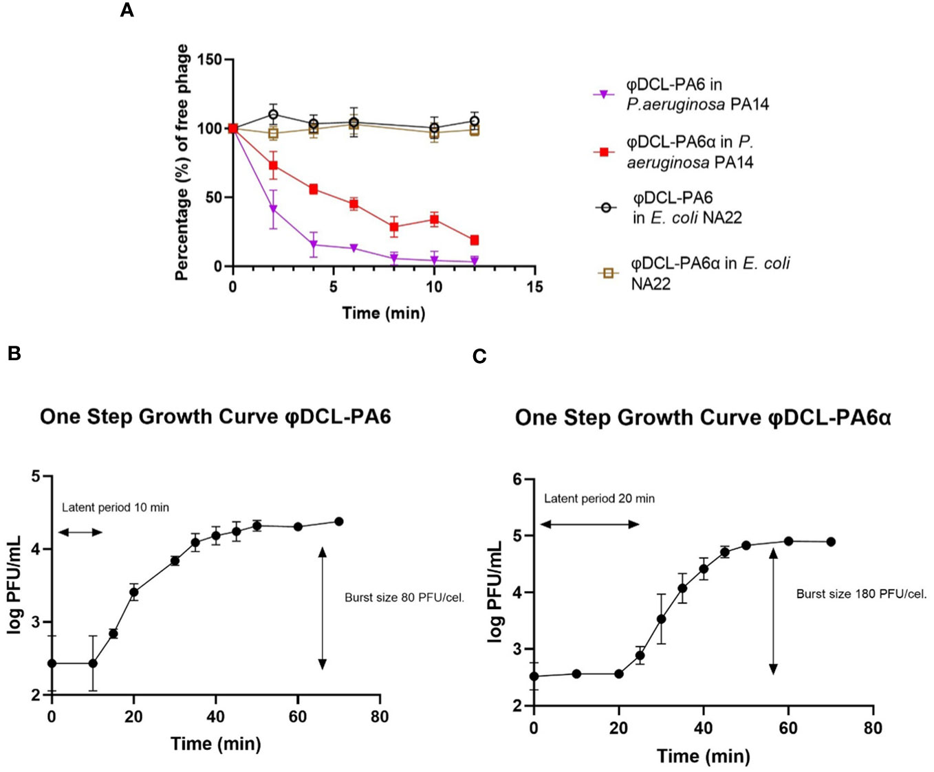
Figure 2 (A) Phage adsorption curves of phage φDCL-PA6 and its variant φDCL-PA6α on P. aeruginosa PA14 and E. coli NA22. The adsorption of the phage variant is less effective than the adsorption of the original phage. For future experiments, the adsorption time for both phages was 10 minutes. One-step growth curves for phage φDCL-PA6 (B) and its variant φDCL-PA6α (C). The latent period and burst size for phage φDCL-PA6 were 10 minutes and 80 PFU/cell, respectively. For the phage variant φDCL-PA6α, the latent period and burst size were 20 minutes and 180 PFU/cell, respectively. The dots represent the mean of three experiments, and the bars represent the standard deviation.

Figure 3 Mutations identified on the P. aeruginosa PA14 resistant clones to phage φDCL-PA6 (A) and phage φDCL-PA6α (B). The mutations of phage φDCL-PA6-resistant clones are located in the wzzB gene involved in the O-antigen synthesis, whereas the mutations of phage φDCL-PA6α-resistant clones are located in the wapH gene involved in the LPS core synthesis. Each yellow square represents a nucleotide with its respective position on the top. Blank spaces represent deletions. Green boxes represent the stop codon. Del stands for deletion and ins stands for insertion.

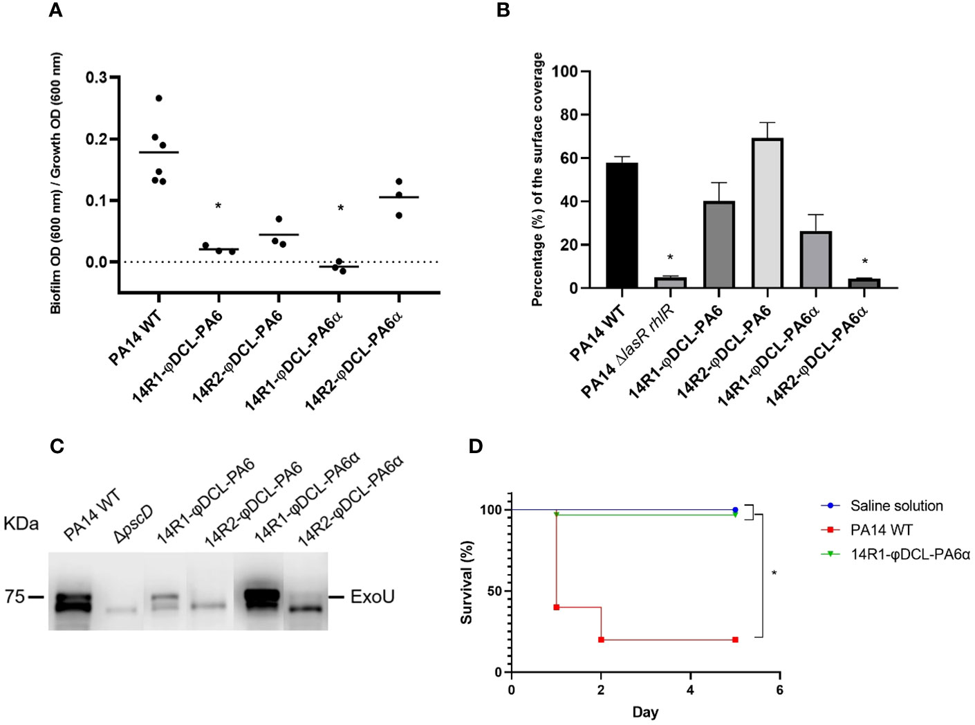
Figure 4 (A) Biofilm production of the P. aeruginosa PA14 phage-resistant clones. The dots represent the mean of three experiments. (B) Swarming motility assessment of the PA14 WT, the PA14 ΔlasRrhlR mutant, and the phage-resistant clones of the PA14 strain. For the statistical analysis of (A, B), Kruskal-Wallis and Dunn’s tests for independent groups were used (p-values < 0.05 were regarded as significant compared to the PA14 WT group). (C) Immunoblotting of the ExoU protein from P. aeruginosa supernatants. The image corresponds to a representative image of three different replicates. (D) Kaplan–Meier survival curve of Galleria mellonella infected with P. aeruginosa PA14 WT and the P. aeruginosa PA14 phage-resistant clone 14R1-φDCL-PA6α. Control groups were administered with saline solution. At least 20 larvae were examined per group. Data were analyzed using the Log Rank Mantel–Cox test in GraphPad Prism 8. Significance was determined by the Mantel–Cox test (*, p 0.05).
The publisher apologizes for this mistake.
The original version of this article has been updated.
Keywords: virulence, tradeoffs, biofilm, phage resistance, phage therapy
Citation: Frontiers Production Office (2024) Erratum: Resistance against two lytic phage variants attenuates virulence and antibiotic resistance in Pseudomonas aeruginosa. Front. Cell. Infect. Microbiol. 14:1391783. doi: 10.3389/fcimb.2024.1391783
Received: 26 February 2024; Accepted: 26 February 2024;
Published: 06 March 2024.
Approved by:
Frontiers Editorial Office, Frontiers Media SA, SwitzerlandCopyright © 2024 Frontiers Production Office. This is an open-access article distributed under the terms of the Creative Commons Attribution License (CC BY). The use, distribution or reproduction in other forums is permitted, provided the original author(s) and the copyright owner(s) are credited and that the original publication in this journal is cited, in accordance with accepted academic practice. No use, distribution or reproduction is permitted which does not comply with these terms.
*Correspondence: Frontiers Production Office, cHJvZHVjdGlvbi5vZmZpY2VAZnJvbnRpZXJzaW4ub3Jn
Disclaimer: All claims expressed in this article are solely those of the authors and do not necessarily represent those of their affiliated organizations, or those of the publisher, the editors and the reviewers. Any product that may be evaluated in this article or claim that may be made by its manufacturer is not guaranteed or endorsed by the publisher.
Research integrity at Frontiers

Learn more about the work of our research integrity team to safeguard the quality of each article we publish.