- 1College of Chemistry and Life Sciences, Chengdu Normal University, Chengdu, China
- 2College of Forestry, Sichuan Agricultural University, Chengdu, China
- 3College of Life Science, Sichuan Agricultural University, Yaan, China
A Corrigendum on
Combined intestinal metabolomics and microbiota analysis for acute endometritis induced by lipopolysaccharide in mice
by Dong Y, Yuan Y, Ma Y, Luo Y, Zhou W, Deng X, Pu J, Hu B and Liu S (2021) Front. Cell. Infect. Microbiol. 11:791373. doi: 10.3389/fcimb.2021.791373
Error in Figure/Table
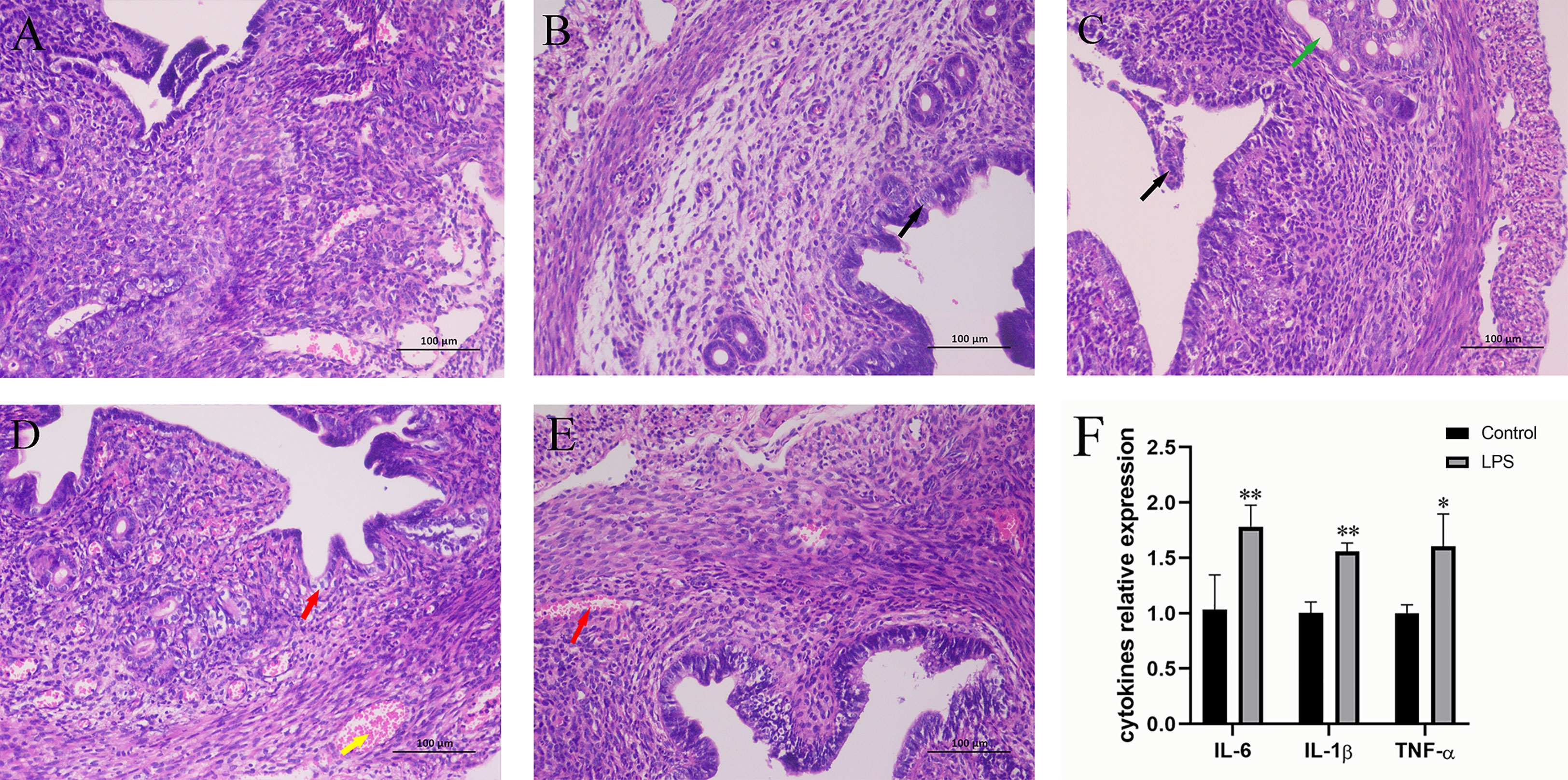
In the published article, there was an error in Figure 1 as published. This Figure was used incorrectly. The corrected Figure 1 and its caption appear below.

Figure 1 Effect of LPS on inflammation of the mouse uterus. (A) Control group. (B) LPS group (3 h). A small amount of endometrial epithelial cells seems swollen, and the cytoplasm is loose and light-stained (black arrow). (C) LPS group (6 h). Bits of endometrial epithelial cells are shed (black arrow), and a small number of uterine glands are slightly dilated (green arrow). (D) LPS group (12 h). The endometrial epithelium and glandular epithelium are swollen, the cytoplasm has loosened and lightly stained (red arrow), and a large number of capillaries in the lamina propriety are congested and dilated (yellow arrow). (E) LPS group (24 h). A spot of blood stasis in the lamina propria (red arrow). (Hematoxylin and eosin staining; magnification, 200×). (F) The expression of inflammatory cytokines IL-6, IL-1β, and TNF-α. Mean ± SD was employed for data processing. Three replicates were processed in each group. *p < 0.05, **p < 0.01 vs. control group.
Caption: Effect of LPS on inflammation of the mouse uterus. (A) Control group. (B) LPS group (3 h). A small amount of endometrial epithelial cells seems swollen, and the cytoplasm is loose and light-stained (black arrow). (C) LPS group (6 h). Bits of endometrial epithelial cells are shed (black arrow), and a small number of uterine glands are slightly dilated (green arrow). (D) LPS group (12 h). The endometrial epithelium and glandular epithelium are swollen, the cytoplasm has loosened and lightly stained (red arrow), and a large number of capillaries in the lamina propriety are congested and dilated (yellow arrow). (E) LPS group (24 h). A spot of blood stasis in the lamina propria (red arrow). (Hematoxylin and eosin staining; magnification, 200×). (F) The expression of inflammatory cytokines IL-6, IL-1β, and TNF-α. Mean ± SD was employed for data processing. Three replicates were processed in each group. *p < 0.05, **p < 0.01 vs. control group.
The authors apologize for this error and state that this does not change the scientific conclusions of the article in any way. The original article has been updated.
Publisher’s note
All claims expressed in this article are solely those of the authors and do not necessarily represent those of their affiliated organizations, or those of the publisher, the editors and the reviewers. Any product that may be evaluated in this article, or claim that may be made by its manufacturer, is not guaranteed or endorsed by the publisher.
Keywords: acute endometritis, intestinal microbiota, metabolomics, lipopolysaccharide, mice
Citation: Dong Y, Yuan Y, Ma Y, Luo Y, Zhou W, Deng X, Pu J, Hu B and Liu S (2023) Corrigendum: Combined intestinal metabolomics and microbiota analysis for acute endometritis induced by lipopolysaccharide in mice. Front. Cell. Infect. Microbiol. 13:1223663. doi: 10.3389/fcimb.2023.1223663
Received: 16 May 2023; Accepted: 30 May 2023;
Published: 09 June 2023.
Edited and Reviewed by:
Benoit Chassaing, Institut National de la Santé et de la Recherche Médicale (INSERM), FranceCopyright © 2023 Dong, Yuan, Ma, Luo, Zhou, Deng, Pu, Hu and Liu. This is an open-access article distributed under the terms of the Creative Commons Attribution License (CC BY). The use, distribution or reproduction in other forums is permitted, provided the original author(s) and the copyright owner(s) are credited and that the original publication in this journal is cited, in accordance with accepted academic practice. No use, distribution or reproduction is permitted which does not comply with these terms.
*Correspondence: Binhong Hu, YmluaG9uZy5odTg2QG1haWwucnU=; Songqing Liu, c29uZ3FpbmdsaXVAY2RudS5lZHUuY24=
 Yuqing Dong
 Yuan Yuan
 Yichuan Ma
 Yuanyue Luo
 Wenjing Zhou
 Xin Deng
 Jingyu Pu
 Binhong Hu
 Songqing Liu