
94% of researchers rate our articles as excellent or good
Learn more about the work of our research integrity team to safeguard the quality of each article we publish.
Find out more
REVIEW article
Front. Cell. Infect. Microbiol. , 26 May 2023
Sec. Microbiome in Health and Disease
Volume 13 - 2023 | https://doi.org/10.3389/fcimb.2023.1177366
This article is part of the Research Topic Reviews in Microbiome in Health & Disease View all 13 articles
 Lidia Korczynska1Natalia Zeber-Lubecka2,3*Magdalena Zgliczynska4Elzbieta Zarychta1Kornelia Zareba5Cezary Wojtyla6Michalina Dabrowska3Michal Ciebiera1
For a long time, the uterus had been considered a sterile organ, meaning that under physiological conditions the uterus would not be colonized by bacteria. Based on available data, it may be concluded that the gut and uterine microbiome are related, and that the role of this microbiome is greater than expected. Despite being the most common pelvic neoplasms in women of reproductive age, uterine fibroids (UFs) are still poorly understood tumors whose etiology has not been fully determined. This systematic review presents the relationship between intestinal and uterine dysbiosis and uterine fibroids. A systematic review of three medical databases was carried out: the MEDLINE/PubMed, Scopus and Cochrane. In this study, 195 titles and abstracts were reviewed, including only original articles and clinical trials of uterine microbiome criteria. Finally, 16 studies were included to the analysis. In recent years, researchers dealing with reproduction in a broad sense have focused on the microbiome in various locations to study its role in the pathogenesis and, consequently, the prevention and treatment of diseases of the genital organ. Conventional microbial detection methods are not suitable for identifying bacteria, which are difficult to culture. Next-generation sequencing (NGS) provides an easier and faster and more informative analysis of bacterial populations. It seems that gut microbiota dysbiosis has the potential to be a risk factor for uterine fibroids or affect the disease process. Some changes were shown in many types of bacteria, such as Firmicutes, Proteobacteria, Actinobacteria and Verrucomicrobia detected in fecal samples in patients with uterine fibroids. In view of the few results on the link between the microbiome and uterine fibroids, further intensive studies in humans and animal models are necessary, including the possible use of different microbiome modulations in the prevention or treatment of uterine fibroids.
It had been believed until recently that the uterine cavity and the upper parts of the female genital organ are a sterile environment. However, subsequent studies provided strong evidence that this view was no longer valid (Chen et al., 2017; Simon, 2018). It is not surprising, especially when considering the fact that bacterial cells in the human body account for 1-3% of its total weight and are equal in number to human cells (Moreno et al., 2016).
The use of next-generation sequencing (NGS) in the analysis of hypervariable fragments of the 16S rRNA bacterial gene showed the presence of a variety of microorganisms within the uterine cavity which were usually found in the vagina and in the colon (Pereira et al., 2016; Baker et al., 2018). According to available data, the difference between the vagina, cervix, and the uterine cavity is that the colonization of the uterine cavity is relatively low (Ichiyama et al., 2021).
Fertility disorders are the driving force behind numerous studies in gynecology and reproductive medicine. For a long time, researchers have been wondering whether there are proper microbiomes for specific parts of the reproductive organ, or whether alteration in those microbiomes might have negative consequences (Brandão and Gonçalves-Henriques, 2020; Vitale et al., 2022). The effect of female genital microflora on the ability to conceive is still unclear due to the scarcity and inconsistency of published data. Nevertheless, it seems that the flora dominated by Lactobacillus spp. plays a key role in determining fertility, and the presence of pathogens in the genitals may disturb issues associated with it (Vitale et al., 2022). Toson et al. have recently proposed that the physiological endometrial microbiota should be considered as a group of microorganisms that allows embryo implantation and live birth, regardless of the minimal presence of pathogenic bacteria (Toson et al., 2022).
Previous studies determined the normal uterine microbiome in women of childbearing age, where Lactobacillus spp. played a particular role (Mitchell et al., 2015). Other types of bacteria commonly detected in endometrial/uterine swabs included Bifidobacterium, Gardnerella, Prevotella and Streptococcus (Moreno et al., 2016). Further research showed that the abundance of Lactobacillus might be negatively correlated with the genus Gardnerella, Bifidobacterium and Atopobium and positively correlated with commensal bacteria, which are very important for the immune system, e.g., Clostridium and Streptomyces (Moreno et al., 2016).
Hormonal changes affect not only the uterine muscle and the uterine mucosa itself, but they also seem to affect the microbiome found in those areas. Current data suggest that the microbiome plays an important role in female reproductive endocrine system throughout the life by interacting with estrogens, androgens, insulin, and other hormones. An imbalance in the composition of the intestinal microflora may modify the course of numerous diseases and conditions, such as pregnancy complications, polycystic ovary syndrome, endometriosis, or reproductive organ tumors (He et al., 2021; Qi et al., 2021).
Available data indicated that exogenous progesterone might significantly alter the microflora of the endometrium (Brooks et al., 2017). Interestingly, according to Brooks et al., the use of oral hormonal contraceptives may positively influence the proper functioning of the reproductive system by increasing the quantity of Lactobacillus spp. and reducing bacterial taxa associated with bacterial vaginosis (BV) (Brooks et al., 2017). Progesterone may increase the α-diversity of both the vaginal and endometrial microbiome. Moreover, the quantity of bacteria that may interfere with proper functioning may increase after hormonal treatment (Toson et al., 2022). Notably, naturally occurring hormonal fluctuations during the menstrual cycle correlate with the instability of the microbial population, and a regular replacement of bacterial species occurs in the vagina during the cycle (Gajer et al., 2012). Significant changes also occur in the endometrial microbiome. The increased abundance of Prevotella spp. and Sneathia spp. may constitute the features of the proliferative and secretory phases, respectively (Gajer et al., 2012).
Despite being the most common pelvic neoplasms in women of reproductive age, uterine fibroids (UFs) are still poorly understood tumors whose etiology has not been fully determined (Ciebiera et al., 2020; Yang et al., 2022). They mainly affect women of childbearing age and are diagnosed in about 70% of with European-American women and in over 80% of African women throughout their lifetime (Giuliani et al., 2020). Uterine fibroids are heterogeneous in number, composition, and size. The risk factors for those tumors seem to include age, obesity, low vitamin D levels, and endogenous and exogenous hormonal factors (Yang et al., 2022). Heavy menstrual bleeding (HMB) is the most commonly mentioned abnormal symptom associated with uterine fibroids. Other symptoms may include anemia due to increased blood loss, pressure in the pelvic cavity and on organs adjacent to the uterus, urinary and digestive complaints, as well as reproductive disorders (Stewart et al., 2016). Uterine fibroids may require surgical treatment and are a major source of gynecological and reproductive dysfunction (Navarro et al., 2021).
Given that little is known about fibroids, it is obvious that a lot is still unknown about the interaction between the endometrial and myometrial microbiome and the immune response modulating inflammatory processes within the uterus. In view of the confirmed anti-inflammatory role of Lactobacillus spp. in the vaginal microenvironment, it is believed that the above may also contribute to disorders of uterine homeostasis by inducing the secretion of anti-inflammatory cytokines and the production of antimicrobial peptides. Therefore, the association between uterine and endometrial dysbiosis and immune dysregulation is highly possible (Toson et al., 2022).
The intestinal flora and host’s body show a variety of reciprocal benefits. Numerous gut microbes affect the physiological functions of the host and affect the synthesis and secretion of hormones, trace elements, growth factors and immune system functions. In turn, the intestinal flora may be modified by hormonal interactions both in vitro and in vivo, thereby affecting the biological balance of the body (Rowland et al., 2018). The glucuronidase of intestinal bacteria improves estrogen reabsorption, and some intestinal bacteria can metabolize estrogen and are referred to as estrobolome (Sui et al., 2021). The high activity of estrobolome bacterial enzymes raises the level of free estrogen in the enterohepatic circulation by promoting an endogenous hormonal environment, leading to an increase in hormone levels, which can have a direct and indirect impact on the risk of developing uterine fibroids (Wang et al., 2020c). The abundance of intestinal microflora is closely correlated with the estrogenic metabolism. Numerous bacteria control estrogen content at the family and species level, with Clostridium and Pneumococcus exerting the most significant effect on estrogen metabolism (Wang et al., 2020a). Therefore, decreased estrogen levels are associated with impaired specific gut microbiota diversity (Fuhrman et al., 2014).
Pilot studies concerning the composition of the uterine cavity microbiome of women who developed fibroids showed a greater diversity of bacteria compared to control group women (Baker et al., 2018; Toson et al., 2022). It seems that fibroids may be associated with minimally altered vaginal and uterine microflora (Chen et al., 2017). For example, the endometrial microflora of patients undergoing hysterectomy for fibroids was dominated by Acinetobacter, Cloacibacterium, Comamonadaceae and Pseudomonas, while the species Lactobacillus were rare in the uterus (Winters et al., 2019). The results suggest that the systemic distribution of intestinal bacteria extends to the patients’ fibroids following dysbiosis or impaired intestinal barrier (Yang et al., 2022). However, the above data were obtained from few studies, and the data need to be systematized.
The purpose of this systematic review is to provide a comprehensive summary of the fibroids and uterine microbiome literature published to date.
A systematic review of three medical databases was carried out: the MEDLINE/PubMed, Scopus and Cochrane. The last search was conducted on January 26, 2023. The details of the search strategy are presented in Table 1. As a result, a total of 263 articles were obtained. Using EndNote X9 automatic duplicate search tool, 42 duplicates were identified, and then another 10 duplicates were found via manual search. Finally, 195 titles and abstracts were reviewed, including only original articles and clinical trials of 281 uterine microbiome criteria (Figure 1).

Table 1 Detailed search strategy in individual databases.
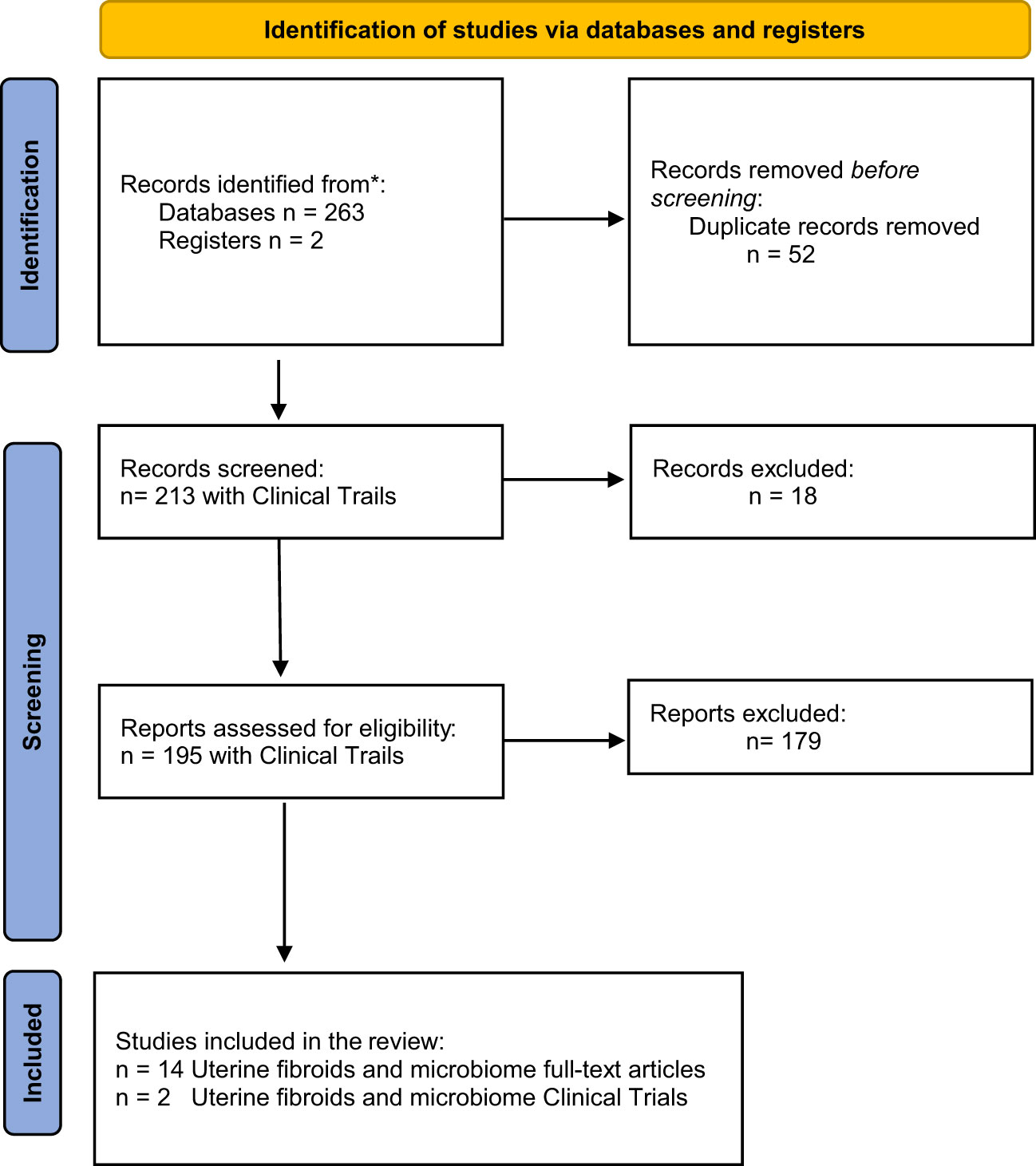
Figure 1 PRISMA 2020 flow diagram for new systematic reviews which included searches of databases and registers only.
Conventional microbial detection methods are not suitable for identifying bacteria, which are difficult to culture. In turn, advanced molecular biology techniques can resolve this problem, but even such methods have limitations. The restrictions are primarily related to the possibility of detecting DNA derived from lifeless bacteria or DNA fragments. In addition, sensitive molecular biology methods, together with the low number of bacteria present in the tissue, carry a risk of contamination, which may very easily falsify the result. Experimental difficulties in the interpretation, determination of the endometrial microbiota composition are the result of conducting studies in various populations, using diverse sampling methods and a large number of research approaches as well as abundance of human DNA also create technical challenges (Abah et al., 2021). Conversely, despite numerous limitations, NGS methods seem to provide the widest variety of insights. The application of advanced sequencing and genome analysis methods resulted in rapid publications enhancing the uterine microbiota field. Today, uterine microbiota is an important research direction in human reproductive studies.
As regards uterine fibroids microbiome research, both conventional culture and NGS studies remain rather rare (Tables 2, 3). Due to the limited ability to cultivate larger quantities of bacterial species in laboratory procedures, metagenomics established a popular and useful tool applied in the analysis of bacterial composition residing in the reproductive system. NGS provides an easier and faster analysis of bacterial populations, without cloning DNA sequences into a vector. Today, metagenomics represents a powerful tool used in microbiology which, via combining various molecular biology techniques, enables the study of the biodiversity of microbial populations and provides a better understanding of the role of individual bacteria in a particular environment, as well as the discovery of new genes (Table 3; Figure 2).
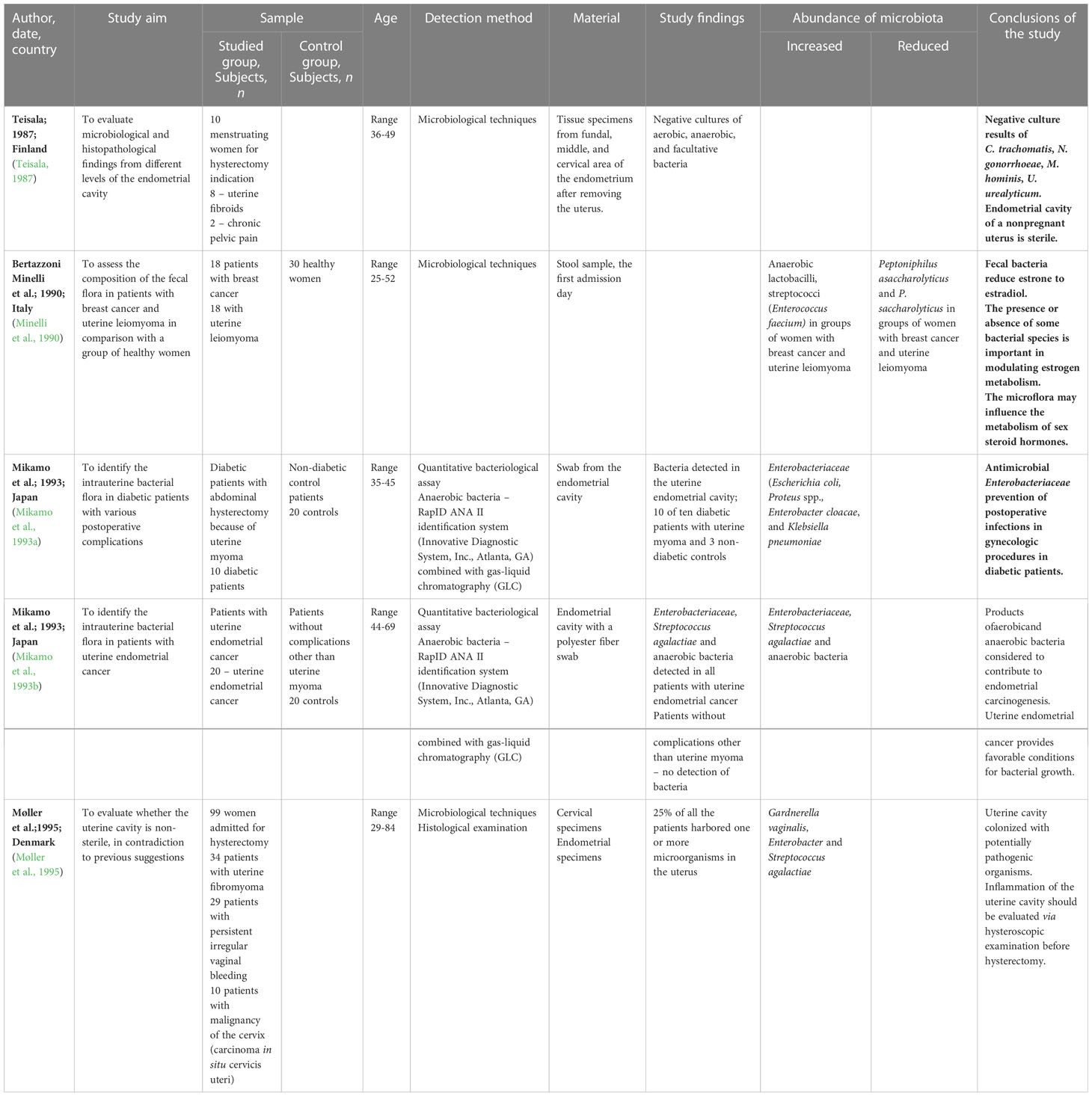
Table 2 Data extracted from studies using culture-based technology to investigate the uterine, vaginal, endocervical, endometrial and gut microbiome and uterine leiomyoma (uterine myoma, uterine fibroid).
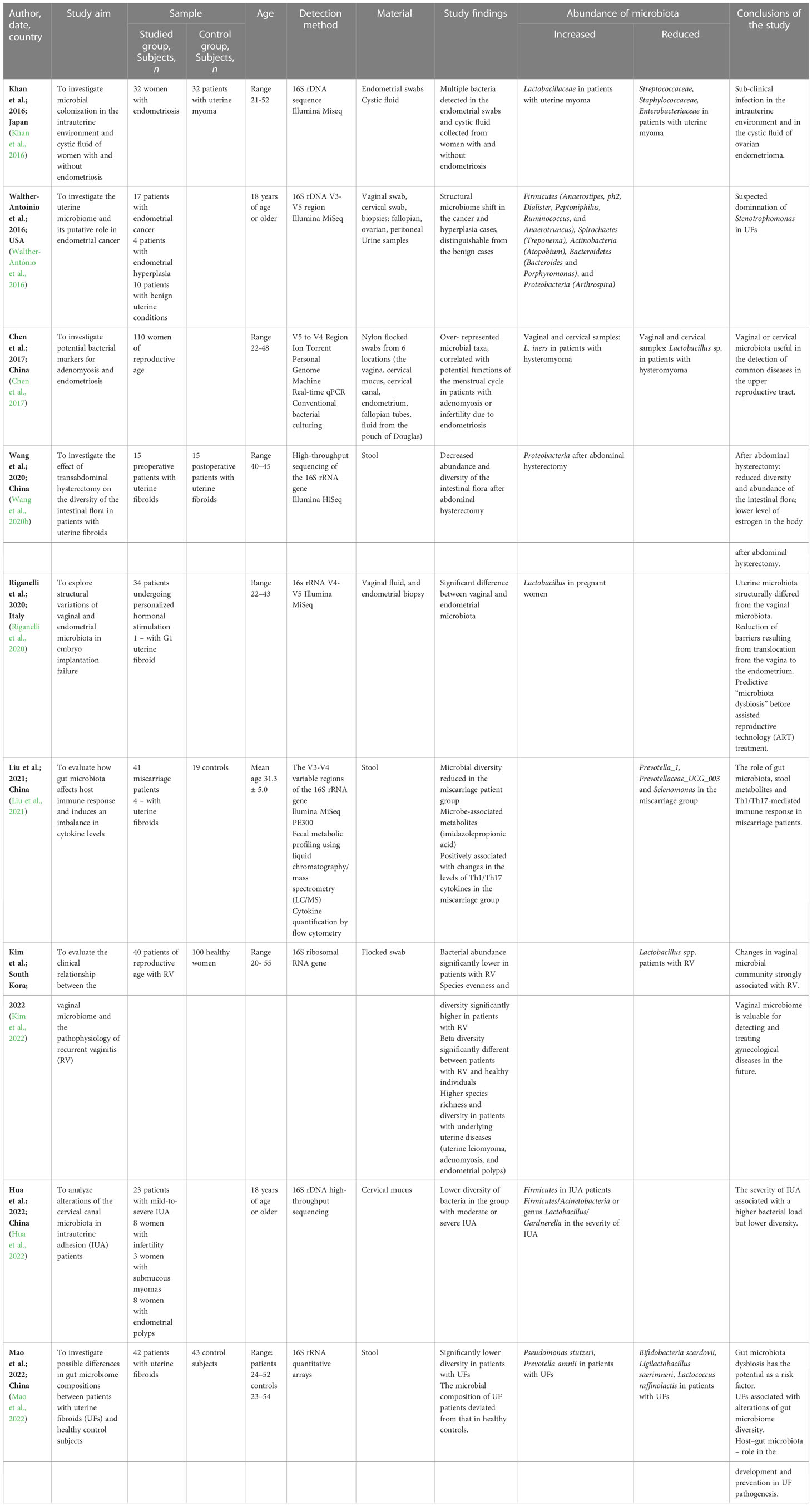
Table 3 Data extracted from studies using sequencing-based technology to investigate the uterine, vaginal, endocervical, endometrial and gut microbiome and uterine leiomyoma (uterine fibroid).
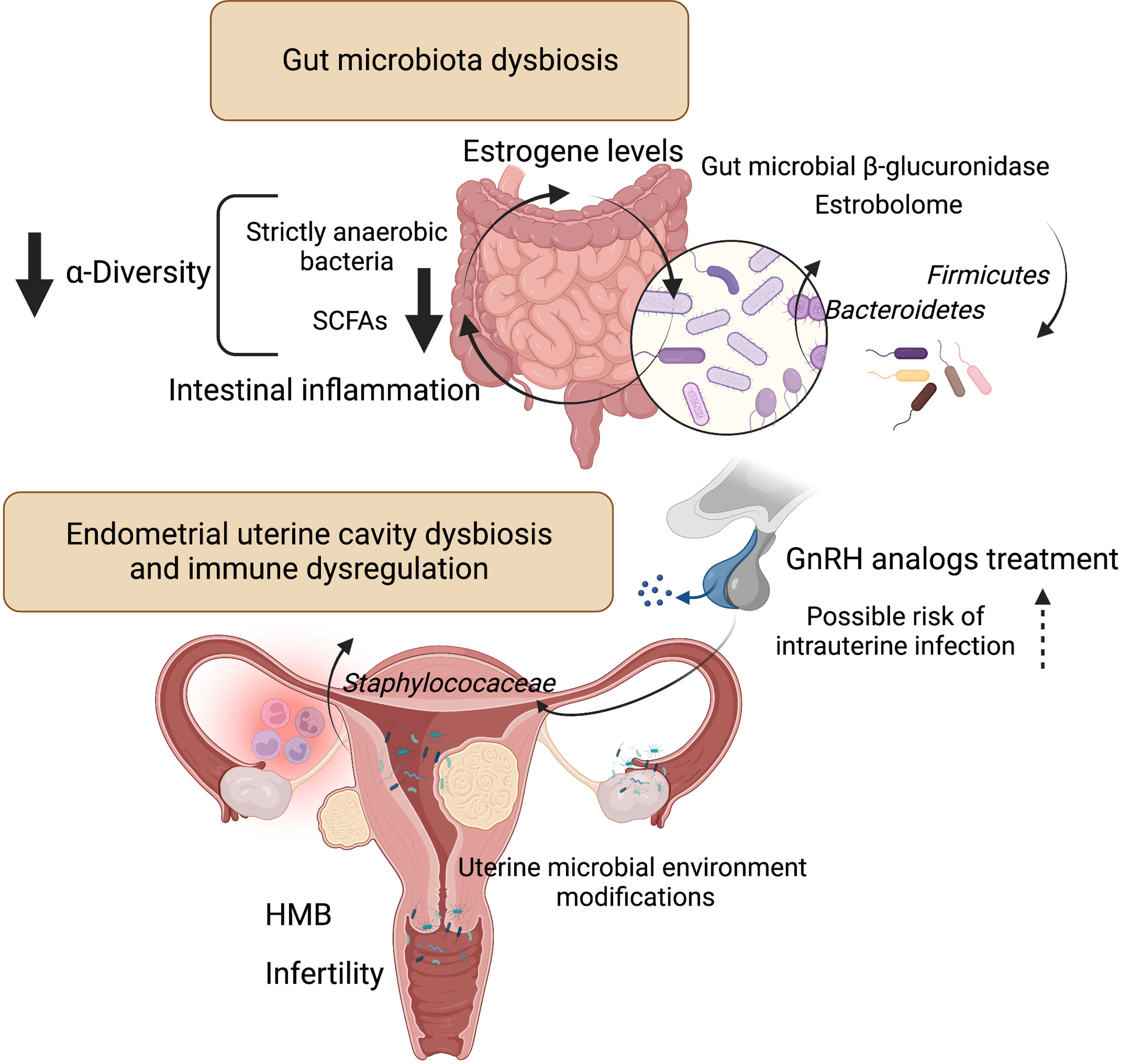
Figure 2 Influence of microbiota on estrogen metabolism and endometrial uterine cavity dysbiosis in patients with uterine fibroids and possible role of GnRH analogs treatment. Liver-conjugated estrogens excreted in the bile into the gastrointestinal tract could be deconjugated by intestinal bacteria that produce ß-glucosidase enzymes involved in estrogen deconjugation. This leads to their absorption into the bloodstream and allows them to bind to estrogen receptors. However, the relationship is a bidirectional - the gut microflora can also be affected by estrogen. The experimental evidence for the key role of gut bacteria in estrogen metabolism was observed decades ago. The estrobolome is defined as the gene repertoire of intestinal microflora with products capable to metabolize estrogens. By influencing and modulating the intestinal and hepatic circulation of estrogens, the estrobolome affects the excretion and circulation of estrogens. Numerous evidence indicates that dysbiosis of intestinal bacteria increases the chance of intestinal inflammation. This is particularly related to a decrease in strictly anaerobic bacteria and a simultaneous increase in facultative anaerobes such as Escherichia coli and Klebsiella. And SCFAs, produced mainly by strict anaerobes, have anti-inflammatory effects. According to one study (Khan et al., 2016), intrauterine higher colonization by Staphylococaceae was revealed in a group of control patients who used GnRH analogs. Patients with fibroids after GnRH treatment had the highest staphylococcal levels compared to other groups. Figure 2 created with BioRender.com. Heavy menstrual bleeding (HMB); gonadotropin-releasing hormone (GnRH); short-chain fatty acids (SCFAs).
Search Results on ClinicalTrials.gov website revealed 426 studies for: Myoma/Fibroid; Uterus terms (Table 4). The issue of the uterine microbiome was tackled in 7 of them, and only 2 without the exclusion criteria of fibroid, leiomyoma or myoma (Table 5).
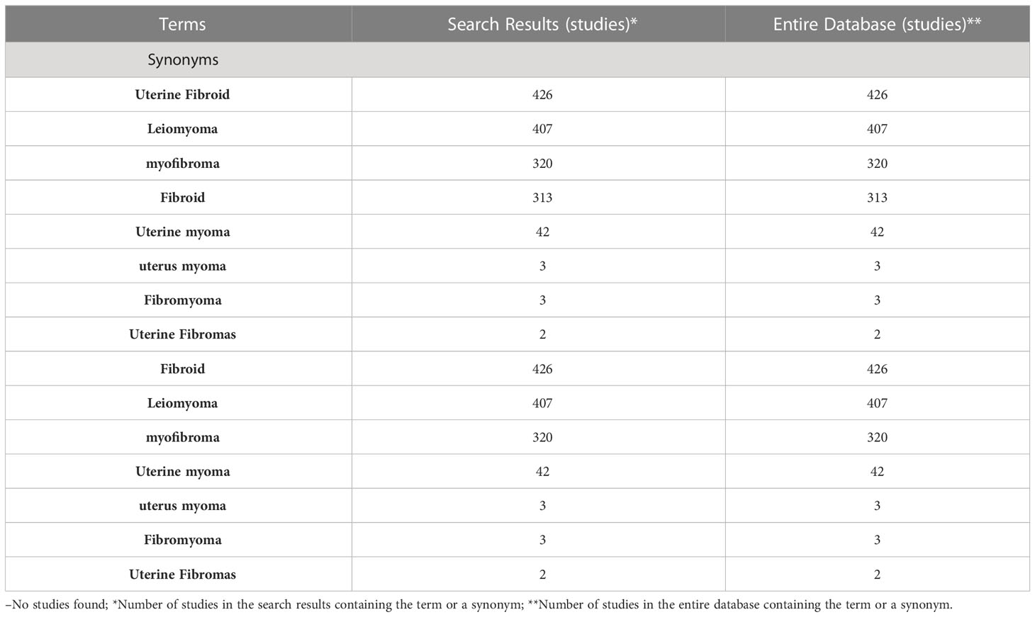
Table 4 Myoma/Fibroid; Uterus terms of 426 studies (ClinicalTrials.gov website, February 2023).
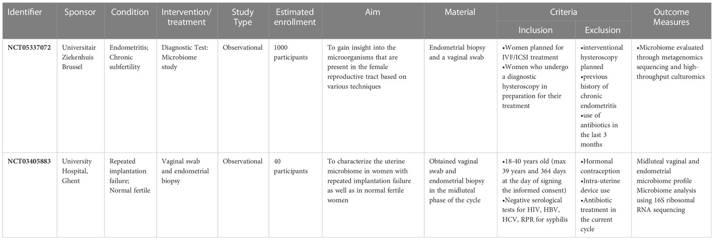
Table 5 Clinical trials of the uterine microbiome, without the exclusion criteria of fibroid, leiomyoma, myoma (ClinicalTrials.gov website, February 2023).
The presence of the microbiota in the body is known to be important for human health. In recent years, advances in molecular biology techniques have not only allowed the confirmation of the occurrence of microorganisms in the human digestive, respiratory and urinary systems, but also prompted the detection of the presence of microorganisms in organs previously considered sterile, such as the uterine cavity (Dekaboruah et al., 2020).
For a long time, the uterus had been considered a sterile organ, meaning that under physiological conditions the uterus would not be colonized by bacteria. In 1950s, research based on bacterial culture methods revealed the first reports suggesting that the uterus was an organ with its own microbiota. Further development of diagnostic techniques and molecular biology methods in early 21st century allowed the reanalysis of the composition of the uterine microbiota, including bacteria which are not easily cultured under laboratory conditions (Franasiak et al., 2021).
Until the late 1980s, the results of most studies related to the microbiome of the myomal uterine cavity had indicated the sterility of the uterus (Teisala, 1987; Mikamo et al., 1993b) (Table 2). Several years later, a quantitative bacteriologic assay of swabs from the endometrial cavity after myoma uteri hysterectomy demonstrated an increased amount of Enterobacteriaceae family, including Escherichia coli, Proteus spp., Enterobacter cloacae, and Klebsiella pneumoniae (Mikamo et al., 1993a). In contrast, the authors expressed a different view of the microbiome of the uterine cavity affected by cancer. The results showed the distribution of Enterobacteriaceae, Streptococcus agalactiae and anaerobic bacteria detected in all patients with uterine endometrial cancer (Mikamo et al., 1993b). In conclusion, the products of aerobic and anaerobic bacteria contributing to endometrial carcinogenesis as well as uterine endometrial cancer provided preferential environment for bacterial growth. A turning point occurred when a study of nearly 100 women was conducted and revealed that myomal uterine cavity was colonized with potentially pathogenic organisms (Møller et al., 1995). In samples from the uterine cavity, a quarter of the women cultured one or more bacteria using microbiological techniques. Gardnerella vaginalis, Enterobacter and Streptococcus agalactiae pathogenic bacteria were identified. The deviations of obtained results were due to non-cultivation of bacteria under laboratory conditions or the presence of only lifeless bacteria in the material.
The modulating effect of the gut microbiome on the uterine cavity was highlighted in the 1990s. Using culture techniques, the composition of the stool microbiome was examined in patients with breast cancer and uterine leiomyoma (Minelli et al., 1990). The study showed fecal bacteria reduction of estrone to estradiol (Minelli et al., 1990). The findings confirmed the decreased amount of Peptoniphilus asaccharolyticus and P. saccharolyticus and an increased number of anaerobic lactobacilli and Enterococcus faecium in the gut microbiome of breast cancer and uterine leiomyoma women. The presence or absence of some bacterial species is important in modulating estrogen metabolism. Thus, the microbiome influenced the metabolism of sex steroid hormones (Hussain et al., 2021).
It has been known for a long time that the genus Lactobacillus represents the dominant component of the vaginal microbiota and plays a protective role for the vaginal microenvironment. The presence of Lactobacilli was also confirmed in the upper female reproductive tract (FRT) (Verstraelen et al., 2016; Chen et al., 2017; Benner et al., 2018). Chen et al. demonstrated the predominance of Lactobacillus bacteria (> 97.56%) in the cervix. In the uterus, Lactobacillus (30.6%), Pseudomonas (9.09%), Acinetobacter (9.07%), Vagococcus (7.29%), and Sphingobium (5.0%) were identified, while the fallopian tubes revealed the presence of Acinetobacter (18.27%), Comamonas (11.49%), Pseudomonas (9.9%), Pseudomonadaceae (9.1%), and Dysgonomonas (5.11%) (Chen et al., 2017). Further studies published in 2018 and 2019 confirmed the occurrence of bacteria from the Lactobacillaceae family not only in the vagina, but also in other segments of the female reproductive tract (Moreno et al., 2016; Koedooder et al., 2019). The protective role of Lactobacillus in the vagina was recognized. However, the precise mechanism of the effect and a possible impact on fertility have not been fully elucidated and require further research.
Today, we know that a gigantic number of human microbes influences the physiological functions of the human body at all times and has a profound effect on the synthesis and secretion of hormones and various growth factors. As a result, changes in the composition of the human gut flora may affect the body in various aspects, including the immune or hormonal system (Thursby and Juge, 2017). It is well known how important hormones (estrogens and progesterone) are in the pathophysiology of fibroids (Bulun, 2013). The associated growth factors are equally important, with transforming beta growth factor probably being the crucial one (Ciebiera et al., 2017).
Leaving aside the above words of introduction concerning new generation research and general disorders related to female reproductive tract colonization, our analyses of publications mostly indicate that almost no data are available on the microbiota and uterine fibroids. It seems almost unlikely assuming that uterine fibroids are the most common problem with which women present to gynecologists (Stewart et al., 2016). This problem has a large clinical impact because most papers that may be reviewed are not analyzed in terms of fibroids. The analyses concentrated on the fact that fibroids were not an exclusion criterion for other studies. Therefore, our conclusions will certainly not be strong.
We identified 4 studies that have attracted our greatest attention in the analysis and that seem to be more significant in terms of the pathophysiology of uterine fibroids and possible clinical situations associated with them. We would like to focus on those publications below.
The first study on the uterine cavity microbiota was published in 2016 by Khan et al. The Japanese authors conducted a molecular analysis of intrauterine colonization with microorganisms of women with endometriosis (Khan et al., 2016). The study included a total of 64 women half of whom had endometriosis and the other half was not affected by the condition. In each group, 16 patients additionally received GnRH analogs for 4-6 months. The results were highly interesting. A high percentage of bacteria was detected in the swabs collected from the endometrium and from the fluid obtained from cysts. Metagenomic tests showed that the proportion of Lactobacillaceae was significantly reduced, while the proportion of Streptococcaceae, Staphylococaceae, and Enterobacteriaceae was significantly increased in the GnRH analog-treated groups of women with endometriosis compared to women who did not receive those drugs. Ordinary tests did not allow for the assessment of cystic fluid for microbial colonization. The results of the 16S metagenomic test were different. A much higher percentage of Streptococcaceae (p<0.01) and Staphylococaceae (p<0.05) was detected in the fluid collected from cysts of women with ovarian endometriosis compared to fluid collected from cysts without an endometriotic etiology. It is difficult to determine what effect those data have on uterine fibroids. However, a significantly higher colonization by Staphylococaceae (p<0.05) and an insignificant statistical difference for colonization by Enterobacteriaceae was revealed in a group of control patients who used GnRH analogs (due to uterine fibroids) compared to samples obtained from control group women who were not pharmacologically treated. Interestingly, according to the data obtained from the study, the group of patients with fibroids after GnRH treatment had the lowest overall percentage of streptococcal colonization and the highest Staphylococcal levels compared to other groups. It is not known what these data mean from the viewpoint of uterine fibroids and what the clinical application may be. However, the role of drugs blocking the pituitary was shown not only to consist in regulating the growth of fibroids, but also to change the entire uterine environment, which may affect other important features such as fertility or a possible relationship with pregnancy. It certainly requires a lot of well-designed research, in which not endometriosis, but uterine fibroids will constitute the focus (Linder et al., 2021; Maxim et al., 2022). The authors concluded that the results indicated the presence of a subclinical infection in the intrauterine environment and, importantly, in the fluid collected from the lesions in the ovary. Furthermore, the authors emphasized the side effect of treatment with GnRH analogs associated with promoting an intrauterine and/or ovarian infection (Khan et al., 2016)
The second of the studies analyzed in the context of uterine fibroids concerned the effect of transabdominal hysterectomy on the intestinal flora in patients with uterine fibroids using high-throughput sequencing (Wang et al., 2020b). According to the authors, as could be predicted, estrogen levels decreased after transabdominal hysterectomy. However, post-hysterectomy high-throughput sequencing showed that the quantity and diversity of intestinal flora decreased.
It is known that total hysterectomy is one of more commonly proposed procedures in women with uterine fibroids who do not have fertility requirements. A group of doctors obviously struggle with such an approach and try to convince their colleagues to switch to more conservative surgical methods, use non-operative methods such as thermoablation (Lozinski et al., 2021) or switch to pharmacological treatment (Niaz et al., 2022). Complete hysterectomy affects the level of sex hormones. According to available data, serum AMH levels might be decreased after hysterectomy, with a greater reduction when total hysterectomy is performed in comparison with supracervical hysterectomy (Yuan et al., 2015). It is due to the fact that the anatomical structure of the uterus and ovary is closely correlated. A significant portion of the blood supplied to the ovary comes from the ascending branch of the uterine artery. Simultaneously, regardless of anatomical changes, thermal damage is common during procedures, which can impair the blood supply to the ovaries after complete hysterectomy, thus leading to a reduction in the secretion of sex hormones (Wang et al., 2020b).
So what is the significance of the results of the study by Wang et al.? Previous research demonstrated that a decrease in estrogen levels leads to a decrease in the diversity of the intestinal flora and a reduction in the abundance of thick-walled bacteria, such as Clostridium (Breban, 2016). Decreased estrogen levels in the study described leads to other situations, because of an increase in the quantity of Firmicutes, a decrease in the diversity of Bacteroidetes and an increase in the species diversity of Proteobacteria. The authors concluded that transabdominal hysterectomy could reduce the level of estrogen in the body and reduce the diversity and abundance of intestinal bacterial flora before and after surgery with the growth of Proteobacteria being the main difference in that case (Wang et al., 2020b). Obviously, it probably cannot be said that the mechanism underlying this regulation has already been determined. Only a deeper analysis can help understand whether the intestinal microflora may affect the risk of uterine fibroids by affecting endogenous estrogens (Yang et al., 2022).
Another analyzed study tackled the issue of interactions between the intestinal microflora and cytokine disorders in women with unexplained causes of miscarriages. Five patients with fibroids were included in this study, according to the data provided. Regrettably, this group did not undergo any additional individual analyses (Liu et al., 2021). Multiple authors suggested that dysregulation within the cytokine and growth factor network is involved in the pathogenesis of unexplained pregnancy loss (Calleja-Agius and Brincat, 2008). As already known from the previous paragraphs, the microbiota affects the host’s immune response. However, the question of how this dysbiosis impairs he cellular immune function in the event of miscarriage remains ambiguous. It is also known that factors related to the Th1 response, such as tumor necrosis factor alpha (TNF-α), increase in the serum of patients affected by miscarriage (Mallmann et al., 1991). TNF-α was also reported to inhibit trophoblast invasion and elevated TNF-α levels were identified in women who experienced miscarriage (Azizieh and Raghupathy, 2015). According to a large amount of data, TNF-α also seems to be an extremely important cytokine in the biology of uterine fibroids, associated symptoms and conditions (Ciebiera et al., 2018b). It was shown that its concentration was increased in women with symptomatic uterine fibroids (Ciebiera et al., 2018a; Kali and Cagiran, 2022). Currently available data suggest the occurrence of an inflammation-like condition in women with uterine fibroids, with TNF-α being a strong inducer of the state (Ciebiera et al., 2018b).
Liu et al. (Liu et al., 2021) demonstrated that the microbiological diversity and a relative abundance of Prevotella_1, Prevotellaceae_UCG_003 and Selenomonas_1 were significantly reduced in cases of miscarriage. Further analyses indicated that some microbial-related metabolites were positively associated with changes in Th1/Th17 cytokine levels in the miscarriage group. The authors concluded that a network between the intestinal microbiota, fecal metabolites and Th1/Th17 might mediate miscarriage recurrences. Moreover, the intestinal microflora in patients with a history of miscarriage showed a much higher concentration compared to controls. Given that only a few patients with uterine fibroids participated in this study, it is not possible to draw unambiguous conclusions about this particular condition. However, a number of factors and pathways, e.g., hormonal-immune ones, fit quite well into the same ones that affect uterine fibroids. Seemingly, it may be a very promising topic of research, e.g., to study why some fibroids may increase the risk of miscarriages or improper implantation.
Finally, the last study was designed for a group of patients with uterine fibroids. The authors used 16 rRNA high-throughput microarrays to compare gut microbiological differences between healthy women and women with uterine fibroids. Moreover, the authors aimed to determine the correlations and interactions between the intestines, microbiome and uterine fibroids (Mao et al., 2022). The study included 42 patients with uterine fibroids and 43 patients without a history of fibroids. The authors demonstrated very interesting correlations. Firstly, microbiome diversity in patients with uterine fibroids was significantly lower than in healthy controls. The microbiological composition of samples collected from patients with uterine fibroids differed from that of healthy women. Some changes were shown in many types of bacteria, such as Firmicutes, Proteobacteria, Actinobacteria and Verrucomicrobia detected in fecal samples in patients with uterine fibroids.
Further analysis of bacterial abundance showed that some species were present to a smaller extent (e.g., Bifidobacteria scardovii, Ligilactobacillus saerimneri and Lactococcus raffinolactis) and some were more abundant (e.g., Pseudomonas stutzeri and Prevotella amnii). Moreover, microbial interactions and specific networks in uterine fibroids exhibited lower connectivity and complexity as well as higher clustering properties compared to the second group. The results indicated it was highly possible that intestinal dysbiosis might be a significant risk factor for the development or growth of uterine fibroids. The data confirmed that uterine fibroids might be strongly associated with changes in the diversity of the gut microbiome. This provides a new direction for further analyses of the intestinal microflora and its links to the development and growth of uterine fibroids. The results will certainly provide an incentive to develop new research and further analyses. Furthermore, as the authors noted, they may also constitute pioneering reports in terms of new therapies for uterine fibroids, such as the development or use of probiotics to protect against uterine fibroids or support therapy (Mao et al., 2022).
Apart from the scarcity of publications on the microbiota and uterine fibroids, it is also quite surprising that there are practically no registered studies that could expand such knowledge. In preparing this systematic review, the authors also reviewed the clinicaltrials.gov database and, to their surprise, it turned out that only 7 out of 426 studies related to the microbiome were related to the uterine microbiome (only 2 were directly linked, and only 2 indirectly addressed the issue of uterine fibroids). As regards those two clinical studies, the first one concerns chronic endometritis and its effects on fertility and pregnancy. This study is particularly important because chronic endometritis is commonly diagnosed in the context of fertility problems, and the patient is often treated blindly with broad-spectrum antibiotics in this case. Uterine fibroids are not simply an exclusion criterion here and such patients can be recruited for the study (Table 5). The topic of the second study is different, i.e., it addresses implantation failures. The study focuses on the characteristics of the uterine microbiome in women with repeated implantation failures and in women with normal fertility. The absence of pregnancy after the transfer of a total of 5 high-quality embryos is the inclusion criterion in the study. As regards uterine fibroids, they are not an exclusion criterion in the study.
Metagenomics and traditional methods share some limitations, while also complement each other. Thus, this creates new diagnostic potential for research. The microbiota in the uterine cavity has been much less characterized. The upper female genital tract, consisting of the uterus, fallopian tubes and ovaries, was once considered a sterile environment. Although this approach has fundamentally changed over the years, there is still no current consensus on the basic female genital tract microbiota occurring in healthy women, nor its exact role in the formation of uterine fibroids. However, an increasing amount of strong evidence is available to support this changing concept.
This systematic review presents the relationship between intestinal and uterine dysbiosis and uterine fibroids. In recent years, researchers dealing with reproduction in a broad sense have focused on the microbiome in various locations to study its role in the pathogenesis and, consequently, the prevention and treatment of diseases of the genital organ. Recent studies of microbiomes of different locations have identified patterns of the bacterial composition of these sites depending on such factors as the population. The next step involves increasingly advanced research on health changes related to specific conditions, especially with regard to the impact of steroid hormone axes, and the interaction of estrogens with the gut microbiome.
Based on available data, it may be concluded that the gut and uterine microbiome are related and that the role of this microbiome is greater than expected. Numerous pathogenetic pathways are shared by the microbiome and fibroids, which allows a conclusion that the presence of fibroids or some symptoms may be dependent on changes in the microbiome. In view of the few results on the link between the microbiome and uterine fibroids, further intensive studies in humans and animal models are necessary, including the possible use of different microbiome modulations in the prevention or treatment of uterine fibroids.
Conceptualization, MC, NZ-L; Methodology, MZ; Search for references, MZ, NZ-L, LK; Writing—original draft preparation, NZ-L, MC, LK; Writing—review and editing, NZ-L, LK, MZ, EZ, KZ, CW, MD; Visualization, NZ-L, MZ; Supervision, MC. All authors contributed to the article and approved the submitted version.
This work was supported by 501-1-022-26-22/MG5 (MC) and 501-1-022-26-23 grants from the Centre of Postgraduate Medical Education.
The authors declare that the research was conducted in the absence of any commercial or financial relationships that could be construed as a potential conflict of interest.
All claims expressed in this article are solely those of the authors and do not necessarily represent those of their affiliated organizations, or those of the publisher, the editors and the reviewers. Any product that may be evaluated in this article, or claim that may be made by its manufacturer, is not guaranteed or endorsed by the publisher.
Abah, K. O., Ogwu, D., Allam, L., Obudu, C. E., Itodo, J. I., Sani, N. A. (2021). Genital lesions in cows naturally infected with trypanosomes in Abuja, Nigeria. Vet. World 14, 1363–1370. doi: 10.14202/vetworld.2021.1363-1370
Azizieh, F. Y., Raghupathy, R. G. (2015). Tumor necrosis factor-α and pregnancy complications: a prospective study. Med. Princ. Pract. 24, 165–170. doi: 10.1159/000369363
Baker, J. M., Chase, D. M., Herbst-Kralovetz, M. M. (2018). Uterine microbiota: residents, tourists, or invaders? Front. Immunol. 9. doi: 10.3389/fimmu.2018.00208
Benner, M., Ferwerda, G., Joosten, I., van der Molen, R. G. (2018). How uterine microbiota might be responsible for a receptive, fertile endometrium. Hum. Reprod. Update 24, 393–415. doi: 10.1093/humupd/dmy012
Brandão, P., Gonçalves-Henriques, M. (2020). The impact of female genital microbiota on fertility and assisted reproductive treatments. J. Fam. Reprod. Health 14, 131–149. doi: 10.18502/jfrh.v14i3.4666
Breban, M. (2016). Gut microbiota and inflammatory joint diseases. Joint Bone Spine 83, 645–649. doi: 10.1016/j.jbspin.2016.04.005
Brooks, J. P., Edwards, D. J., Blithe, D. L., Fettweis, J. M., Serrano, M. G., Sheth, N. U., et al. (2017). Effects of combined oral contraceptives, depot medroxyprogesterone acetate and the levonorgestrel-releasing intrauterine system on the vaginal microbiome. Contraception 95, 405–413. doi: 10.1016/j.contraception.2016.11.006
Calleja-Agius, J., Brincat, M. P. (2008). Recurrent miscarriages: what is the role of cytokines? Gynecol. Endocrinol. 24, 663–668. doi: 10.1080/09513590802288275
Chen, C., Song, X., Wei, W., Zhong, H., Dai, J., Lan, Z., et al. (2017). The microbiota continuum along the female reproductive tract and its relation to uterine-related diseases. Nat. Commun. 8, 875. doi: 10.1038/s41467-017-00901-0
Ciebiera, M., Ali, M., Prince, L., Jackson-Bey, T., Atabiekov, I., Zgliczyński, S., et al. (2020). The evolving role of natural compounds in the medical treatment of uterine fibroids. J. Clin. Med. 9, 1479. doi: 10.3390/jcm9051479
Ciebiera, M., Włodarczyk, M., Wrzosek, M., Męczekalski, B., Nowicka, G., Łukaszuk, K., et al. (2017). Role of transforming growth factor β in uterine fibroid biology. Int. J. Mol. Sci. 18, 2435. doi: 10.3390/ijms18112435
Ciebiera, M., Włodarczyk, M., Wrzosek, M., Wojtyła, C., Męczekalski, B., Nowicka, G., et al. (2018a). TNF-α serum levels are elevated in women with clinically symptomatic uterine fibroids. Int. J. Immunopathol. Pharmacol. 32, 2058738418779461. doi: 10.1177/2058738418779461
Ciebiera, M., Włodarczyk, M., Zgliczyńska, M., Łukaszuk, K., Męczekalski, B., Kobierzycki, C., et al. (2018b). The role of tumor necrosis factor α in the biology of uterine fibroids and the related symptoms. Int. J. Mol. Sci. 19, 3869. doi: 10.3390/ijms19123869
Dekaboruah, E., Suryavanshi, M. V., Chettri, D., Verma, A. K. (2020). Human microbiome: an academic update on human body site specific surveillance and its possible role. Arch. Microbiol. 202, 2147–2167. doi: 10.1007/s00203-020-01931-x
Franasiak, J. M., Alecsandru, D., Forman, E. J., Gemmell, L. C., Goldberg, J. M., Llarena, N., et al. (2021). A review of the pathophysiology of recurrent implantation failure. Fertil. Steril. 116, 1436–1448. doi: 10.1016/j.fertnstert.2021.09.014
Fuhrman, B. J., Feigelson, H. S., Flores, R., Gail, M. H., Xu, X., Ravel, J., et al. (2014). Associations of the fecal microbiome with urinary estrogens and estrogen metabolites in postmenopausal women. J. Clin. Endocrinol. Metab. 99, 4632–4640. doi: 10.1210/jc.2014-2222
Gajer, P., Brotman, R. M., Bai, G., Sakamoto, J., Schütte, U. M. E., Zhong, X., et al. (2012). Temporal dynamics of the human vaginal microbiota. Sci. Transl. Med. 4, 132ra52. doi: 10.1126/scitranslmed.3003605
Giuliani, E., As-Sanie, S., Marsh, E. E. (2020). Epidemiology and management of uterine fibroids. Int. J. Gynecol. Obstet. 149, 3–9. doi: 10.1002/ijgo.13102
He, Y., Fu, L., Li, Y., Wang, W., Gong, M., Zhang, J., et al. (2021). Gut microbial metabolites facilitate anticancer therapy efficacy by modulating cytotoxic CD8+ T cell immunity. Cell Metab. 33, 988–1000.e7. doi: 10.1016/j.cmet.2021.03.002
Hua, X., Xu, L., Li, Q., Zhang, M., Chen, X., Zhu, Y., et al. (2022). Deep grouping analysis of the altered cervical canal microbiota in intrauterine adhesion patients. Reprod. Sci. 29 (12), 3494–3507. doi: 10.1007/s43032-022-01006-w
Hussain, T., Murtaza, G., Kalhoro, D. H., Kalhoro, M. S., Metwally, E., Chughtai, M. I., et al. (2021). Relationship between gut microbiota and host-metabolism: emphasis on hormones related to reproductive function. Anim. Nutr. 7, 1–10. doi: 10.1016/j.aninu.2020.11.005
Ichiyama, T., Kuroda, K., Nagai, Y., Urushiyama, D., Ohno, M., Yamaguchi, T., et al. (2021). Analysis of vaginal and endometrial microbiota communities in infertile women with a history of repeated implantation failure. Reprod. Med. Biol. 20, 334–344. doi: 10.1002/rmb2.12389
Kali, Z., Cagiran, F. T. (2022). Surgical removal of intramural fibroids improves the TNF-α induced inflammatory events in endometrium. Eur Rev Med Pharmacol Sci. 26 (24), 9180–9186. doi: 10.26355/eurrev_202212_30669
Khan, K. N., Fujishita, A., Masumoto, H., Muto, H., Kitajima, M., Masuzaki, H., et al. (2016). Molecular detection of intrauterine microbial colonization in women with endometriosis. Eur. J. Obstet. Gynecol. Reprod. Biol. 199, 69–75. doi: 10.1016/j.ejogrb.2016.01.040
Kim, M. J., Lee, S., Kwon, M. Y., Kim, M. (2022). Clinical significance of composition and functional diversity of the vaginal microbiome in recurrent vaginitis. Front. Microbiol. 13. doi: 10.3389/fmicb.2022.851670
Koedooder, R., Singer, M., Schoenmakers, S., Savelkoul, P. H. M., Morré, S. A., de Jonge, J. D., et al. (2019). The vaginal microbiome as a predictor for outcome of in vitro fertilization with or without intracytoplasmic sperm injection: a prospective study. Hum. Reprod. 34, 1042–1054. doi: 10.1093/humrep/dez065
Linder, K. E., Burnett, T. L., Weng, C.-S., Khan, Z., Mara, K., Cope, A. G. (2021). Treatment patterns in women seeking care for endometriosis at an endometriosis center. J. Endometr. Pelvic Pain Disord. 13, 244–249. doi: 10.1177/22840265211045606
Liu, Y., Chen, H., Feng, L., Zhang, J. (2021). Interactions between gut microbiota and metabolites modulate cytokine network imbalances in women with unexplained miscarriage. NPJ Biofilms Microbiomes 7, 24. doi: 10.1038/s41522-021-00199-3
Lozinski, T., Filipowska, J., Pyka, M., Baczkowska, M., Ciebiera, M. (2021). Magnetic resonance-guided high-intensity ultrasound (MR-HIFU) in the treatment of symptomatic uterine fibroids - five-year experience. Ginekol. Pol. 93 (3), 185–194. doi: 10.5603/GP.a2021.0098
Mallmann, P., Mallmann, R., Krebs, D. (1991). Determination of tumor necrosis factor alpha (TNF alpha) and interleukin 2 (IL 2) in women with idiopathic recurrent miscarriage. Arch. Gynecol. Obstet. 249, 73–78. doi: 10.1007/BF02390365
Mao, X., Peng, X., Pan, Q., Zhao, X., Yu, Z., Xu, D. (2022). Uterine fibroid patients reveal alterations in the gut microbiome. Front. Cell. Infect. Microbiol. 12. doi: 10.3389/fcimb.2022.863594
Maxim, M., Dason, E. S., Chan, C., Luketic, L., Li, Q., Huszti, E., et al. (2022). Current diagnosis and management of adenomyosis in Canada: a survey of Canadian gynaecologists. J. Endometr. Pelvic Pain Disord. 14, 98–105. doi: 10.1177/22840265221093263
Mikamo, H., Izumi, K., Ito, K., Watanabe, K., Ueno, K., Tamaya, T. (1993a). Bacterial flora detected of the uterine endometrial cavity of diabetic patients with myoma uteri. Kansenshogaku Zasshi 67, 654–658. doi: 10.11150/kansenshogakuzasshi1970.67.654
Mikamo, H., Izumi, K., Ito, K., Watanabe, K., Ueno, K., Tamaya, T. (1993b). Endometrial bacterial flora detected in patients with uterine endometrial cancer. Kansenshogaku Zasshi 67, 712–717. doi: 10.11150/kansenshogakuzasshi1970.67.712
Minelli, E. B., Beghini, A. M., Vesentini, S., Marchiori, L., Nardo, G., Cerutti, R., et al. (1990). Intestinal microflora as an alternative metabolic source of estrogens in women with uterine leiomyoma and breast cancer. Ann N Y Acad Sci 595, 473–479. doi: 10.1111/j.1749-6632.1990.tb34337.x
Mitchell, C. M., Haick, A., Nkwopara, E., Garcia, R., Rendi, M., Agnew, K., et al. (2015). Colonization of the upper genital tract by vaginal bacterial species in nonpregnant women. Am. J. Obstet. Gynecol. 212, 611.e1–611.e9. doi: 10.1016/j.ajog.2014.11.043
Møller, B. R., Kristiansen, F. V., Thorsen, P., Frost, L., Mogensen, S. C. (1995). Sterility of the uterine cavity. Acta Obstet. Gynecol. Scand. 74, 216–219. doi: 10.3109/00016349509008942
Moreno, I., Codoñer, F. M., Vilella, F., Valbuena, D., Martinez-Blanch, J. F., Jimenez-Almazán, J., et al. (2016). Evidence that the endometrial microbiota has an effect on implantation success or failure. Am. J. Obstet. Gynecol. 215, 684–703. doi: 10.1016/j.ajog.2016.09.075
Navarro, A., Bariani, M. V., Yang, Q., Al-Hendy, A. (2021). Understanding the impact of uterine fibroids on human endometrium function. Front. Cell Dev. Biol. 9. doi: 10.3389/fcell.2021.633180
Niaz, R., Saeed, M., Khan, H., Ali, M. A., Irshad, A., Faiz, M., et al. (2022). Efficacy and safety of oral GnRh antagonists in patients with uterine fibroids: a systematic review. J. Obstet. Gynaecol. Can. JOGC J. Obstet. Gynecol. Can. JOGC 44, 1279–1288. doi: 10.1016/j.jogc.2022.10.012
Pereira, N., Hutchinson, A. P., Lekovich, J. P., Hobeika, E., Elias, R. T. (2016). Antibiotic prophylaxis for gynecologic procedures prior to and during the utilization of assisted reproductive technologies: a systematic review. J. Pathog. 2016, e4698314. doi: 10.1155/2016/4698314
Qi, X., Yun, C., Pang, Y., Qiao, J. (2021). The impact of the gut microbiota on the reproductive and metabolic endocrine system. Gut Microbes 13, 1894070. doi: 10.1080/19490976.2021.1894070
Riganelli, L., Iebba, V., Piccioni, M., Illuminati, I., Bonfiglio, G., Neroni, B., et al. (2020). Structural variations of vaginal and endometrial microbiota: hints on female infertility. Front. Cell. Infect. Microbiol. 10. doi: 10.3389/fcimb.2020.00350
Rowland, I., Gibson, G., Heinken, A., Scott, K., Swann, J., Thiele, I., et al. (2018). Gut microbiota functions: metabolism of nutrients and other food components. Eur. J. Nutr. 57, 1–24. doi: 10.1007/s00394-017-1445-8
Simon, C. (2018). Introduction: do microbes in the female reproductive function matter? Fertil. Steril. 110, 325–326. doi: 10.1016/j.fertnstert.2018.06.041
Stewart, E. A., Laughlin-Tommaso, S. K., Catherino, W. H., Lalitkumar, S., Gupta, D., Vollenhoven, B. (2016). Uterine fibroids. Nat. Rev. Dis. Primer 2, 1–18. doi: 10.1038/nrdp.2016.43
Sui, Y., Wu, J., Chen, J. (2021). The role of gut microbial β-glucuronidase in estrogen reactivation and breast cancer. Front. Cell Dev. Biol. 9. doi: 10.3389/fcell.2021.631552
Teisala, K. (1987). Endometrial microbial flora of hysterectomy specimens. Eur. J. Obstet. Gynecol. Reprod. Biol. 26, 151–155. doi: 10.1016/0028-2243(87)90050-5
Thursby, E., Juge, N. (2017). Introduction to the human gut microbiota. Biochem. J. 474, 1823–1836. doi: 10.1042/BCJ20160510
Toson, B., Simon, C., Moreno, I. (2022). The endometrial microbiome and its impact on human conception. Int. J. Mol. Sci. 23, 485. doi: 10.3390/ijms23010485
Verstraelen, H., Vilchez-Vargas, R., Desimpel, F., Jauregui, R., Vankeirsbilck, N., Weyers, S., et al. (2016). Characterisation of the human uterine microbiome in non-pregnant women through deep sequencing of the V1-2 region of the 16S rRNA gene. PeerJ 4, e1602. doi: 10.7717/peerj.1602
Vitale, S. G., Ferrari, F., Ciebiera, M., Zgliczyńska, M., Rapisarda, A. M. C., Vecchio, G. M., et al. (2022). The role of genital tract microbiome in fertility: a systematic review. Int. J. Mol. Sci. 23, 180. doi: 10.3390/ijms23010180
Walther-António, M. R. S., Chen, J., Multinu, F., Hokenstad, A., Distad, T. J., Cheek, E. H., et al. (2016). Potential contribution of the uterine microbiome in the development of endometrial cancer. Genome Med. 8, 122. doi: 10.1186/s13073-016-0368-y
Wang, P.-H., Chen, Y.-L., Wei, S. T.-S., Wu, K., Lee, T.-H., Wu, T.-Y., et al. (2020a). Retroconversion of estrogens into androgens by bacteria via a cobalamin-mediated methylation. Proc. Natl. Acad. Sci. 117, 1395–1403. doi: 10.1073/pnas.1914380117
Wang, W., Li, Y., Wu, Q., Pan, X., He, X., Ma, X. (2020b). High-throughput sequencing study of the effect of transabdominal hysterectomy on intestinal flora in patients with uterine fibroids. BMC Microbiol. 20, 98. doi: 10.1186/s12866-020-01779-7
Wang, Y., Zhang, S., Li, C., Li, B., Ouyang, L. (2020c). Minimally invasive surgery for uterine fibroids. Ginekol. Pol. 91, 149–157. doi: 10.5603/GP.2020.0032
Winters, A. D., Romero, R., Gervasi, M. T., Gomez-Lopez, N., Tran, M. R., Garcia-Flores, V., et al. (2019). Does the endometrial cavity have a molecular microbial signature? Sci. Rep. 9, 9905. doi: 10.1038/s41598-019-46173-0
Yang, Q., Ciebiera, M., Bariani, M. V., Ali, M., Elkafas, H., Boyer, T. G., et al. (2022). Comprehensive review of uterine fibroids: developmental origin, pathogenesis, and treatment. Endocr. Rev. 43, 678–719. doi: 10.1210/endrev/bnab039
Yuan, H., Wang, C., Wang, D., Wang, Y. (2015). Comparing the effect of laparoscopic supracervical and total hysterectomy for uterine fibroids on ovarian reserve by assessing serum anti-mullerian hormone levels: a prospective cohort study. J. Minim. Invasive Gynecol. 22, 637–641. doi: 10.1016/j.jmig.2015.01.025
Keywords: microbiome, microbiota, 16S rRNA, NGS, bacteria, uterine fibroid, leiomyoma, pathophysiology
Citation: Korczynska L, Zeber-Lubecka N, Zgliczynska M, Zarychta E, Zareba K, Wojtyla C, Dabrowska M and Ciebiera M (2023) The role of microbiota in the pathophysiology of uterine fibroids – a systematic review. Front. Cell. Infect. Microbiol. 13:1177366. doi: 10.3389/fcimb.2023.1177366
Received: 01 March 2023; Accepted: 16 May 2023;
Published: 26 May 2023.
Edited by:
Soumya Panigrahi, Case Western Reserve University, United StatesReviewed by:
Monika Abramiuk, Medical University of Lublin, PolandCopyright © 2023 Korczynska, Zeber-Lubecka, Zgliczynska, Zarychta, Zareba, Wojtyla, Dabrowska and Ciebiera. This is an open-access article distributed under the terms of the Creative Commons Attribution License (CC BY). The use, distribution or reproduction in other forums is permitted, provided the original author(s) and the copyright owner(s) are credited and that the original publication in this journal is cited, in accordance with accepted academic practice. No use, distribution or reproduction is permitted which does not comply with these terms.
*Correspondence: Natalia Zeber-Lubecka, natalia.zeber-lubecka@cmkp.edu.pl
Disclaimer: All claims expressed in this article are solely those of the authors and do not necessarily represent those of their affiliated organizations, or those of the publisher, the editors and the reviewers. Any product that may be evaluated in this article or claim that may be made by its manufacturer is not guaranteed or endorsed by the publisher.
Research integrity at Frontiers
Learn more about the work of our research integrity team to safeguard the quality of each article we publish.