An Immunomodulatory Transcriptional Signature Associated With Persistent Listeria Infection in Hepatocytes
 Natalie Descoeudres1
 Luc Jouneau2
 Céline Henry1Kevin Gorrichon3
 Aurélie Derré-Bobillot1
 Pascale Serror1Laura Lee Gillespie4
 Cristel Archambaud1
 Alessandro Pagliuso1
 Hélène Bierne1*
- 1Université Paris-Saclay, INRAE, AgroParisTech, Micalis Institute, Jouy-en-Josas, France
- 2Université Paris-Saclay, INRAE, Virologie et Immunologie Moléculaires, Jouy-en-Josas, France
- 3Université Paris-Saclay, Institut de Biologie Intégrative de la Cellule, CEA, CNRS UMR 9198, Université Paris-Sud, Gif-sur-Yvette, France
- 4Terry Fox Cancer Research Laboratories, Division of BioMedical Sciences, Faculty of Medicine, Memorial University of Newfoundland, St. John’s, NL, Canada
A Corrigendum on
An Immunomodulatory Transcriptional Signature Associated With Persistent Listeria Infection in Hepatocytes
By Descoeudres N, Jouneau L, Henry C, Gorrichon K, Derre´-Bobillot A, Serror P, Gillespie LL, Archambaud C, Pagliuso A and Bierne H (2021) Front. Cell. Infect. Microbiol. 11:761945. doi: 10.3389/fcimb.2021.761945
In the original article, there was a mistake in Supplementary Figure S1, as published. Supplementary Figure S1Bwas mistakenly replaced by Figure S2, which appears thus duplicated, and the word “Hoetschst” was mispelled, the correct spelling of this word being “Hoechst”. The corrected Supplementary Figure S1B is shown below.
In addition, there was a mistake in Figure 1 and Supplementary S2, as published. The word “Hoetscht” was misspelled. The correct spelling of this word is “Hoechst”. The corrected Figure 1 and Supplementary S2 are shown below.
The authors apologize for these errors and state that this does not change the scientific conclusions of the article in any way. The original article has been updated.
FIGURE 1
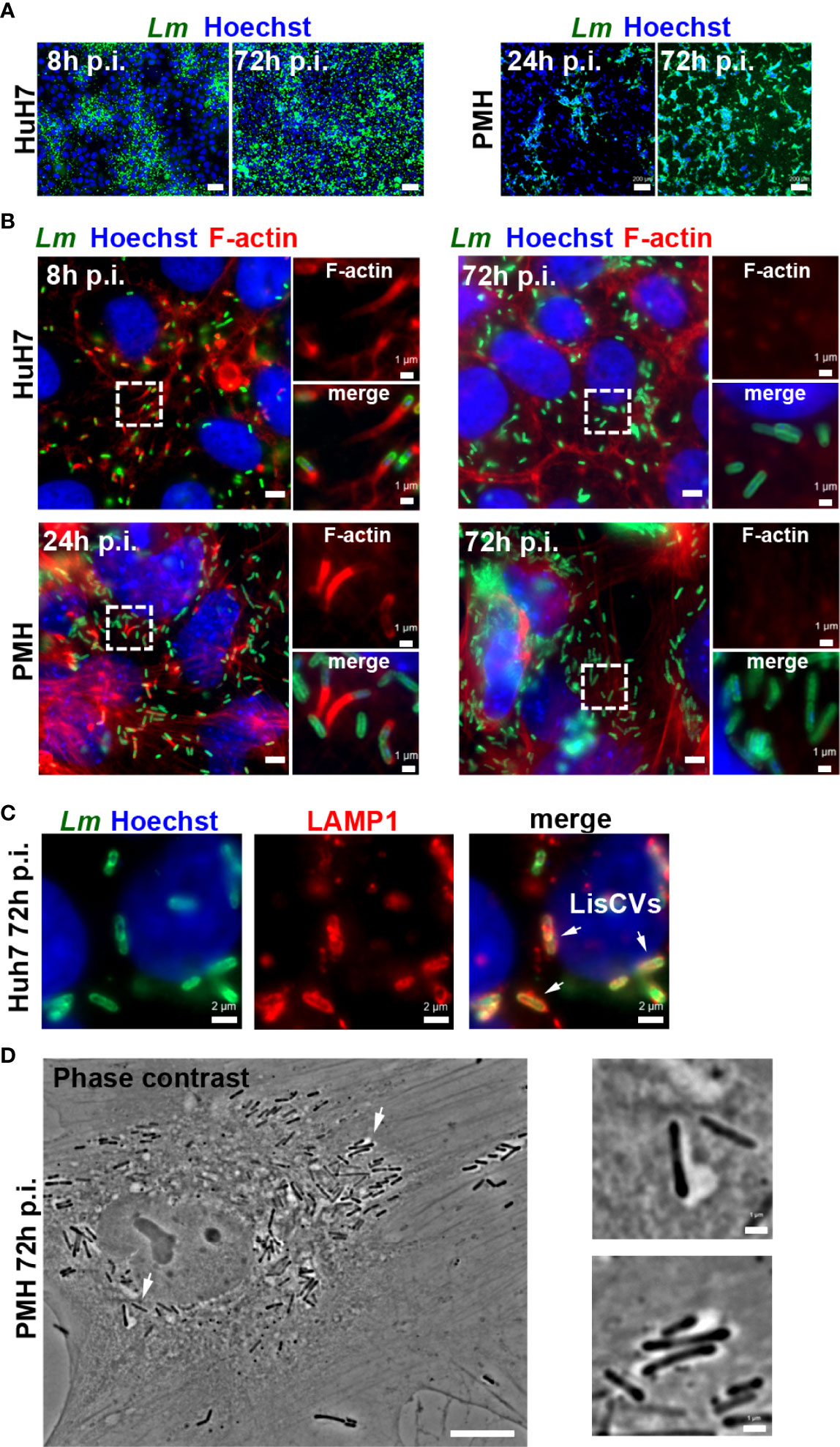
Figure 1 Optimization of hepatocyte culture systems for modeling persistent Listeria infection. Different cell seeding conditions, MOI and Listeria strains (EGDe or 10403S) were tested to obtain optimal long-term Listeria infection of HepG2 (see Supplementary Figure S1), Huh7 or PMH. Infected cells were examined at day 1 (d1) and at day 3 (d3) by immunofluorescence microscopy: representative examples under optimized conditions are shown. (A) Low magnification micrographs of Huh7 cells infected with EGDe strain (MOI=1-5) or PMH infected with 10403S strain (MOI=10) for the indicated time. Images are overlays of Listeria (green) and Hoechst (blue) signals (bars: 50 μm, Huh7, or 200 μm, PMH). (B) High magnification micrographs of infected Huh7 or PMH showing Listeria (green), F-actin (red) and Hoechst (blue) signals. Bars: 5 μm. Boxed regions enlarged on the right show F-actin (top) or merged signals (bottom), highlighting actin-positive bacteria at d1 and actin-negative bacteria at d3 (bars: 1 μm). (C) Micrographs of an infected Huh7 cell at d3, showing Listeria (green), LAMP1 (red) and Hoechst (blue) signals. Arrows indicate 3 examples of LisCVs. (D) Phase contrast image of an infected PMH at d3 (bars: 10 μm). Arrows indicate 2 examples of bacteria within vacuoles, shown at a higher magnification on the right (bars: 1 μm).
Publisher’s Note
All claims expressed in this article are solely those of the authors and do not necessarily represent those of their affiliated organizations, or those of the publisher, the editors and the reviewers. Any product that may be evaluated in this article, or claim that may be made by its manufacturer, is not guaranteed or endorsed by the publisher.
Supplementary Material
The Supplementary Material for this article can be found online at: https://www.frontiersin.org/articles/10.3389/fcimb.2022.911320/full#supplementary-material
Keywords: Listeria monocytogenes, liver, acute phase response, interferon, persistence, innate immunity, cholesterol, transcriptomics
Citation: Descoeudres N, Jouneau L, Henry C, Gorrichon K, Derré-Bobillot A, Serror P, Gillespie LL, Archambaud C, Pagliuso A and Bierne H (2022) Corrigendum: An Immunomodulatory Transcriptional Signature Associated With Persistent Listeria Infection in Hepatocytes. Front. Cell. Infect. Microbiol. 12:911320. doi: 10.3389/fcimb.2022.911320
Received: 02 April 2022; Accepted: 25 April 2022;
Published: 14 June 2022.
Edited and Reviewed by:
Changyong Cheng, Zhejiang A & F University, ChinaCopyright © 2022 Descoeudres, Jouneau, Henry, Gorrichon, Derré-Bobillot, Serror, Gillespie, Archambaud, Pagliuso and Bierne. This is an open-access article distributed under the terms of the Creative Commons Attribution License (CC BY). The use, distribution or reproduction in other forums is permitted, provided the original author(s) and the copyright owner(s) are credited and that the original publication in this journal is cited, in accordance with accepted academic practice. No use, distribution or reproduction is permitted which does not comply with these terms.
*Correspondence: Hélène Bierne, helene.bierne@inrae.fr