Micro-Nano Bioactive Glass Particles Incorporated Porous Scaffold for Promoting Osteogenesis and Angiogenesis in vitro
 Ting Tian1,2,3
 Weihan Xie2,3,4
 Wendong Gao2,3,4
 Gang Wang2,3,4
 Lei Zeng2,3,4Guohou Miao5
 Bo Lei6*Zhanyi Lin1,7*
 Xiaofeng Chen2,3,4*
- 1Guangzhou Higher Education Mega Center, School of Medicine, South China University of Technology, Guangzhou, China
- 2Department of Biomedical Engineering, School of Materials Science and Engineering, South China University of Technology, Guangzhou, China
- 3National Engineering Research Center for Tissue Restoration and Reconstruction, Guangzhou, China
- 4Key Laboratory of Biomedical Materials and Engineering, South China University of Technology, Ministry of Education, Guangzhou, China
- 5Key Laboratory of Oral Medicine, Guangzhou Institute of Oral Disease, Stomatology Hospital of Guangzhou Medical University, Guangzhou, China
- 6Instrument Analysis Center, Frontier Institute of Science and Technology, Xi’an Jiaotong University, Xi’an, China
- 7Department of Cardiology, Guangdong General Hospital, School of Medicine, South China University of Technology, Guangdong, China
A Corrigendum on
Micro-Nano Bioactive Glass Particles Incorporated Porous Scaffold for Promoting Osteogenesis and Angiogenesis In Vitro
by Tian, T, Xie, W, Gao, W, Wang, G, Zeng, L, Miao, G, Lei, B, Lin, Z and Chen, X (2019). Front. Chem. 7:186. doi: 10.3389/fchem.2019.00186
In the original article, there were errors. In the section In Vitro Cellular Evaluation of Composite Scaffold, “Cell Culture”, page 3, an acronym was not given in full at the first mention. The corrected sentence is as follows:
“The scaffold (2 mm height and 8 mm diameter) were placed into the 48-well plates, sterilized by immersing in 75% ethanol overnight and washed with phosphate-buffered saline (PBS) for three times by 30 min interval.”
Consequently, in the next section “Cell attachment”, the first sentence is corrected as follows:
“For cell attachment testing, the scaffolds were harvested at 3 days and washed with PBS for twice, fixed with 2.5% glutaraldehyde at 4°C for 4 h”
In Conclusions, page 9, there was a typo in which “was” was used instead of “were,” The corrected sentence is as follows:
“The mechanical property and pore diameter of PLGA-MNBG scaffold were significantly improved due to the incorporation of MNBG particles.”
In the next sentence, the word “cell” was used incorrectly in “the in vitro cell experiments.” The corrected sentence is as follows:
“In addition, the in vitro experiments demonstrated that PLGA-MNBG scaffolds significantly enhanced the mBMSCs attachment, proliferation and osteogenic differentiation at a low MNBG concentration.”
Figure 4, page 6, was incomplete. The corrected figure is below.
FIGURE 4
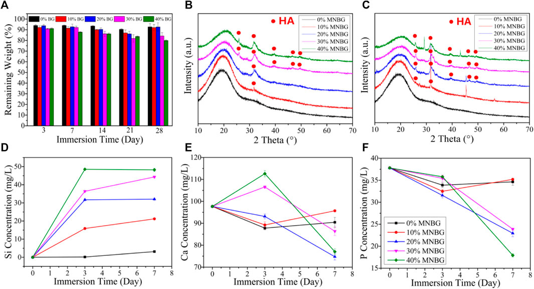
FIGURE 4. Biodegradation and apatite-forming ability of composite scaffolds in SBF. (A) Mass loss behaviors of scaffolds in SBF during 28 days immersing. (B,C) XRD patterns of scaffolds after soaking in SBF for 3 days (B) and 7 days (C). (D–F) Ions release curves of scaffolds for (D) Si; (E) Ca; (F) P after soaking in SBF for 3 and 7 days.
In Figure 5C, the fluorescent image on sample 40% MNBG was provided incorrectly. The corrected figure is above.
FIGURE 5
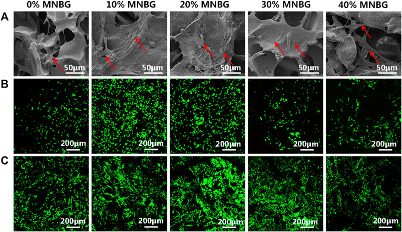
FIGURE 5. Cell attachment and cell viability evaluation on scaffolds. (A) SEM images showing the mBMSCs attachment and spreading at day 1 (Red row in SEM images). (B,C) Cell viability detected by Live-Dead assay suggesting the good cell viability on the surface of scaffolds at (B) day 1 and (C) day 5. Green represents living cells and red represents dead cells.
The authors apologize for these errors and state that this does not change the scientific conclusions of the article in any way. The original article has been updated.
Publisher’s Note
All claims expressed in this article are solely those of the authors and do not necessarily represent those of their affiliated organizations, or those of the publisher, the editors and the reviewers. Any product that may be evaluated in this article, or claim that may be made by its manufacturer, is not guaranteed or endorsed by the publisher.
Keywords: bioactive glass, micro-nano particles, nanocomposites scaffolds, bone regeneration, osteogenesis
Citation: Tian T, Xie W, Gao W, Wang G, Zeng L, Miao G, Lei B, Lin Z and Chen X (2022) Corrigendum: Micro-Nano Bioactive Glass Particles Incorporated Porous Scaffold for Promoting Osteogenesis and Angiogenesis in vitro. Front. Chem. 10:891108. doi: 10.3389/fchem.2022.891108
Received: 07 March 2022; Accepted: 22 March 2022;
Published: 26 April 2022.
Edited by:
Hani Nasser Abdelhamid, Assiut University, EgyptReviewed by:
Walid Sharmoukh, National Research Centre, EgyptFeng Wang, Shanghai Jiao Tong University, China
Copyright © 2022 Tian, Xie, Gao, Wang, Zeng, Miao, Lei, Lin and Chen. This is an open-access article distributed under the terms of the Creative Commons Attribution License (CC BY). The use, distribution or reproduction in other forums is permitted, provided the original author(s) and the copyright owner(s) are credited and that the original publication in this journal is cited, in accordance with accepted academic practice. No use, distribution or reproduction is permitted which does not comply with these terms.
*Correspondence: Bo Lei, rayboo@xjtu.edu.cn; Zhanyi Lin, linzhanyi@hotmail.com; Xiaofeng Chen, chenxf@scut.edu.cn