
94% of researchers rate our articles as excellent or good
Learn more about the work of our research integrity team to safeguard the quality of each article we publish.
Find out more
EDITORIAL article
Front. Cell Dev. Biol., 23 June 2023
Sec. Molecular and Cellular Pathology
Volume 11 - 2023 | https://doi.org/10.3389/fcell.2023.1228803
This article is part of the Research TopicUnravelling Human Placental (Patho-)Physiology at the Epigenetic and Transcriptome LevelView all 5 articles
 Athina Samara1,2,3*
 Asma Khalil4,5,6*
Editorial on the Research Topic
Unravelling human placental (patho-) physiology at the epigenetic and transcriptome level
One of the paradoxes of human development lies in its temporary dependence on an extracorporeal organ, the placenta. The placenta is this complex entity that interfaces with the mother, assuming a multitude of structural and functional roles to sustain the fetus throughout its intrauterine existence. Furthermore, the placenta is the master regulator of nutrient and oxygen supply, efficiently removing waste products from the developing fetus. It also provides critical hormonal support and sustains the developing vasculature. Disruption of any of these roles may have an adverse impact on fetal development.
The complex embryology of the placenta translates into an organ composed of distinct cell lineages arising early in development from a single fertilized oocyte. Research has shown that the normal fetal and placental development rely on epigenetic and spatiotemporal processes (Burton and Fowden, 2015). These mechanisms regulate gene expression and cell-to-cell signaling. In order to predict, prevent, diagnose, or treat pregnancy disorders, such as fetal growth restriction, preeclampsia and preterm birth, it is essential that we understand these mechanisms in humans (Bian et al., 2022; Chen et al., 2022; Liao et al., 2022; Wei et al., 2023).
In tissues with heterogeneous cell composition and multiple sources of stem cell populations such as the placenta (Figure 1A–C, E), each cell type has a distinct and dynamic epigenetic and transcriptional signature. Thus, the bulk of placental tissue analyses, including bulk sequencing methods, simply profile an amalgamation of the individual signatures of constituent cell types, masking real cell resolution and compromising interpretation. This challenge has been the topic of various studies, with increasing interest and focusing on single cell RNA sequencing to identify the transcriptome and interactions of cells in the placenta (Wächter et al., 2022; Bao et al., 2023; Campbell et al., 2023; Dong et al., 2023; Garcia-Flores et al., 2023).
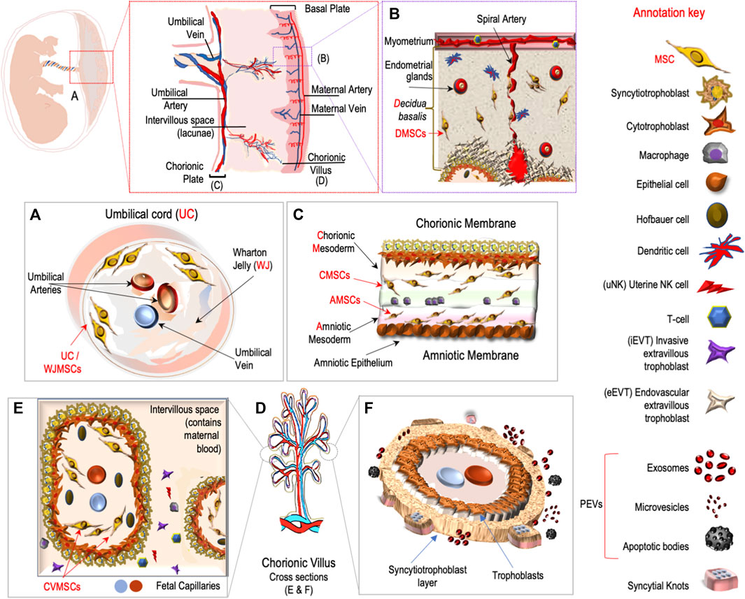
FIGURE 1. Main placental cell types, sources of mesenchymal stem cells and extracellular vesicles.
This Research Topic also aimed to comprehensively address the heterogeneous composition of the placenta in healthy and pathological pregnancies. Liu et al. presented the findings of their investigation into the spatial transcriptome of first trimester human placental villi (Figure 1D, E). They employed a modified spatial transcriptomics approach that was optimized for analyzing loosely structured tissue. One advantage of using spatial transcriptomics in addition to placental single-cell RNA sequencing (scRNA-seq) is the identification of specific cell types within the placental tissue. This enables the mapping and characterization of cell functions, interactions, and the microenvironment in a spatial context, providing valuable insights. Furthermore, the authors emphasized on the use of the modified Stereo-seq method for paraformaldehyde (PFA) fixed samples. This approach allowed them to overcome the challenges associated with collecting placental tissue under special conditions and within a limited time window, which is typically not prioritized. The results demonstrated that PFA fixation significantly enhanced tissue morphology and the specificity of RNA signals compared to using fresh villi embedded in OCT compound.
By employing the spatial transcriptome approach Liu et al. successfully identified the primary cell types present in chorionic villi, including syncytiotrophoblasts, villous cytotrophoblasts, fibroblasts, and extravillous trophoblasts (Figure 1B, E). Additionally, the less abundant cell types, such as Hofbauer cells (Figure 1E) and endothelial cells (Figure 1C), were spatially located through the deconvolution of scRNA-seq dataset. The work of Wang et al. aimed to characterize placental susceptibility to ferroptosis after SARS-CoV-2 placental infection using RNAScope in situ hybridization. The technique provided imaging background control combined with less nonspecific hybridization. They further showed that SARS-CoV-2 functioned as a potent trigger for activation of ferroptosis in human placenta, and that the initiation of the placental pathophysiological events resulting from SARS-CoV-2 intraplacental transfer was linked to the pregnancy-related sequelae.
Maternal obesity has been associated with a range of obstetrics outcomes, such as stillbirth, preeclampsia, gestational diabetes, and an increased risk of congenital heart defects in fetuses (Reynolds et al., 2013; Rubens et al., 2022). Obesity during pregnancy significantly contributes to disruptions in these critical metabolic processes, which might trigger endoplasmic reticulum (ER) stress (Burton and Yung, 2011). However, the specific mechanisms linking the obesogenic metabolic environment to adverse pregnancy outcomes remain poorly understood. Shen et al. investigated whether obesity activates the ER stress pathways, also referred to as the unfolded protein response (UPR), in the placenta and evaluated ER stress and UPR activation in the placentas of pregnancies complicated by maternal obesity. Their results revealed that in the obese placenta, p-IRE1α and XBP1s were significantly increased, whereas CHOP and nine UPR chaperone genes were upregulated. By shedding light on these mechanisms, they partially delineated the underlying processes contributing to adverse pregnancy outcomes associated with obesity.
Ortega et al. conducted a timely and comprehensive review that highlights the significance of placental-derived extracellular vesicles (PEVs). The review emphasizes the clinical relevance of EVs (Figure 1F) in various obstetric pathologies, including preeclampsia and gestational diabetes. In recent years, the investigation of changes in PEVs in different obstetric complications has emerged as a promising area of study (Couch et al., 2021). The concentration and cargo of PEVs have been implicated in the development of diverse pathologies, such as preeclampsia, gestational diabetes mellitus, fetal growth restrictions, and placental infections. These changes in PEVs provide valuable insights into the underlying mechanisms of these conditions. The review presents the latest advancements in both basic research and translational aspects regarding the role of PEVs in both normal and pathological pregnancies. The authors advocate for further investigation to explore the potential of PEVs as pathophysiological biomarkers for these diseases. By addressing the importance of PEVs and their association with obstetric complications, this review contributes to the understanding of their clinical significance.
In summary, these studies strongly advocate for the utilization of spatial, bulk, and single-cell analysis methods in future research endeavors to gain a comprehensive understanding of the role and impact of the different ‘omes’ within the placenta. By employing these integrative approaches, researchers can delve deeper into the complexities of placental pathophysiology, uncovering novel insights and potential applications for the exploration of diagnostic or prognostic biomarkers related to various pregnancy disorders. These findings highlight the significance of continued investigation in this field to advance our understanding and improve clinical management of pregnancy-related conditions.
All authors listed have made a substantial, direct, and intellectual contribution to the work and approved it for publication.
The authors declare that the research was conducted in the absence of any commercial or financial relationships that could be construed as a potential conflict of interest.
All claims expressed in this article are solely those of the authors and do not necessarily represent those of their affiliated organizations, or those of the publisher, the editors and the reviewers. Any product that may be evaluated in this article, or claim that may be made by its manufacturer, is not guaranteed or endorsed by the publisher.
Bao, S., Chen, Z., Qin, D., Xu, H., Deng, X., Zhang, R., et al. (2023). Single-cell profiling reveals mechanisms of uncontrolled inflammation and glycolysis in decidual stromal cell subtypes in recurrent miscarriage. Hum. Reprod. 38 (1), 57–74. doi:10.1093/humrep/deac240
Bian, Y., Li, J., Shen, H., Li, Y., Hou, Y., Huang, L., et al. (2022). WTAP dysregulation-mediated HMGN3-m6A modification inhibited trophoblast invasion in early-onset preeclampsia. FASEB J. 36 (12), e22617. doi:10.1096/fj.202200700RR
Burton, G. J., and Fowden, A. L. (2015). The placenta: A multifaceted, transient organ. Philos. Trans. R. Soc. Lond. B Biol. Sci. 370, 20140066. doi:10.1098/RSTB.2014.0066
Burton, G. J., and Yung, H. W. (2011). Endoplasmic reticulum stress in the pathogenesis of early-onset pre-eclampsia. Pregnancy Hypertens. 1 (1-2), 72–78. doi:10.1016/j.preghy.2010.12.002
Campbell, K. A., Colacino, J. A., Puttabyatappa, M., Dou, J. F., Elkin, E. R., Hammoud, S. S., et al. (2023). Placental cell type deconvolution reveals that cell proportions drive preeclampsia gene expression differences. Commun. Biol. 6 (1), 264. doi:10.1038/s42003-023-04623-6
Chen, F., Fei, X., Zhu, W., Zhang, Z., Shen, Y., Mao, Y., et al. (2022). Placental DNA methylation changes in gestational diabetes mellitus. Epigenetics 17 (13), 2109–2121. doi:10.1080/15592294.2022.2110193
Couch, Y., Buzàs, E. I., Vizio, D. D., Gho, Y. S., Harrison, P., Hill, A. F., et al. (2021). A brief history of nearly EV-erything - the rise and rise of extracellular vesicles. J. Extracell. vesicles 2021, e12144. doi:10.1002/JEV2.12144
Dong, L., Pang, D., Li, Y., Li, S., Wang, Y., Cui, B., et al. (2023). Effect of maternal body mass index on the transcriptomic network of human first-trimester chorionic villi. Reprod. Sci. 30 (4), 1324–1334. doi:10.1007/s43032-022-01088-6
Garcia-Flores, V., Xu, Y., Pusod, E., Romero, R., Pique-Regi, R., and Gomez-Lopez, N. (2023). Preparation of single-cell suspensions from the human placenta. Nat. Protoc. 18 (3), 732–754. doi:10.1038/s41596-022-00772-w
Liao, L., Liu, M., Gao, Y., Wei, X., Yin, Y., Gao, L., et al. (2022). The long noncoding RNA TARID regulates the CXCL3/ERK/MAPK pathway in trophoblasts and is associated with preeclampsia. Reprod. Biol. Endocrinol. 20 (1), 159. doi:10.1186/s12958-022-01036-8
Reynolds, R. M., Allan, K. M., Raja, E. A., Bhattacharya, S., Mcneill, G., Hannaford, P. C., et al. (2013). Maternal obesity during pregnancy and premature mortality from cardiovascular event in adult offspring: Follow-up of 1 323 275 person years. BMJ 347, f4539. doi:10.1136/bmj.f4539
Rubens, M., Ramamoorthy, V., Saxena, A., Mcgranaghan, P., Veledar, E., and Hernandez, A. (2022). Obstetric outcomes during delivery hospitalizations among obese pregnant women in the United States. Sci. Rep. 12, 6862. doi:10.1038/s41598-022-10786-9
Wächter, J., Shannon, M. J., and Beristain, A. G. (2022). Transcriptomic mapping of the metzincin landscape in human trophoblasts. Gene Expr. Patterns 46, 119283. doi:10.1016/j.gep.2022.119283
Keywords: placenta, scRNAseq, epigenetics, transcriptomics, spatial transcriptomics, exosomes
Citation: Samara A and Khalil A (2023) Editorial: Unravelling human placental (patho-) physiology at the epigenetic and transcriptome level. Front. Cell Dev. Biol. 11:1228803. doi: 10.3389/fcell.2023.1228803
Received: 25 May 2023; Accepted: 16 June 2023;
Published: 23 June 2023.
Edited and reviewed by:
Ramani Ramchandran, Medical College of Wisconsin, United StatesCopyright © 2023 Samara and Khalil. This is an open-access article distributed under the terms of the Creative Commons Attribution License (CC BY). The use, distribution or reproduction in other forums is permitted, provided the original author(s) and the copyright owner(s) are credited and that the original publication in this journal is cited, in accordance with accepted academic practice. No use, distribution or reproduction is permitted which does not comply with these terms.
*Correspondence: Athina Samara, YXRoaW5hLnNhbWFyYUBraS5zZQ==; Asma Khalil, YWtoYWxpbEBzZ3VsLmFjLnVr
Disclaimer: All claims expressed in this article are solely those of the authors and do not necessarily represent those of their affiliated organizations, or those of the publisher, the editors and the reviewers. Any product that may be evaluated in this article or claim that may be made by its manufacturer is not guaranteed or endorsed by the publisher.
Research integrity at Frontiers
Learn more about the work of our research integrity team to safeguard the quality of each article we publish.