
94% of researchers rate our articles as excellent or good
Learn more about the work of our research integrity team to safeguard the quality of each article we publish.
Find out more
ORIGINAL RESEARCH article
Front. Cell Dev. Biol. , 13 July 2022
Sec. Cancer Cell Biology
Volume 10 - 2022 | https://doi.org/10.3389/fcell.2022.910749
 Wanying Liao1†
 Junyu Long1†
 Yiran Li1†
 Fucun Xie1
 Ziyu Xun1
 Yanyu Wang1
 Xu Yang1
 Yunchao Wang1
 Kang Zhou2*
 Xinting Sang1*
 Haitao Zhao1*
N6-methyladenosine (m6A) and lncRNAs have been implicated in the development of colon cancer, including tumorigenesis, migration, and invasion. However, the specific effect of m6A regulators on lncRNAs is not clear, and m6A-related lncRNAs may be new prognostic biomarkers and may help direct treatment and medication. We identified 29 prognostic m6A-related lncRNAs and constructed a risk model using 12 lncRNAs. The model was an independent prognostic factor and could accurately predict the prognosis. A stable and robust nomogram that combined the model and pathologic stage was constructed. A total of 2,424 differentially expressed genes (DEGs) were identified based on the model. Functional analysis of the DEGs showed that they were associated with tumor progression, helping investigate the underlying biological functions and signaling pathways of the risk model. In addition, the low-risk group based on the risk model had more sensitivity to afatinib, metformin, and GW.441756, and patients with low risk would more likely respond to immunotherapy. Moreover, patients with higher risk were more sensitive to olaparib, bexarotene, and doxorubicin.
The incidence of colorectal cancer (CRC) is 10.0%, ranking third, and the mortality is 9.4%, ranking second among all types of cancers worldwide according to the newest published cancer statistics (Sung et al., 2021). Colon adenocarcinoma (COAD) is the most frequent subtype (>90%) of CRC (Keum and Giovannucci, 2019). Treatment options for COAD include colectomy, chemotherapy, radiation therapy, and immunotherapy-targeted therapy (Dienstmann et al., 2015). Due to widespread colonoscopy testing and improved therapeutic strategies, the incidence and mortality have been reduced (Siegel et al., 2020). However, the recurrence and metastasis rates within 5 years of treatment of COAD are as high as 30%–50%, and nearly all patients develop drug resistance (Hu et al., 2016). Therefore, reliable molecular biomarkers are urgently needed to provide therapeutic and prognostic information.
N6-methyladenosine (m6A) is a reversible process involving methylation at position N6 of adenosine. It is the most prevalent internal modification in mRNA and long noncoding RNA (lncRNA) in eukaryotes (Zaccara et al., 2019). m6A modification involves methyltransferases (writers), signal transducers (readers), and demethylases (erasers), which are important for controlling many cellular and biological processes (Zaccara et al., 2019). The regulators of m6A have been recently implicated in cancer development, including proliferation, migration, and invasion (Liu et al., 2018). For example, METTL14 is reported to suppress the tumorigenicity and migration of CRC by negatively regulating the lncRNA XIST (Yang et al., 2020). METTL3 enhances CRC progression by stabilizing HK2 and SLC2A1 (GLUT1) and activating the glycolytic pathway (Shen et al., 2020). YTHDF3 downregulates the m6A-modified lncRNA GAS5, which disrupts the interaction between the lncRNA GAS5, leading to CRC progression (Ni et al., 2019). Moreover, lncRNAs were associated with the development of colon cancer, such as regulating cell growth, cell proliferation, cell migratory and invasive abilities, cancer stem cells, and drug resistance (Lan et al., 2018; Chen and Shen, 2020). However, the specific effect of m6A regulators on lncRNAs is not clear, and m6A-related lncRNAs may be new prognostic biomarkers and help find new therapeutic drugs.
In this study, we obtained the expression data of 23 m6A genes and 14,086 lncRNAs from The Cancer Genome Atlas (TCGA) dataset. Then, lncRNAs related to m6A were screened out through Spearman’s correlation analysis. Prognostic m6A-related lncRNAs were screened and used to establish a prognostic signature. A nomogram combining the model and pathologic stage was established as a quantitative scale that could be easily used in the clinic to predict the prognosis of COAD patients. Moreover, the model was used to discover potential chemotherapeutic drugs and predict immunotherapeutic effects.
First, the sequencing data and clinical information (age, sex, pathologic stage, T stage, M stage, N stage, grade, and survival time and state) of COAD patient samples were downloaded from the TCGA database (March 2021, https://cancergenome.nih.gov/), including 398 tumor tissues and 39 adjacent normal colon tissues. A total of 379 tumor samples with complete survival information (survival time and state) were randomly grouped into a training dataset and test dataset and used in subsequent analyses. All the data we used were publicly available and had open access, so there was no need for approval from the local ethics committee. The publication guidelines of the TCGA were followed.
According to the previously published literature, we identified 23 m6A genes, including eight methyltransferases (METTL3, METTL14, METTL16, WTAP, VIRMA, ZC3H13, RBM15, and RBM15B), 13 signal transducers (YTHDC1, YTHDC2, YTHDF1, YTHDF2, YTHDF3, HNRNPC, FMR1, LRPPRC, HNRNPA2B1, IGFBP1, IGFBP2, IGFBP3, and RBMX) and two demethylases (FTO and ALKBH5). (Arguello et al., 2017; Liu et al., 2017; He et al., 2019). The expression levels of these 23 genes and all 14,086 lncRNAs were extracted from the TCGA database. As the expression data did not fit a normal distribution, Spearman’s correlation analysis was used to screen out m6A-related lncRNAs (absolute correlation coefficient > 0.4 and p value < 0.001).
Univariate Cox proportional hazards regression (PHR) analysis was implemented to identify prognostic m6A-related lncRNAs according to the survival information of the COAD samples. A p value < 0.05 was the threshold indicating that a lncRNA was associated with the prognosis. Next, prognosis-associated lncRNAs were applied to construct an m6A-related lncRNA prognostic signature (m6A-LPS) using samples from the training dataset via least absolute shrinkage and selection operator (LASSO) Cox regression analysis. LASSO Cox analysis identified lncRNAs mostly correlated with overall survival with the minimum criteria, and 10-fold cross-validation was performed to prevent overfitting. The constructed formula of the risk model was as follows:
In this formula, Coefi represents the regression coefficient, and Expi represents the expression level of each lncRNA included in the model. Then, we applied the formula to obtain the risk scores for patients in the test set. The median risk score of the training set was identified, based on which all the 379 patients were divided into high- and low-risk groups. Thereafter, the results of Kaplan–Meier survival analysis for COAD patients in the two groups and receiver operating characteristic (ROC) curve analysis of the 1-, 2-, and 3-year survival rates were used as indicators for evaluating the predictive ability and accuracy of m6A-LPS.
To evaluate the independence of m6A-LPS in predicting the overall survival (OS), univariate and multivariate Cox regression analyses were implemented. Age, sex, and pathologic stage were all considered. Two-sided p values, hazard ratios (HRs), and 95% confidence intervals (CIs) were calculated.
To establish a quantitative scale that could be easily used in the clinic to predict the prognosis, a nomogram was established. It combined two independent risk factors, m6A-LPS and pathologic stage. Calibration plots were visualized using the R package “rms”. To validate the nomogram in terms of its predictive accuracy, ROC analysis was performed.
With sex and age applied as confounding factors, we identified the differentially expressed genes (DEGs) between the two groups via the R package “limma”. The adjusted p value was calculated using the Benjamini–Hochberg method. Next, DEGs with an absolute log2-fold change (FC) > 1 and an adjusted p value of <0.05 were used in Gene Ontology (GO) and Kyoto Encyclopedia of Genes and Genomes (KEGG) pathway enrichment analyses. This process utilized the R package “clusterProfiler” to annotate the GO and KEGG functions, with an adjusted p value <0.05 and an overlapping gene number ≥3 as the thresholds for indicating that the DEGs were significantly enriched in a functional term.
To explore the potential chemotherapeutic drugs for COAD treatment, we calculated the half-maximal inhibitory concentration (IC50) of compounds obtainable in the Genomics of Drug Sensitivity in Cancer (GDSC) website for all the COAD samples using the R package “pRRophetic”. We compared the IC50 values between the low- and high-risk groups and explored the correlation between the IC50 value and each prognostic m6A-related lncRNA via linear regression analysis.
The expression levels of 47 common immune checkpoint genes in patients with low and high risk were extracted and compared, including the two commonly used therapies, which targets programmed cell death protein 1 (PD-1) and cytotoxic T-lymphocyte antigen 4 (CTLA-4). To explore the role of m6A-LPS in predicting immune treatment, we used the Tumor Immune Dysfunction and Exclusion (TIDE) algorithm (Jiang et al., 2018) and the immunophenoscore (IPS) from The Cancer Immunome Atlas (https://tcia.at/)
The whole study design is visualized in Figure 1. A total of 379 samples from COAD patients with the survival state and time information were included and randomly divided into the training and testing sets. Table 1 shows the age and sex characteristics and clinical information of the 379 patients. A total of 536 lncRNAs were identified to be m6A-related (|Spearman R|>0.4 and p < 0.001) and 29 were prognosis-related (p < 0.05, Figure 2A). Detailed information on the 29 prognosis-related lncRNAs is shown in Table 2, and their correlation with the 23 m6A genes is shown in Figure 2B. All the 29 prognosis-related lncRNAs had markedly different expression levels between the tumor and normal samples (Figure 2C).
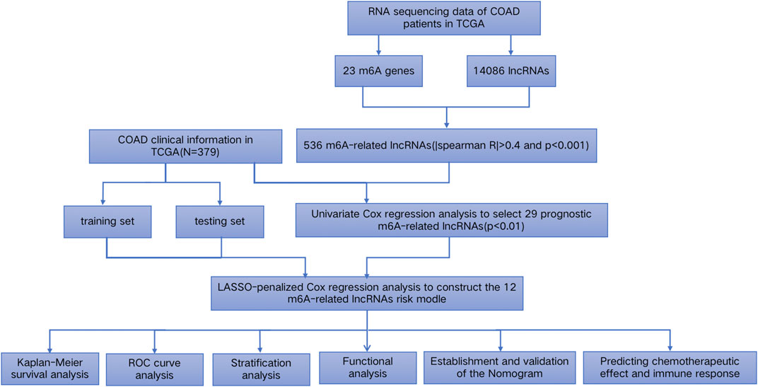
FIGURE 1. Study workflow. First, the sequencing data and clinical information of COAD patient samples were downloaded from the TCGA database. Second, the expression data of 23 m6A genes and 14,086 lncRNAs were extracted, and 536 lncRNAs related to m6A were screened out via Spearman’s correlation analysis. Third, 379 patients with complete survival information were randomly grouped into a training dataset and a test dataset. Fourth, 29 prognostic m6A-related lncRNAs were identified through the univariate Cox PHR analysis according to the survival information of the COAD samples. Then, the 29 lncRNAs were applied to construct the 12 m6A-LPS. Finally, we performed the Kaplan–Meier survival analysis, ROC curve analysis, stratification analysis, and functional analysis, established and validated the nomogram, and predicted the chemotherapeutic effect and immune response of the high- and low-risk groups.

FIGURE 2. Identification of prognostic m6A-related lncRNAs. (A) Forest plot of the results of univariate Cox PHR analysis, including the HR and p values of 29 m6A-related prognostic lncRNAs. (B) Heatmap of the correlations between m6A genes and the 29 prognostic m6A-related lncRNAs. The color represents the coefficient. p < 0.05 (*). (C) Heatmap of the expression levels of the 29 m6A-related prognostic lncRNAs in the COAD samples and adjacent normal samples. p < 0.05 (*), p < 0.01 (**), and p < 0.001 (***).
LASSO-penalized Cox regression analysis was used to build a prognostic signature based on the 29 m6A-related prognostic lncRNAs in the training set. An m6A-LPS involving 12 lncRNAs was developed (Figures 3A,B). The relationship between the 12 lncRNAs used in m6A-LPS and their related m6A genes is shown in Supplementary Figure S1. The coefficient of each lncRNA used in the m6A-LPS is shown in Table 3. Then, the patients of the entire set were divided into high- and low-risk groups according to the median risk score (Figures 3C,D). AC005288.1, ZKSCAN2-DT, AP002449.1, NKILA, AC018647.2, AL391422.4, BOLA3-AS1, AC005229.4, PCED1B-AS1, and LINC01106 were the risk factors associated with high risk. In contrast, the other two, SNHG25 and FAM160A1-DT, tended to be favorable factors in COAD patients. Kaplan–Meier survival analysis was performed and indicated that the OS (Figures 3E,F) and progression-free survival (PFS) (Supplementary Figure S2) of patients with high risk were significantly worse. The results of ROC curve analysis of the 1-, 2-, and 3-year survival rates of the training and testing sets are shown in Figure 3G (1-, 2-, and 3-year AUCs = 0.704, 0.783, and 0.823) and Figure 3H (1-, 2-, and 3-year AUCs = 0.709, 0.743, and 0.751), respectively.
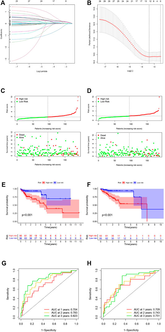
FIGURE 3. Construction and validation of the risk model. (A,B) LASSO Cox analysis identified the best parameters and optimal coefficients. (C,E,G) Analysis results of the training set. (D,F,H) Analysis results of the testing set. (C,D) Risk score distribution of m6A-LPS (panel above) and patient survival status (panel below). The green dots represent surviving patients, and the red dots represent non-surviving patients (E,F) Survival analysis revealed that the low-risk group patients had a better prognosis. (G,H) ROC curves of m6A-LPS for predicting 1-, 2-, and 3-year survival.
We investigated the differences in OS of all the COAD patients stratified by the clinicopathological variables between the two groups. Patients with high risk always had a worse OS when stratified by age, sex, and stage (Figures 4A–F). Moreover, to better explore the potential functions of m6A-LPS in COAD, 2,424 DEGs were identified (Figure 4G) and GO functional analysis and KEGG pathway analysis for biological functions were implemented (Figures 4H,I). The DEGs were enriched in some GO terms, including oxidative phosphorylation, focal adhesion, growth factor binding, and positive regulation of cell growth (Supplementary Table S1). They were also enriched in the NADH dehydrogenase activity signaling pathway (Supplementary Table S2). Functional analysis revealed that m6A-LPS is essential in the complex functions of tumors.
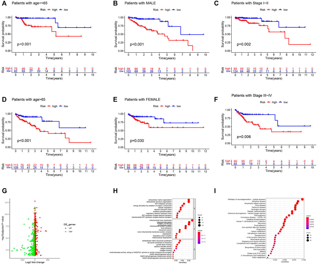
FIGURE 4. Stratification analysis and functional analysis. (A–F) Kaplan–Meier survival analysis showed that patients with high risk always had a worse OS when stratified by age (A,D), sex (B,F), and stage (C,F) in the entire dataset. (G) Volcano plot showing the DEGs. (H,I) GO (H) and KEGG pathway (I) analyses (p < 0.05) of the 2424 DEGs. The size of the circles indicates the gene count, and the color indicates the p value.
Univariate Cox regression and multivariate Cox regression analyses were utilized with the TCGA training and testing sets to detect whether m6A-LPS or clinical characteristics were significantly associated with OS and determine their independence in predicting the prognosis (Figures 5A,B). Then, the pathologic stage and m6A-LPS, two independent prognostic factors, were selected to construct a prognostic nomogram in the entire TCGA cohort (Figure 5C). Calibration plots showed that the observed 1-, 2-, and 3-year OS rates had good concordance with the predicted rates (Figures 5D–F). Then, the area under the curve (AUC) of the 1-, 2-, and 3-year survival rates of the nomogram, risk scores, and pathologic stage was assessed (Figures 5G–I), which revealed that the nomogram had the best predictive ability.
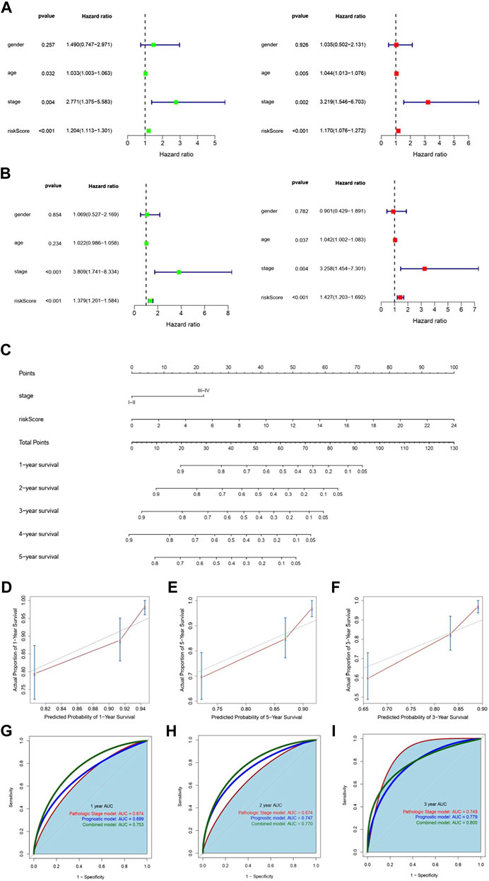
FIGURE 5. Establishment and validation of the nomogram. (A,B) Univariate and multivariate regression analyses showed that m6A-LPS and pathological stage were independent prognostic predictors in both the training set (A) and testing set (B). The left and right pictures represent the univariate analysis and multivariate analysis, respectively. (C) Nomogram combining m6A-LPS and stage. (D–F) Calibration curves for predicting 1-, 2-, and 3-year OS for patients with COAD. (D–F) Smooth ROC curves of 1-, 2-, and 3-year OS based on the pathological stage, m6A-LPS, and the nomogram.
The IC50 values of compounds available on the GDSC website were calculated to explore potential chemotherapeutic drugs for COAD treatment. Seventy-eight compounds had significantly different IC50 values, and most of them had a higher IC50 in low-risk patients, including olaparib (AZD.2281), bexarotene, and doxorubici (Supplementary Figures S3A–C), indicating that patients with higher risk were more sensitive to these drugs. In addition, afatinib (BIBW2992), metformin, and GW.441756 were more useful for low-risk patients (Supplementary Figures S3D–F). The correlation coefficients between the IC50 values of olaparib, bexarotene, doxorubicin, afatinib, metformin, and GW.441756 and each prognostic m6A-related lncRNA were calculated. Correlation coefficients with a p value <0.05 are summarized in Supplementary Table S3.
The expression levels of 47 immune checkpoint genes, including PD-1 and CTLA-4, were compared in patients with low and high risk. Thirty-seven genes with significantly different expression levels were identified and visualized (Figure 6A). All the 37 immune checkpoint genes were expressed more actively in the high-risk patients. To explore the role of m6A-LPS in predicting the immune response, the TIDE algorithm (Figures 6B–D) and the IPS from TCIA (Figures 6E–H) were used. The results showed that patients in the low-risk group may have a better response to immunotherapy and had a higher IPS to CTLA4 inhibitor therapy.
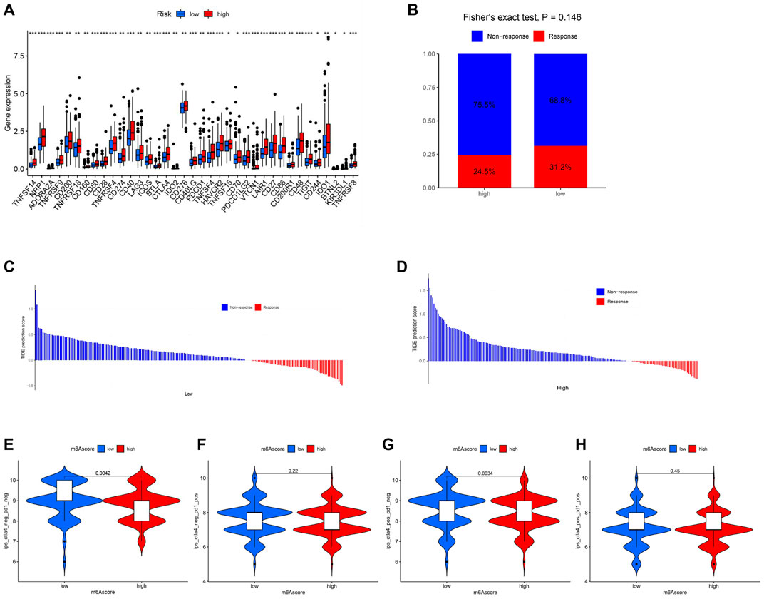
FIGURE 6. Prediction of immunotherapeutic treatment. (A) Expression of 37 immune checkpoint genes was significantly different between high- and low-risk patients. p < 0.05 (*), p < 0.01 (**), and p < 0.001 (***). (B) Box plot of the TIDE prediction scores of COAD patients. (C,D) Waterfall plot of the TIDE prediction scores of COAD patients. (E–H) Immunophenoscore of the high- and low-risk groups from TCIA.
m6A regulators were reported to be critical players in cancer tumorigenesis, progression, and metastasis by regulating lncRNAs (Chen et al., 2020; Yi et al., 2020). m6A modification of the lncRNA NEAT1-1 plays a critical role in promoting bone metastasis of prostate cancer (Wen et al., 2020). m6A modification upregulates the expression level of the lncRNA RP11, resulting in the upregulation of Zeb1, which promotes the metastasis of CRC (Wu et al., 2019). METTL3 facilitates hepatocellular carcinoma (HCC) lipogenesis and progression through the m6A modification of LINC00958 (Zuo et al., 2020). The demethylation of the lncRNA NEAT1 caused by ALKBH5, an m6A demethylase, promotes the progression of colon cancer (Guo et al., 2020a). In summary, m6A modification of lncRNAs is critical in the occurrence and progression of tumors, and m6A-related lncRNAs should be considered to discover reliable and robust molecular biomarkers for COAD.
Our study identified 29 prognosis-associated, m6A-related lncRNAs based on data from 379 COAD patients, and 12 of them were used to construct the m6A-LPS. NKILA and BOLA3-AS1 have been identified as unfavorable factors for colon cancer (Xu et al., 2021a; Guo et al., 2021; Chen et al., 2022; Qiu et al., 2022), which is consistent with the findings of this study. PCED1B-AS1 was reported to promote the proliferation of COAD by regulating the miR-633/HOXA9 axis, which was proven by in vitro and in vivo experiments (Liu et al., 2022). LINC01106 was also reported to drive the growth of CRC and lead to poor prognosis (Guo et al., 2020b). The other eight lncRNAs have not been reported in CRC, and further research and experiments on these lncRNAs might provide new insights.
According to the median risk score, the 379 COAD patients were divided into high- and low-risk groups. Kaplan–Meier survival analysis was performed and indicated that patients in the high-risk group had significantly worse OS. The results of ROC curve analysis of the 1-, 2-, and 3-year survival rates proved the high accuracy of m6A-LPS. Univariate and multivariate Cox regression analyses identified m6A-LPS and pathologic stage as independent indicators in predicting the prognosis. However, the outcomes of COAD patients at the same stage could be very different clinically. We performed the stratification analysis and found that m6A-LPS could perfectly identify the prognosis of patients in the same stage. Moreover, to establish a quantitative scale that could be easily used in the clinic, we constructed a nomogram that combined m6A-LPS and pathologic stage, two independent indicators. Calibration plots and AUC proved that the nomogram was stable and robust and had high accuracy.
To search for the underlying signaling pathways and biological processes of the lncRNAs in the model, 2,424 DEGs were identified and used for functional analysis. The DEGs were enriched in oxidative phosphorylation, focal adhesion, growth factor binding, positive regulation of cell growth, and the NADH dehydrogenase activity signaling pathway. In recent years, oxidative phosphorylation has become a novel target in cancer therapy (Ashton et al., 2018), and combination with targeting oxidative phosphorylation can improve the efficacy of radiotherapy and immunotherapy (Boreel et al., 2021). Focal adhesion plays a key role in tumor invasiveness and metastasis, drug resistance, and radioresistance (Eke and Cordes, 2015). The NADH dehydrogenase complex has been reported to be overexpressed in metastatic mouse colon tumor cells (Marquez et al., 2019). In conclusion, functional analysis provided new insights into the process and mechanism of m6A-lncRNAs in COAD.
In addition, m6A-LPS was used to investigate potential chemotherapeutic drugs and immunotherapeutic targets. The IC50 values of chemotherapeutic drugs available on the GDSC website were calculated, which indicated that the low-risk group was more sensitive to almost all the drugs, including olaparib, bexarotene, and doxorubicin, which were reported to be helpful for preventing or treating COAD (Bowen et al., 2021; de Castro e Gloria et al., 2021; Pan et al., 2021). Afatinib, metformin, and GW.441756 are more useful for low-risk patients. Afatinib, a drug mainly used for non–small-cell lung cancer (NSCLC) patients (Park et al., 2016), has a good effect in treating HER2-mutant CRC cell lines. (Kloth et al., 2016). Metformin, an antidiabetic drug, has been proven to be an effective anticancer agent for CRC (Kamarudin et al., 2019). GW441756, a TRKA kinase inhibitor being researched in vitro, has been reported to reduce the proliferation and metastasis of pancreatic cancer (Bapat et al., 2016) but has not been studied in COAD. The expression levels of PD-1 and CTLA-4, two immune checkpoint genes, were significantly different between the patients with high and low risk. Low-risk COAD patients were predicted to have a significantly higher IPS to CTLA4 target therapy. Consistently, the prediction results from TIDE showed that patients with low risk would more likely respond to immunotherapeutic therapy, although the difference was not significant.
In conclusion, we constructed a 12 m6A-related lncRNA risk model and a nomogram combining m6A-LPS and pathologic stage, which could help predict the OS and PFS of COAD patients and provide a direction on treatment and medication. In addition, we investigated the underlying biological functions and signaling pathways of m6A-LPS, which provided new insights into the process and mechanism of m6A-lncRNAs in COAD and helped identify new therapeutic targets.
In recent years, with the development of biotechnology and the emergence of public databases, bioinformation technology has played an increasingly important role. There have been several previously established lncRNA-based prognostic models for patients with COAD (Sun et al., 2020; Chen et al., 2021; Ren et al., 2021; Huang et al., 2022). This study was the first to establish an m6A-related lncRNA risk model to predict the prognosis, guide immunotherapy, and discover potential chemotherapeutic drugs. Furthermore, the specific effect of m6A regulators on lncRNAs is not clear, and m6A-related lncRNAs may be new effective prognostic biomarkers and may help direct treatment and medication. Xu et al. (2021b) identified 83 protective and 18 risk m6A-related tissue-elevated lncRNAs across all cancer types. They reported that HCC patients with low expression levels of F11-AS1 and LINC01018 and low-grade glioma and glioblastoma multiforme patients with low expression levels of MIR325HG had a worse prognosis (Xu et al., 2021b). However, Xu et al. (2021b) did not identify the specific m6A-lncRNAs associated with the prognosis of COAD patients. Moreover, this study established a nomogram, which is a quantitative scale that can be easily used in the clinic to predict the prognosis of COAD patients.
The risk model has some limitations. First, the association between lncRNAs and m6A genes was determined by a computational method, lacking experimental evidence. We found valuable published m6A and lncRNA data for normal colon tissues (Liu et al., 2020), but data from the COAD samples may be preferable. Second, further experimental and clinical evidence is needed to explore the molecular mechanisms and verify our prediction results. Nonetheless, in consideration of the limited number of samples, the model needs further validation in other cohorts.
The datasets presented in this study can be found in online repositories. The names of the repository/repositories and accession number(s) can be found in the article/Supplementary Material.
HZ, XS, and KZ led the entire project, and all authors participated in the discussion and interpretation of the data and results. WL, JL, and YL performed bioinformatics analysis of the sequencing data, wrote the manuscript, and were involved in planning the project. FX, ZX, YYW, XY, and YCW generated the figures and tables.
This work was supported by the CAMS Innovation Fund for Medical Sciences (CIFMS) (2021-I2M-1-003 and 2021-I2M-1-061 and 2018-I2M-3-001), CAMS Clinical and Translational Medicine Research Funds (2019XK320006), Beijing Natural Science Foundation (7192158), CSCO-Hengrui Cancer Research Fund (Y-HR2019-0239) and National Ten-thousand Talent Program.
The authors declare that the research was conducted in the absence of any commercial or financial relationships that could be construed as a potential conflict of interest.
All claims expressed in this article are solely those of the authors and do not necessarily represent those of their affiliated organizations, or those of the publisher, the editors, and the reviewers. Any product that may be evaluated in this article, or claim that may be made by its manufacturer, is not guaranteed or endorsed by the publisher.
The Supplementary Material for this article can be found online at: https://www.frontiersin.org/articles/10.3389/fcell.2022.910749/full#supplementary-material
Supplementary Table S1 | Results of Gene Ontology (GO) pathway enrichment analysis.
Supplementary Table S2 | Results of Kyoto Encyclopedia of Genes and Genomes (KEGG) pathway enrichment analysis.
Supplementary Table S3 | Correlation coefficients between IC50 value and each prognostic m6A-related lncRNA. p < 0.05 (*), p < 0.01 (**), and p < 0.001 (***).
Arguello, A. E., DeLiberto, A. N., and Kleiner, R. E. (2017). RNA Chemical Proteomics Reveals the N6-Methyladenosine (m6A)-Regulated Protein-RNA Interactome. J. Am. Chem. Soc. 139, 17249–17252. doi:10.1021/jacs.7b09213
Ashton, T. M., McKenna, W. G., Kunz-Schughart, L. A., and Higgins, G. S. (2018). Oxidative Phosphorylation as an Emerging Target in Cancer Therapy. Clin. Cancer Res. 24, 2482–2490. doi:10.1158/1078-0432.ccr-17-3070
Bapat, A. A., Munoz, R. M., Von Hoff, D. D., and Han, H. (2016). Blocking Nerve Growth Factor Signaling Reduces the Neural Invasion Potential of Pancreatic Cancer Cells. PLoS One 11, e0165586. doi:10.1371/journal.pone.0165586
Boreel, D. F., Span, P. N., Heskamp, S., Adema, G. J., and Bussink, J. (2021). Targeting Oxidative Phosphorylation to Increase the Efficacy of Radio- and Immune-Combination Therapy. Clin. Cancer Res. 27, 2970–2978. doi:10.1158/1078-0432.ccr-20-3913
Bowen, C. M., Walter, L., Borras, E., Wu, W., Ozcan, Z., Chang, K., et al. (2021). Combination of Sulindac and Bexarotene for Prevention of Intestinal Carcinogenesis in Familial Adenomatous Polyposis. Cancer Prev. Res. (Phila) 14, 851–862. doi:10.1158/1940-6207.capr-20-0496
Chen, S., Li, X., Guo, L., Zhang, J., Li, L., Wang, X., et al. (2021). Characterization of the m6A-Related lncRNA Signature in Predicting Prognosis and Immune Response in Patients with Colon Cancer. J. buon 26, 1931–1941.
Chen, S., and Shen, X. (2020). Long Noncoding RNAs: Functions and Mechanisms in Colon Cancer. Mol. Cancer 19, 167. doi:10.1186/s12943-020-01287-2
Chen, W., Chen, Y., Liu, L., Wu, Y., Fu, P., Cao, Y., et al. (2022). Comprehensive Analysis of Immune Infiltrates of Ferroptosis-Related Long Noncoding RNA and Prediction of Colon Cancer Patient Prognoses. J. Immunol. Res. 2022, 9480628. doi:10.1155/2022/9480628
Chen, Y., Lin, Y., Shu, Y., He, J., and Gao, W. (2020). Interaction between N6-Methyladenosine (m6A) Modification and Noncoding RNAs in Cancer. Mol. Cancer 19, 94. doi:10.1186/s12943-020-01207-4
de Castro e Gloria, H., Jesuíno Nogueira, L., Bencke Grudzinski, P., da Costa Ghignatti, P. V., Guecheva, T. N., Motta Leguisamo, N., et al. (2021). Olaparib-mediated Enhancement of 5-fluorouracil Cytotoxicity in Mismatch Repair Deficient Colorectal Cancer Cells. BMC Cancer 21, 448. doi:10.1186/s12885-021-08188-7
Dienstmann, R., Salazar, R., and Tabernero, J. (2015). Personalizing Colon Cancer Adjuvant Therapy: Selecting Optimal Treatments for Individual Patients. Jco 33, 1787–1796. doi:10.1200/jco.2014.60.0213
Eke, I., and Cordes, N. (2015). Focal Adhesion Signaling and Therapy Resistance in Cancer. Seminars Cancer Biol. 31, 65–75. doi:10.1016/j.semcancer.2014.07.009
Guo, J.-N., Xia, T.-Y., Deng, S.-H., Xue, W.-N., Cui, B.-B., and Liu, Y.-L. (2021). Prognostic Immunity and Therapeutic Sensitivity Analyses Based on Differential Genomic Instability-Associated LncRNAs in Left- and Right-Sided Colon Adenocarcinoma. Front. Mol. Biosci. 8, 668888. doi:10.3389/fmolb.2021.668888
Guo, K., Gong, W., Wang, Q., Gu, G., Zheng, T., Li, Y., et al. (2020). LINC01106 Drives Colorectal Cancer Growth and Stemness through a Positive Feedback Loop to Regulate the Gli Family Factors. Cell Death Dis. 11, 869. doi:10.1038/s41419-020-03026-3
Guo, T., Liu, D. F., Peng, S. H., and Xu, A. M. (2020). ALKBH5 Promotes Colon Cancer Progression by Decreasing Methylation of the lncRNA NEAT1. Am. J. Transl. Res. 12, 4542–4549.
He, L., Li, H., Wu, A., Peng, Y., Shu, G., and Yin, G. (2019). Functions of N6-Methyladenosine and its Role in Cancer. Mol. Cancer 18, 176. doi:10.1186/s12943-019-1109-9
Hu, T., Li, Z., Gao, C.-Y., and Cho, C. H. (2016). Mechanisms of Drug Resistance in Colon Cancer and its Therapeutic Strategies. Wjg 22, 6876–6889. doi:10.3748/wjg.v22.i30.6876
Huang, Y., Huang, H., Wang, Y., Liu, H., and Huang, Y. (2022). 5mC-Related lncRNAs as Potential Prognostic Biomarkers in Colon Adenocarcinoma. Biol. (Basel) 11, 231. doi:10.3390/biology11020231
Jiang, P., Gu, S., Pan, D., Fu, J., Sahu, A., Hu, X., et al. (2018). Signatures of T Cell Dysfunction and Exclusion Predict Cancer Immunotherapy Response. Nat. Med. 24, 1550–1558. doi:10.1038/s41591-018-0136-1
Kamarudin, M. N. A., Sarker, M. M. R., Zhou, J.-R., and Parhar, I. (2019). Metformin in Colorectal Cancer: Molecular Mechanism, Preclinical and Clinical Aspects. J. Exp. Clin. Cancer Res. 38, 491. doi:10.1186/s13046-019-1495-2
Keum, N., and Giovannucci, E. (2019). Global Burden of Colorectal Cancer: Emerging Trends, Risk Factors and Prevention Strategies. Nat. Rev. Gastroenterol. Hepatol. 16, 713–732. doi:10.1038/s41575-019-0189-8
Kloth, M., Ruesseler, V., Engel, C., Koenig, K., Peifer, M., Mariotti, E., et al. (2016). Activating ERBB2/HER2 Mutations Indicate Susceptibility to Pan-HER Inhibitors in Lynch and Lynch-like Colorectal Cancer. Gut 65, 1296–1305. doi:10.1136/gutjnl-2014-309026
Lan, Y., Xiao, X., He, Z., Luo, Y., Wu, C., Li, L., et al. (2018). Long Noncoding RNA OCC-1 Suppresses Cell Growth through Destabilizing HuR Protein in Colorectal Cancer. Nucleic Acids Res. 46, 5809–5821. doi:10.1093/nar/gky214
Liu, J. e., Li, K., Cai, J., Zhang, M., Zhang, X., Xiong, X., et al. (2020). Landscape and Regulation of m6A and m6Am Methylome across Human and Mouse Tissues. Mol. Cell 77, 426–440. e426. doi:10.1016/j.molcel.2019.09.032
Liu, J., Qian, J., Mo, Q., Tang, L., and Xu, Q. (2022). Long Non-coding RNA PCED1B-AS1 Promotes the Proliferation of Colorectal Adenocarcinoma through Regulating the miR-633/HOXA9 axis. Bioengineered 13, 5407–5420. doi:10.1080/21655979.2022.2037225
Liu, N., Zhou, K. I., Parisien, M., Dai, Q., Diatchenko, L., and Pan, T. (2017). N 6-methyladenosine Alters RNA Structure to Regulate Binding of a Low-Complexity Protein. Nucleic Acids Res. 45, 6051–6063. doi:10.1093/nar/gkx141
Liu, Z.-X., Li, L.-M., Sun, H.-L., and Liu, S.-M. (2018). Link Between m6A Modification and Cancers. Front. Bioeng. Biotechnol. 6, 89. doi:10.3389/fbioe.2018.00089
Marquez, J., Kratchmarova, I., Akimov, V., Unda, F., Ibarretxe, G., Clerigué, A. S., et al. (2019). NADH Dehydrogenase Complex I Is Overexpressed in Incipient Metastatic Murine Colon Cancer Cells. Oncol. Rep. 41, 742–752. doi:10.3892/or.2018.6892
Ni, W., Yao, S., Zhou, Y., Liu, Y., Huang, P., Zhou, A., et al. (2019). Long Noncoding RNA GAS5 Inhibits Progression of Colorectal Cancer by Interacting with and Triggering YAP Phosphorylation and Degradation and Is Negatively Regulated by the m6A Reader YTHDF3. Mol. Cancer 18, 143. doi:10.1186/s12943-019-1079-y
Pan, D. C., Krishnan, V., Salinas, A. K., Kim, J., Sun, T., Ravid, S., et al. (2021). Hyaluronic Acid-Doxorubicin Nanoparticles for Targeted Treatment of Colorectal Cancer. Bioeng. Transl. Med. 6, e10166. doi:10.1002/btm2.10166
Park, K., Tan, E.-H., O'Byrne, K., Zhang, L., Boyer, M., Mok, T., et al. (2016). Afatinib versus Gefitinib as First-Line Treatment of Patients with EGFR Mutation-Positive Non-small-cell Lung Cancer (LUX-Lung 7): a Phase 2B, Open-Label, Randomised Controlled Trial. Lancet Oncol. 17, 577–589. doi:10.1016/s1470-2045(16)30033-x
Qiu, Y., Li, H., Zhang, Q., Qiao, X., and Wu, J. (2022). Ferroptosis-Related Long Noncoding RNAs as Prognostic Marker for Colon Adenocarcinoma. Appl. Bionics Biomech. 2022, 5220368. doi:10.1155/2022/5220368
Ren, Z., Wang, Z., Gu, D., Ma, H., Zhu, Y., Cai, M., et al. (2021). Genome Instability and Long Noncoding RNA Reveal Biomarkers for Immunotherapy and Prognosis and Novel Competing Endogenous RNA Mechanism in Colon Adenocarcinoma. Front. Cell Dev. Biol. 9, 740455. doi:10.3389/fcell.2021.740455
Shen, C., Xuan, B., Yan, T., Ma, Y., Xu, P., Tian, X., et al. (2020). m6A-dependent Glycolysis Enhances Colorectal Cancer Progression. Mol. Cancer 19, 72. doi:10.1186/s12943-020-01190-w
Siegel, R. L., Miller, K. D., Goding Sauer, A., Fedewa, S. A., Butterly, L. F., Anderson, J. C., et al. (2020). Colorectal Cancer Statistics, 2020. CA A Cancer J. Clin. 70, 145–164. doi:10.3322/caac.21601
Sun, Y.-L., Zhang, Y., Guo, Y.-C., Yang, Z.-H., and Xu, Y.-C. (2020). A Prognostic Model Based on the Immune-Related Genes in Colon Adenocarcinoma. Int. J. Med. Sci. 17, 1879–1896. doi:10.7150/ijms.45813
Sung, H., Ferlay, J., Siegel, R. L., Laversanne, M., Soerjomataram, I., Jemal, A., et al. (2021). Global Cancer Statistics 2020: GLOBOCAN Estimates of Incidence and Mortality Worldwide for 36 Cancers in 185 Countries. CA A Cancer J. Clin. 71, 209–249. doi:10.3322/caac.21660
Wen, S., Wei, Y., Zen, C., Xiong, W., Niu, Y., and Zhao, Y. (2020). Long Non-coding RNA NEAT1 Promotes Bone Metastasis of Prostate Cancer through N6-Methyladenosine. Mol. Cancer 19, 171. doi:10.1186/s12943-020-01293-4
Wu, Y., Yang, X., Chen, Z., Tian, L., Jiang, G., Chen, F., et al. (2019). m6A-induced lncRNA RP11 Triggers the Dissemination of Colorectal Cancer Cells via Upregulation of Zeb1. Mol. Cancer 18, 87. doi:10.1186/s12943-019-1014-2
Xu, G., Yang, M., Wang, Q., Zhao, L., Zhu, S., Zhu, L., et al. (2021a). A Novel Prognostic Prediction Model for Colorectal Cancer Based on Nine Autophagy-Related Long Noncoding RNAs. Front. Oncol. 11, 613949. doi:10.3389/fonc.2021.613949
Xu, K., Cai, Y., Zhang, M., Zou, H., Chang, Z., Li, D., et al. (2021b). Pan-cancer Characterization of Expression and Clinical Relevance of m6A-Related Tissue-Elevated Long Non-coding RNAs. Mol. Cancer 20, 31. doi:10.1186/s12943-021-01324-8
Yang, X., Zhang, S., He, C., Xue, P., Zhang, L., He, Z., et al. (2020). METTL14 Suppresses Proliferation and Metastasis of Colorectal Cancer by Down-Regulating Oncogenic Long Non-coding RNA XIST. Mol. Cancer 19, 46. doi:10.1186/s12943-020-1146-4
Yi, Y.-C., Chen, X.-Y., Zhang, J., and Zhu, J.-S. (2020). Novel Insights into the Interplay between m6A Modification and Noncoding RNAs in Cancer. Mol. Cancer 19, 121. doi:10.1186/s12943-020-01233-2
Zaccara, S., Ries, R. J., and Jaffrey, S. R. (2019). Reading, Writing and Erasing mRNA Methylation. Nat. Rev. Mol. Cell Biol. 20, 608–624. doi:10.1038/s41580-019-0168-5
Keywords: colon adenocarcinoma, N6-methyladenosine, long noncoding RNA, TCGA, prognostic signature
Citation: Liao W, Long J, Li Y, Xie F, Xun Z, Wang Y, Yang X, Wang Y, Zhou K, Sang X and Zhao H (2022) Identification of an m6A-Related Long Noncoding RNA Risk Model for Predicting Prognosis and Directing Treatments in Patients With Colon Adenocarcinoma. Front. Cell Dev. Biol. 10:910749. doi: 10.3389/fcell.2022.910749
Received: 01 April 2022; Accepted: 26 May 2022;
Published: 13 July 2022.
Edited by:
Emanuela Grassilli, University of Milano Bicocca, ItalyReviewed by:
Zishan Wang, Icahn School of Medicine at Mount Sinai, United StatesCopyright © 2022 Liao, Long, Li, Xie, Xun, Wang, Yang, Wang, Zhou, Sang and Zhao. This is an open-access article distributed under the terms of the Creative Commons Attribution License (CC BY). The use, distribution or reproduction in other forums is permitted, provided the original author(s) and the copyright owner(s) are credited and that the original publication in this journal is cited, in accordance with accepted academic practice. No use, distribution or reproduction is permitted which does not comply with these terms.
*Correspondence: Kang Zhou, emhvdWthbmdwdW1jaEBob3RtYWlsLmNvbQ==; Xinting Sang, U2FuZ1hUQHB1bWNoLmNu; Haitao Zhao, Wmhhb0hUQHB1bWNoLmNu
†These authors have contributed equally to this work
Disclaimer: All claims expressed in this article are solely those of the authors and do not necessarily represent those of their affiliated organizations, or those of the publisher, the editors and the reviewers. Any product that may be evaluated in this article or claim that may be made by its manufacturer is not guaranteed or endorsed by the publisher.
Research integrity at Frontiers

Learn more about the work of our research integrity team to safeguard the quality of each article we publish.