- 1Center for Precision Medicine, School of Medicine and School of Biomedical Sciences, Huaqiao University, Xiamen, China
- 2College of Materials Science and Engineering, Huaqiao University, Xiamen, China
- 3Shenzhen Key Laboratory of Marine Bioresource and Eco- Environmental Science, Shenzhen Engineering Laboratory for Marine Algal Biotechnology, Guangdong Provincial Key Laboratory for Plant Epigenetics, College of Life Sciences and Oceanography, Shenzhen University, Shenzhen, China
- 4Genergy Bio-Technology (Shanghai) Co., Ltd, Shanghai, China
- 5Department of Cell and Developmental Biology, Cornell University Weill Medical College, New York, NY, United States
- 6Department of Radiation Oncology, First Hospital of Quanzhou, Fujian Medical University, Quanzhou, China
- 7Department of Clinical Laboratory, First Hospital of Quanzhou, Fujian Medical University, Quanzhou, China
- 8Neurochemical Biology and Genetics Laboratory, Department of Physiology, Faculty of Life Sciences, Government College University, Faisalabad, Pakistan
- 9Department of Neurology, School of Medicine, Zhongshan Hospital, Xiamen University, Xiamen, China
- 10United Diagnostic and Research Center for Clinical Genetics, School of Medicine and School of Public Health, Women and Children’s Hospital, Xiamen University, Xiamen, China
Trisomy 18, commonly known as Edwards syndrome, is the second most common autosomal trisomy among live born neonates. Multiple tissues including cardiac, abdominal, and nervous systems are affected by an extra chromosome 18. To delineate the complexity of anomalies of trisomy 18, we analyzed cultured amniotic fluid cells from two euploid and three trisomy 18 samples using single-cell transcriptomics. We identified 6 cell groups, which function in development of major tissues such as kidney, vasculature and smooth muscle, and display significant alterations in gene expression as detected by single-cell RNA-sequencing. Moreover, we demonstrated significant gene expression changes in previously proposed trisomy 18 critical regions, and identified three new regions such as 18p11.32, 18q11 and 18q21.32, which are likely associated with trisomy 18 phenotypes. Our results indicate complexity of trisomy 18 at the gene expression level and reveal genetic reasoning of diverse phenotypes in trisomy 18 patients.
Introduction
Trisomy 18, also known as Edwards syndrome (Edwards et al., 1960), is characterized by the presence of an extra copy of chromosome 18, and is the most common form of autosomal trisomy in live births beside of Down syndrome (trisomy 21) (Carey, 2005; Savva et al., 2010; Cavadino and Morris, 2017). The incidence of full trisomy 18 is about 94%, compared to all types of trisomy 18 (Cereda and Carey, 2012; Mirmohammadsadeghi et al., 2017). Trisomy 18 is a severe birth defect, 80% of all trisomy 18 pregnancies results in miscarriage, almost half of trisomy 18 infants dies within the first week of life, and the majority of the remaining dies in the next 12 months (Batees and Altirkawi, 2014). Case reports have shown that craniofacial, musculoskeletal, cardiac, abdominal, and nervous systems are most likely to be affected by trisomy 18, even though many organs and tissues display defects (Cereda and Carey, 2012; Nagamuthu and Neelaveni, 2014; Roberts et al., 2016; Turbiville et al., 2017; Fan et al., 2018). Risk factors for the disease include a positive family history in close relatives and the increasing maternal age (Ginsberg et al., 1968; Calderone et al., 1983; Mirmohammadsadeghi et al., 2017). The prognosis for this genetic defect is poorly understood due to multiple system involvement (Norwitz and Levy, 2013; Imataka et al., 2016). Restriction fragment length polymorphisms (RFLP) and short-sequence repeats (SSR) have been exercised to determine the parental origin of the extra chromosome 18 (Kondoh et al., 1988; Kupke and Müller, 1989; Nöthen et al., 1993; Ya-gang et al., 1993).
The cause of an extra chromosome 18 is unclear. Studies have shown that nondisjunction predominating at the maternal meiosis I in acroxentric chromosomes results in the addition of an extra chromosome (Eggermann et al., 1996; Bugge et al., 1998). Distal q11.2 at the long arm of chromosome 18 has been proposed as a critical region for the trisomy 18 phenotype, but some controversial views have been reported (Wilson, 1993; Boghosian-Sell et al., 1994). The other two critical regions, one within 18q12.1-18q21.2 and one within 18q22.3-18qter, have been proposed (Boghosian-Sell et al., 1994; Koide et al., 2011; Roberts et al., 2016). Due to limited samples, pathogenesis of trisomy 18 is poorly studied (Tobin et al., 2019). Because amniocytes are derived from embryonic and extra-embryonic tissues and originate from all three germ layers, amniotic fluid cells are becoming an efficient source of samples for the study of genetic diseases (Underwood et al., 2005; Montemurro et al., 2008). A transcriptomic profile of induced pluripotent stem cells from human amniotic fluid cells with trisomy 18 has shown several gene clusters associated with clinical manifestations of trisomy 18 (FitzPatrick et al., 2002; Li et al., 2017; Xing et al., 2018). However, the extremely high mortality rate and complexity of phenotypes in trisomy 18 have hindered genetic and molecular understanding of its etiology (Babu and Verma, 1986; Rasmussen et al., 2003; Cereda and Carey, 2012; Cavadino and Morris, 2017).
The advance of single-cell sequencing technology has allowed comprehensive analyses of gene expression profiles of tens of thousands of single cells at once (Grun and van Oudenaarden, 2015; Wen and Tang, 2016; Potter, 2018). Single-cell sequencing has been used to highlight gene expression in 1 cell resolution, and to reveal complex and diverse cell types in a tissue and organ (Kim et al., 2018; Park et al., 2018; Raj et al., 2018; Suijuan et al., 2018). Thus, it has been expeditiously applied in understanding heterogeneity of organ formation, and complexity of disease conditions (Puram et al., 2017; Young et al., 2018; Zeng et al., 2018; Zhao et al., 2018; Velmeshev et al., 2019; Yu et al., 2021).
In this study, to better understand genetic variations of the trisomy 18 phenotype at 1 cell resolution, we generate gene expression profiles of cultured amniotic fluid cells from euploid and trisomy 18 fetuses using 10X Genomics (Zheng et al., 2017). We show that an extra chromosome 18 has a broad and distinct impact on expression of genes involved in the development of multiple systems such as vascular, renal and nervous systems. We confirm that up-regulation of genes in previously proposed two critical regions (18q12.1-22.1, 18q22.3-18qter) may contribute to the trisomy 18 phenotype. Moreover, we identify three new regions such as 18p11.32, 18q11 and 18q21.32, which are likely associated with trisomy 18 defects. Our study demonstrates complexity of trisomy 18 at the gene expression level, and highlights alterations of multiple genes that may contribute to trisomy 18 phenotypes.
Results
Single-Cell RNA Sequencing Profiles of Cultured Trisomy 18 Amniotic Fluid Cells
Trisomy 18 is a hereditary disorder with an extra copy of chromosome 18, which may cause changes in expression levels of many genes, and in turn affects multiple organs (Antonarakis et al., 2004; Antonarakis, 2017). To understand complexity of such anomalies, we analyzed single-cell transcriptomics of cultured amniotic fluid cells associated with euploid and trisomy 18 fetuses using 10X Genomics. This approach allowed bypass of direct fetal tissue collection and facilitated deciphering diversity in amniotic fluid cells from trisomy 18 fetuses, which may reflect complexity of this disorder.
Human amniotic fluid cells were collected from amniocentesis screen for hereditary disorders using the karyotype analysis. Based on karyotypes, we selected amniotic fluid samples from two euploid and three trisomy 18 fetuses at the second trimester and cultured them to gather enough cells for single cell transcriptomics analyses (Supplementary Figures S1A–C and Supplementary Table S1). We sequenced and analyzed 56,517 cells, in which 18,281 cells were from euploid samples and 38,236 cells from trisomy 18 (Supplementary Table S1). We further grouped these cells into 13 transcriptionally distinct clusters using non-linear dimensionality reduction with spectral t-distributed stochastic neighbor embedding (tSNE) (Supplementary Figure S1D). In details, two euploid samples were grouped into 12 clusters, while three trisomy 18 samples were grouped into 11, 9 and 11 clusters, respectively (Supplementary Figures S1E–H). These results indicate that amniotic fluid cells from euploid and trisomy 18 fetuses display a diverse transcriptional identity.
To further classify major cellular functions, we annotated genes from the 13 clusters according to their major functions using gene ontology (GO) analyses (Supplementary Figure S2A, Supplementary Table S2). We identified 6 functional categories, and subsequently classified two euploid samples into 6 sub-clusters, which play a role in kidney function, vasculature development, smooth muscle cell proliferation, protein synthesis, nuclear division and metabolic process (Figures 1A,B). Trisomy 18 samples were grouped into similar 6 functional sub-clusters, with different cell numbers in each sub-cluster. For example, compared to euploid individuals, trisomy 18 sample-1 (T18-1) displayed very few cells with the function in nuclear division and protein synthesis, and T18-2 and T18-3 had fewer cells in metabolic process (Figures 1B,C and Supplementary Figures S2C,D). These data suggests that trisomy 18 amniotic fluid cells display similar functional groups but altered cell components in each group, compared to those in euploid ones.
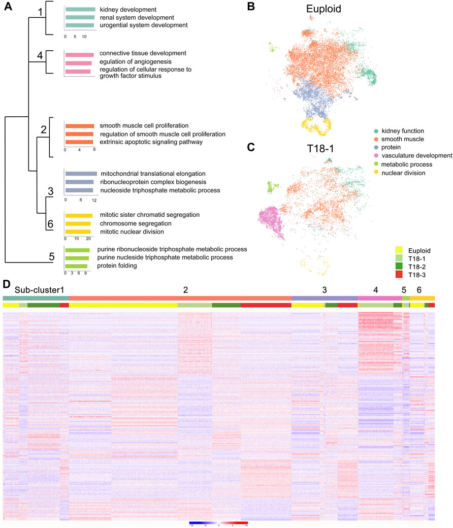
FIGURE 1. Functional grouping of amniotic fluid cells from euploid and trisomy 18 (T18) samples. (A) Different functional sub-clusters were annotated according to expression of significantly altered genes for two euploid samples. Gene ontology (GO) analyses of differentially expressed genes in three trisomy 18 samples were compared with two euploid samples. (B,C) t-distributed Stochastic Neighbor Embedding (t-SNE) projection of cells and typical gene expression in euploid and T18-1 cells. (D) Heatmap of differentially expressed genes in two euploid and three trisomy 18 samples.
To reveal gene expression patterns in 6 sub-clusters, we randomly selected 2,000 cells per sub-clusters, and top 100 differentially expressed genes in each sub-cluster by using a random forest classification. Compared to euploid samples, genes in each trisomy 18 sample displayed either up- or down-regulated expression in each functional sub-clusters (Figure 1D). For example, the sub-cluster 4 was functionally grouped as vasculature development, and genes in T18-1 and T18-2 displayed higher expression in this sub-cluster. Moreover, in each sub-cluster, genes in T18-3 were also noticed with a differential expression compared to those in T18-1 and T18-2. For instance, compared to euploid samples, genes grouped in the sub-cluster 1 showed up-regulation in T18-1, -2 and -3, but the up-regulated genes were distinct in the T18-3 sample. These results suggest that an extra copy of chromosome 18 causes altered gene expression in human amniotic fluid cells, and this alteration shows variations in each sample.
Differentially Expressed Genes in Trisomy 18 Amniotic Fluid Cells
Studies have shown that multiple organs are affected in trisomy 18 (Cereda and Carey, 2012). We next wanted to identify major genes and pathways that might be altered in each functional sub-cluster at a single cell level. According to numbers of cells, detected genes (p < 0.01), and unique molecular identifiers (UMIs) in every sub-clusters, we identified 2,523 specific genes in 6 sub-clusters (Figure 2A and Supplementary Table S3). Representative genes in each sub-clusters were identified, for example STAT1, DLG1, and EDN1 in the sub-cluster-1 of kidney development, IGFBP2 and PDGFB in the sub-cluster-2 of smooth muscle cell proliferation, VEGFA, VEGFC, FLT1, and ZNF703 in the sub-cluster-4 of regulation of vascularization, and CDC20, PRC1, NUSAP1, and CCNB1 in the sub-cluster-6 of mitotic nuclear division (Figure 2B).
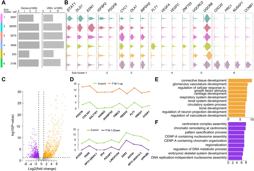
FIGURE 2. Altered genes in trisomy 18 amniotic fluid cells. (A) Dendrogram of 6 sub-clusters, cell number per sub-cluster, number of genes in each sub-cluster (p < 0.01), and unique molecular identifiers (UMIs). (B) Violin plots of representative genes identified in 6 sub-clusters. (C) Volcano plot of differentially expressed genes by comparing the T18-1 with euploid samples. Up-regulated genes: orange dots; down-regulated genes: purple dots. (D) Top 10 (−log2fold change > 1 and FDR corrected p ≤ 0.05) up-regulated genes (orange), down-regulated genes (purple) in the T18-1 sample compared with two euploid samples (green). (E) Gene ontology (GO) analysis of up-regulated genes in T18-1 cells compared with two euploid samples. (F) GO analysis of down-regulated genes in 18T-1 cells compared with two euploid samples.
We then compared differentially expressed genes (p ≤ 0.01) between every trisomy 18 sample and two euploid samples (Figure 2C and Supplementary Figure S3). In the T18-1 sample, a majority of differentially expressed genes was up-regulated (Figure 2C). Among all differentially expressed genes (−log2fold change > 1 and FDR corrected p ≤ 0.05), top 10 up-regulated and down-regulated genes in T18-1 cells were listed (Figure 2D and Supplementary Table S4). Moreover, we performed GO analyses for differentially expressed genes in T18-1 cells. GO terms for up-regulated genes were enriched in regulation of glomerulus vasculature development, circulatory system development, and neuron projection development (Figure 2E). GO terms for down-regulated genes were enriched in regulation of centromere complex assembly, pattern specification, and DNA metabolic process (Figure 2F and Supplementary Tables S5, S6). These analyses suggest that an extra chromosome 18 alters expression of genes that are associated with major organ developmental processes and fundamental cellular functions.
Furthermore, we analyzed and compared differentially expressed genes in T18-2 and T18-3 samples with two euploid samples (Supplementary Figures S3A,B and Supplementary Tables S7, S8). Relative expression levels of top 10 up-regulated and down-regulated genes from T18-2 and T18-3 samples were listed (Supplementary Figures S3C,D). Interestingly, POSTN, COL3A1, FLT1, ADAMTS2 were up-regulated in both T18-1 and T18-2 cells, while UPK3BL was down-regulated in both T18-1 and T18-3 cells (Figure 2 and Supplementary Figures S3C,D).
Finally, the GO term of renal system development was enriched in up-regulated genes from all three trisomy 18 samples (Supplementary Tables S9, S11), while neuron fate commitment was enriched for down-regulated genes in 18T-2 and 18T-3 (Supplementary Tables S10, S12). In addition, among up-regulated genes, the GO term of stem cell development was enriched in T18-3 cells. And among down-regulated genes, GO terms of neuron fate commitment, negative regulation of oligodendrocyte differentiation, peripheral nervous system neuron development and spinal cord motor neuron cell fate specification were enriched in the T18-2 sample, and they were enriched in kidney development and embryonic digestive tract development in the T18-3 sample (Supplementary Figures S3E,F). These results further indicate that while the extra chromosome 18 has a broad impact on development of renal and nervous systems, it also preferentially affects different genes associated with either stem cells, neurons, or digestive track cells in each trisomy 18 sample.
Trisomy 18 Alters Expression of Genes Located on all Chromosomes
We further investigated the effect of an extra copy of chromosome 18 on expression of genes located on all chromosomes. We merged expression levels of all genes detected by single-cell sequencing on each chromosome, and compared their differential expression in euploid and trisomy 18 samples using Deseq2. Box plots showed increased expression of genes located on chromosome 18 in trisomy 18 samples (Figure 3A). Moreover, gene expression levels (−log10P-value) in each chromosome also showed an up-regulated gene expression in trisomy 18 samples (Figure 3B). These data indicates that an extra chromosome 18 indeed causes up-regulated expression of genes located on chromosome 18.
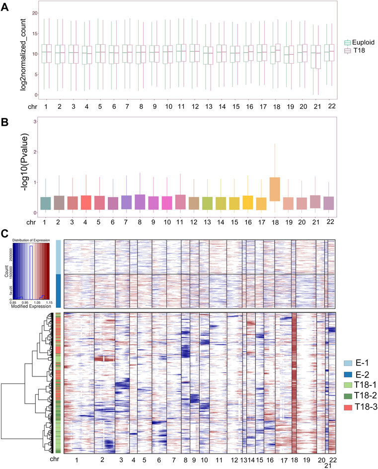
FIGURE 3. Trisomy 18 alters gene expression on chromosomes. (A) Merged expression levels of genes, showing log2 euploidized counts, on each chromosome. (B) Up-regulated gene expression (−log10p-value) on chromosome 18 from trisomy 18 samples. (C) Heatmap of large-scale chromosomal copy-number variations (CNVs). Euploid cells served as reference cells, and trisomy 18 samples (T18-1, T18-2 and T 18-3) as observed cells.
We next analyzed single-cell transcriptomics data including gene numbers and expression levels, based on randomly selected 2,000 cells in each euploid (E-1 and E2) and trisomy 18 samples. We inferred large-scale chromosomal copy-number variations (CNVs) in every single cell that is based on averaged expression profiles across chromosome intervals (Patel et al., 2014; Muller et al., 2016; Filbin et al., 2018). CNV analyses and inference of haplotypes uncovered distinct genetic variations in trisomy 18 samples, and CNVs in each trisomy 18 samples showed distinct alterations (Figure 3C, Supplementary Figure S4). As a consequence of the third chromosome 18, CNVs were greatly increased in trisomy 18 samples (Figure 3C). In addition, CNVs from chromosome 1, 19 and 20 showed an increase, and those from chromosome 3, 8, 9, 10, 15 and 21 displayed a decrease in trisomy 18 samples (Figure 3C and Supplementary Figures S4B–D). These results suggest that the third chromosome 18 affects the expression of genes on all chromosomes, and causes altered copy numbers of genes on them.
Gene Expression Patterns in Proposed Trisomy 18 Critical Regions
Previous studies proposed a few chromosomal regions as critical regions for trisomy 18 phenotypes, such as 18q12.1-21.2 and 18q22.3-18qter (Boghosian-Sell et al., 1994). We thus examined whether altered expression of genes within these regions might be responsible for trisomy 18 phenotypes.
The human chromosome 18 consists of 984 genes (Homo sapiens. GRCh38). Our single-cell transcriptomics identified 292 up-regulated genes out of 984 genes in all trisomy 18 samples (p < 0.05) (Supplementary Table S13). Within the proposed 18q12.1-21.2 critical region, we identified 78 genes out of 292 genes based on single-cell transcriptomics (Figure 4A). Among these 78 genes, 58 genes were significantly up-regulated (p < 0.05) and displayed higher copy numbers detected by the CNV analysis (Figures 4B,C). The up-regulated genes were enriched in ventricular cardiac muscle tissue development and mesonephric tubules in the GO term analysis (Figure 4G).
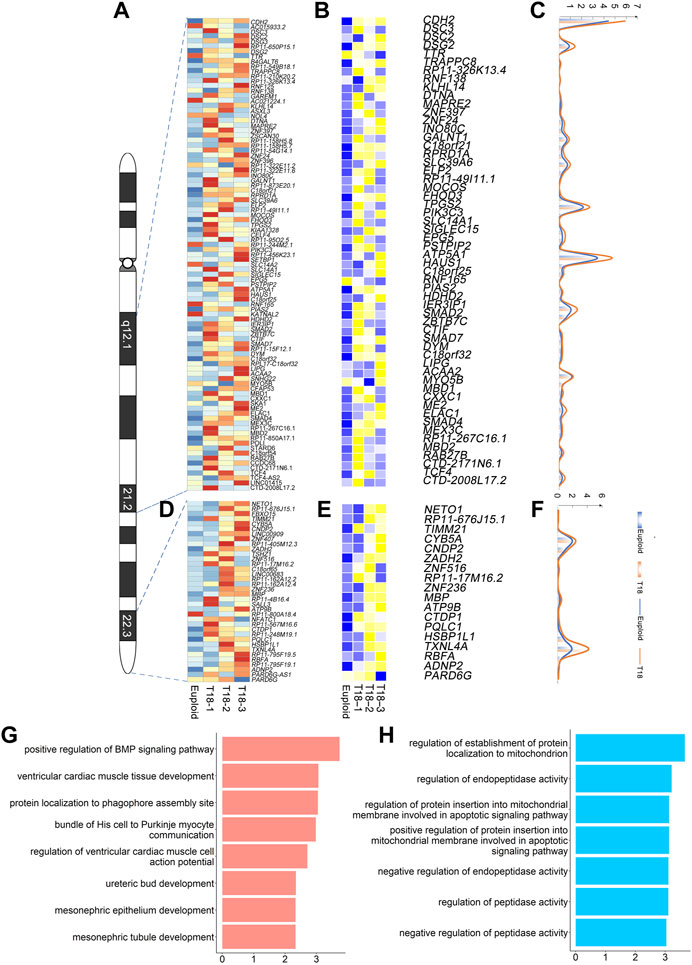
FIGURE 4. Gene expression analyses in previously proposed trisomy 18 critical regions: 18q12.1-21.2 and 18q22.3-18qter. (A) Heatmap of expression levels of genes in the 18q12.1-21.2 region from two euploid and three trisomy 18 samples. (B) Heatmap of differentially expressed genes (p ≤ 0.01) in the 18q12.1-21.2 region. (C) Unique molecular identifiers (UMIs) counts of differentially expressed genes in euploid (blue line) and trisomy 18 samples (orange line). (D) Heatmap of expression levels of genes in the 18q22.3-18qter region from two euploid and three trisomy 18 samples. (E) Heatmap of differentially expressed genes (p ≤ 0.01) in the 18q22.3-18qter. (F) UMIs counts of differentially expressed genes in euploid (blue line) and trisomy 18 samples (orange line). (G) Top gene ontology (GO) terms of differentially expressed genes in the 18q12.1-21.2 region. (H) Top GO terms enriched for differentially expressed genes in the 18q22.1-18qter region.
Moreover, 110 genes were identified in the proposed 18q22.3-qter critical region. Among these 110 genes, we identified 36 genes with altered expression (Figure 4D). Of them, 18 genes showed a significantly up-regulated expression (p < 0.05), and a higher copy number detected by the CNV analysis (Figures 4E,F). Up-regulated genes were enriched in regulation of establishment of protein localization to mitochondrion and negative regulation of endopeptidase activity in the GO term analysis (Figure 4H).
Overall, our results indicate that single-cell transcriptomics confirms the previously reported critical regions, and identifies crucial genes that are involved in development of cardiac muscle and mesonephric tubule, and in basic cellular function such as protein localization.
Prediction of New Chromosome 18 Critical Regions
Because of the complexity of phenotypes in trisomy 18 patients, we suspected that there might be some other trisomy 18 critical regions, which contribute to these defects. Based on the sequencing data of single-cell transcriptomics, we screened out differentially expressed genes (p < 0.05) in all five euploid and trisomy 18 samples by DESeq2. We merged all differentially expressed genes from trisomy 18 samples, and mapped them onto chromosome 18. We identified three regions, in which genes are significantly up-regulated (p < 0.05) and have higher copy numbers as detected by the CNV analysis. These regions included 18p11.32 on the short arm, and 18q11 and 18q21.32 on the long arm of chromosome 18.
Single-cell transcriptomics showed that 15 genes out of 57 genes on 18p11.32, 33 genes out of 101 genes on 18q11, and 12 genes out of 47 genes on 18q21.32 are significantly up-regulated (Figures 5A–C). The degree of up-regulation varied among these genes. Moreover, in the 18p11.32 region, up-regulated genes were enriched in negative regulation of mitotic metaphase plate congression, centrosome duplication, and nuclear division in the GO term analysis (Figure 5D). In the 18q11 region, up-regulated genes were enriched in endodermal cell differentiation, in utero embryonic development, and cardiac muscle hypertrophy (Figure 5E). Finally, in the 18q21.32 region, up-regulated genes were enriched in protein localization to mitochondrion, lymphangiogensis, and positive regulation of T cell cytokine production (Figure 5F). These analyses suggest that these new regions in chromosome 18 might contribute to the complexity of phenotypes in trisomy 18 patients, including cardiac muscle development, immune response and basic cell division regulation.
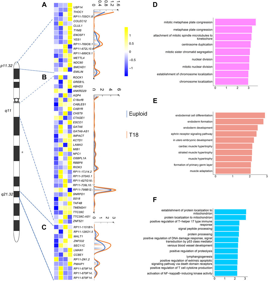
FIGURE 5. Gene expression analyses in 18p11.32, 18q11 and 18q11.32 regions. (A) Heatmap of differentially expressed genes (p ≤ 0.01) in the 18p11.32 region from two euploid and three trisomy 18 samples. Unique molecular identifiers (UMIs) counts of differentially expressed genes in euploid (blue line) and trisomy 18 samples (orange line). (B) Heatmap of differentially expressed genes (p ≤ 0.01) in the 18q11 region. UMIs counts of differentially expressed genes in euploid (blue line) and trisomy 18 samples (orange line). (C) Heatmap of differentially expressed genes (p ≤ 0.01) in the18q21.32 region. UMIs counts of differentially expressed genes in euploid (blue line) and trisomy 18 samples (orange line). (D–F) Top gene ontology (GO) terms of differentially expressed genes in the 18q11.32 (D), 18p11 (E) and 18q21.32 (F) regions.
Discussion
Trisomy 18 pregnancies, characterized by an extra copy of chromosome 18, have a high risk of fetal loss and stillbirth (Won et al., 2005; Morris and Savva, 2008). Insightful studies on pathophysiology and genetic causes of trisomy 18 have been sparse, because of the difficulty of sample collection, unavailability of disease models and extremely high mortality rate among neonates (Cavadino and Morris, 2017; Xing et al., 2018). Here, we have analyzed three amniotic fluid cell samples from trisomy 18 fetuses and two from euploid using the single-cell sequencing technology. We find that the extra chromosome 18 alters expression of genes functioning in multiple systems such as vascular, renal and nervous systems. We demonstrate striking gene expression changes in previously proposed trisomy 18 critical regions, and identify three new regions such as 18p11.32, 18q11 and 18q21.32, which are likely associated with trisomy 18 phenotypes. Our study indicates complexity of trisomy 18 at the gene expression level revealed by the single-cell sequencing.
A decrease in the frequency of live born with trisomy 18 has been observed because of increased use of prenatal diagnosis and the high rate of pregnancy termination based on such diagnosis (Crider et al., 2008; Irving et al., 2011). For instance, in the second and the third trimester, antenatal trisomy 18 recognition by ultrasound and genetic screen can detect almost all cases of trisomy 18 (DeVore, 2000; Oyelese and Vintzileos, 2005). The amniotic fluid has been collected as an untapped source of cells with broad potential, such as prenatal diagnosis and genetic analysis, which don’t have the ethical and legal limitations of embryonic stem cells (De Coppi et al., 2007; Loukogeorgakis and De Coppi, 2016; 2017). In this study, we thus used amniotic fluid cells as a source to study complexity of the trisomy 18 phenotype based on gene expression. While RNA-sequencing has been widely used to generate gene expression profiles in a tissue and organ, it cannot distinguish diversity and complexity of cell composition. Single-cell sequencing can reveal comprehensive transcriptomics at an individual cell resolution, overcome limited tissue supply, identify novel cell types, and uncover diverse contributions of individual cells to organ formation and disease conditions (Tang et al., 2009; Tang et al., 2010; Haque et al., 2017; Hochgerner et al., 2018; Li et al., 2018; Potter, 2018). Therefore, in this study, we have applied the single-cell sequencing approach to examine the composition of cultured amniotic fluid cells from euploid and trisomy 18 fetuses.
Clinical studies indicate that among trisomy 18 phenotypes, 75% cases show malformations in heart, 25–75% in genitourinary system, and 5–25% in gastrointestinal system, central nervous system, craniofacial and eye and limb, suggesting that multiple systems are affected by the extra chromosome 18 (Hodes et al., 1978; Lyons Jones, 1988; Balderston et al., 1990; Root and Carey, 1994; Cereda and Carey, 2012; Hoon et al., 2018). Our single-cell sequencing has identified 6 functional sub-clusters, in particular cell groups functioning in the development of kidney, vasculature and smooth muscle, which display significant alterations in gene expression. Our finding has provided genetic reasoning that is associated with multiple and as well specific trisomy 18 defects. Moreover, we have found that up- and down-regulated genes are similar but also distinct among three trisomy 18 samples. While variations of amniotic fluid cells from three individual fetuses may contribute to diversity of altered genes, distinct alterations of gene expression in each single cells in three individuals, which has been captured by single-cell sequencing, is likely associated with the complexity of trisomy 18 defects. Nevertheless, our GO term analyses indicate that up-regulated genes detected in three trisomy 18 samples are commonly enriched in development of the renal system, bone, and urogenital system. Single-cell sequencing of amniotic fluid cells from trisomy 18 fetuses not only reveals diverse gene expression programs in each cell from each individual, but also identifies common genes that are associated with major defects in trisomy 18.
Identifying critical regions in a chromosome is an effective way to uncover genetic reasoning of autosomal trisomy disorders (Mrozek et al., 2002; Olson et al., 2004; Chang and Min, 2005; Li et al., 2014; Mowery et al., 2018). 18q12.1-18q21.2 and 18q22.33-18qter have been proposed as trisomy 18 critical regions (Boghosian-Sell et al., 1994). We have found that genes in these two regions are indeed significantly up-regulated in trisomy 18 amniotic fluid cells. Moreover, GO analyses of up-regulated genes also support functional association with cardiac ventricular development and protein localization, which highlights tight relation of these two regions with trisomy 18 defects. Furthermore, because of the involvement of multiple tissues in trisomy 18 anomalies, it appears unlikely only two trisomy 18 critical regions contribute to the phenotypes. Our results suggest that three new regions (18p11.32, 18q11 and 18q21.32) are tightly associated with trisomy 18 defects. Expression levels of genes located in these regions are significantly altered, and these genes are functionally involved in cell division regulation, endoderm formation, and immune response. Our finding indicates that trisomy 18 affects immune system and fundamental cellular function such as cell division, and in turn has a broad impact on proper development and function of multiple systems.
Human amniotic fluid cells have been broadly used for genetic screen. In this study, in order to collect sufficient cells for single-cell RNA sequencing, amniotic fluid cells were cultured and expanded. Thus, while our single-cell transcriptomics has identified most crucial genes associated with trisomy 18 defects, it may miss out some genes or pathways due to the cell culture itself, which can be detected by conventional techniques (Koide et al., 2011; Hui et al., 2012; Kamath-Rayne et al., 2015; Zwemer and Bianchi, 2015; Volk et al., 2017; Cho et al., 2018; Albizua et al., 2020).
Overall, our single-cell sequencing analysis reveals complexity of gene expression alterations in amniotic fluid cells from trisomy 18 fetuses. The previously proposed and newly identified trisomy 18 critical regions highlight genetic contributions to the aggravation of symptoms in the trisomy 18 condition.
Materials and Methods
Amniotic Fluid Cells Acquisition, Culturing and Karyotype Analyses
Amniotic fluid cell samples were recruited from pregnant women at the second trimester, who underwent amniocentesis for prenatal diagnoses at Women and Children’s Hospital, School of Medicine, Xiamen University. Signed informed consents had been obtained from pregnant women who received amniocentesis for prenatal diagnosis to agree the research use of the remainder amniotic fluid samples. The cultured cells used in this study were those remainder samples. There was no additional sampling or any health-related intervention performed. Moreover, except for karyotypes, other information including names of patients was withheld from the study group.
Cells were cultured according to standard protocols of using the flask method and commercially available complete medium for amniotic fluid cells (Amniopan, Biotech) at 37°C in 5% CO2 environment. The karyotype analysis was conducted according to standard protocols for the chromosomal Giemsa (G)-banding. A total of 100 metaphases of each passage were evaluated. Following a routine cytogenetic analysis, the second passage of amniotic cell culture was grown in the same condition as for the primary cell culture, and was used for single-cell sequencing.
Preparation of Single-Cell Suspension
Cultured amniotic fluid cells were digested with 0.05% trypsin-EDTA (Gibco, Life Technologies) at 37°C for 3 min. The reaction was terminated by adding the complete medium. Cell suspension was pipetted to dissociate it into single cells, and centrifuged at 1,000 rpm for 5 min to obtain a cell pellet. The cell pellet was suspended in 5 ml of Phosphate Buffered Saline (PBS), and was centrifuged again at 1,000 rpm to remove cell debris. The cell pellet was re-suspended into single cells in 100 μl ice-cold PBS for single-cell sequencing analysis.
Single-Cell RNA Sequencing (ScRNA-Seq) Library Construction Using the 10X Genomics Chromium Platform
ScRNA-seq libraries were prepared per the Single Cell 30 Reagent Kit User Guide v2 (10X Genomics). Briefly, cell suspensions were loaded on a Chromium Controller instrument (10X Genomics) to generate single-cell Gel Bead-In-EMulsions (GEMs). GEM-reverse transcriptions (GEM-RTs) were performed in a Veriti 96-well thermal cycler (Thermo Fisher Scientific). After reverse transcription, GEMs were harvested, and cDNAs were amplified and cleaned up with the SPRIselect Reagent Kit (Beckman Coulter). Indexed sequencing libraries were constructed using the Chromium Single-Cell 30 Library Kit (10X Genomics) for enzymatic fragmentation, end-repair, A-tailing, adaptor ligation, ligation cleanup, sample index PCR, and PCR cleanup. The barcoded sequencing libraries were quantified by quantitative PCR using the KAPA Library Quantification Kit (KAPA Biosystems). Sequencing libraries were loaded on a NextSeq500 (Illumina) with a custom sequencing setting (26 bp for read 1 and 98 bp for read 2) to obtain a sequencing depth of 80,000 reads per cell (Xie et al., 2018).
Data Processing of Single-Cell RNA-Seq From the Chromium System
Mapping to the GRCh38 human genome, quality control and read counting of Ensembl genes were performed using Cellranger software with default parameter (v2.1.0). Cells with ≥200 genes and ≤10% percentage of detected mitochondrial genes were retained for subsequent analyses. Normalization, dimensionality reduction and clustering of single cells were also performed by the Cellranger.
Identification of Differentially Expressed Genes
Differentially expressed genes (DEGs) were first identified by comparing cells in a specific cluster with cells in all other clusters (Seurat package Wilcox: average expression difference >0.5 natural log with an FDR corrected p < 0.01). Next, cells in a specific cluster were compared to cells in every other cluster in a pairwise manner to identify a second set of DEGs (DESeq2 package: FDR ≤0.05 and |log2 Fold Change| ≥ 2).
Identification of Cell Types and Subtypes by Nonlinear Dimensional Reduction and Random Forests
Batch effects were corrected by matching mutual nearest neighbors (Haghverdi et al., 2018), and the dimensionality was reduced by running a principal-component analysis (PCA) on the most highly variable genes. Graph-based clustering was performed on significant principal components (PCs).
The Seurat package (v.3.0.0) implemented in R was applied to identify major cellular functions of 56,517 single cells from amniotic fluid cells. The top 1,500 most variable genes selected by Seurat were used to compute the PCs. The first 50 significant (p ≤ 0.01) PCs selected based on the built-in jackstraw analysis were used for t-distributed stochastic neighbor Embedding (t-SNE) visualization. Dimensionality was reduced by running a PCA on the most highly variable genes, and the graph-based clustering was performed on the significant PCs. Finally, the distinct sub-groups of cells were visualized by using t-distributed stochastic neighbor embedding tSNE.
Analysis of Copy-Number Variations (CNVs) in Single-Cell Data
Analysis of CNVs with single-cell data was conducted using the R package inferCNV (https://github.com/broadinstitute/inferCNV) with default parameters. Genome-wide expression of genes in the trisomy 18 samples was compared with that in the euploid cells in order to identify large-scale chromosomal copy number variations. The raw expression matrix generated from Seurat was used as input data for the inferCNV analysis.
Data Availability Statement
The datasets presented in this study can be found in online repositories. The names of the repository/repositories and accession number(s) can be found below: https://www.ncbi.nlm.nih.gov/geo/ GSE141074
Ethics Statement
The studies involving human participants were reviewed and approved by Ethics committee of Women and Children’s Hospital, School of Medicine, Xiamen University. The patients/participants provided their written informed consent to participate in this study.
Author Contributions
TS and JW conceived and designed the study. JW performed major experiments and analyzed the data. ZC performed single cell transcriptome analysis. FH assisted analyzing the data. TL and GH analyzed the data and edited the article. WjC, WhC, NM, ZZ, QY, and QG assisted experimental sample collection and processing. JW and TS interpreted the data and wrote the paper, and all authors provided feedback.
Funding
This work was supported by the Scientific Research Funds of Huaqiao University (Z17Y0026 and Z16Y0017), Innovation Awards of Quanzhou Talents (2018C057R), Natural Science Foundation of Fujian Province, China (2019J01071), China Postdoctoral Science Foundation (2017M622053), and National Natural Science Foundation of China (31771141 and 32100775).
Conflict of Interest
Author FH was employed by the company Genergy Bio-Technology (Shanghai) Co., Ltd.
The remaining authors declare that the research was conducted in the absence of any commercial or financial relationships that could be construed as a potential conflict of interest.
Publisher’s Note
All claims expressed in this article are solely those of the authors and do not necessarily represent those of their affiliated organizations, or those of the publisher, the editors and the reviewers. Any product that may be evaluated in this article, or claim that may be made by its manufacturer, is not guaranteed or endorsed by the publisher.
Acknowledgments
We thank all Sun Lab members for their help in our experiments.
Supplementary Material
The Supplementary Material for this article can be found online at: https://www.frontiersin.org/articles/10.3389/fcell.2022.825345/full#supplementary-material
References
Albizua, I., Chopra, P., Sherman, S. L., Gambello, M. J., and Warren, S. T. (2020). Analysis of the Genomic Expression Profile in Trisomy 18: Insight into Possible Genes Involved in the Associated Phenotypes. Hum. Mol. Genet. 29 (2), 238–247. doi:10.1093/hmg/ddz279
Antonarakis, S. E. (2017). Down Syndrome and the Complexity of Genome Dosage Imbalance. Nat. Rev. Genet. 18 (3), 147–163. doi:10.1038/nrg.2016.154
Antonarakis, S. E., Lyle, R., Dermitzakis, E. T., Reymond, A., and Deutsch, S. (2004). Chromosome 21 and Down Syndrome: from Genomics to Pathophysiology. Nat. Rev. Genet. 5 (10), 725–738. doi:10.1038/nrg1448
Babu, A., and Verma, R. S. (1986). The Heteromorphic Marker on Chromosome 18 Using Restriction Endonuclease AluI. Am. J. Hum. Genet. 38 (4), 549–554.
Balderston, S. M., Shaffer, E. M., Washington, R. L., and Sondheimer, H. M. (1990). Congenital Polyvalvular Disease in Trisomy 18: Echocardiographic Diagnosis. Pediatr. Cardiol. 11 (3), 138–142. doi:10.1007/bf02238843
Batees, H., and Altirkawi, K. A. (2014). Trisomy 18 Syndrome: Towards a Balanced Approach. Sudan J. Paediatr. 14 (2), 76–84.
Boghosian-Sell, L., Mewar, R., Harrison, W., Shapiro, R. M., Zackai, E. H., Carey, J., et al. (1994). Molecular Mapping of the Edwards Syndrome Phenotype to Two Noncontiguous Regions on Chromosome 18. Am. J. Hum. Genet. 55 (3), 476–483.
Bugge, M., Collins, A., Petersen, M. B., Fisher, J., Brandt, C., Hertz, J. M., et al. (1998). Non-disjunction of Chromosome 18. Hum. Mol. Genet. 7 (4), 661–669. doi:10.1093/hmg/7.4.661
Calderone, J. P., Chess, J., Borodic, G., and Albert, D. M. (1983). Intraocular Pathology of Trisomy 18 (Edwards's Syndrome): Report of a Case and Review of the Literature. Br. J. Ophthalmol. 67 (3), 162–169. doi:10.1136/bjo.67.3.162
Carey, J. C. (2005). Trisomy 18 and Trisomy 13 syndromesManagement of Genetic Syndromes. Wiley Online Library.doi:10.1002/9781119432692.ch58
Cavadino, A., and Morris, J. K. (2017). Revised Estimates of the Risk of Fetal Loss Following a Prenatal Diagnosis of Trisomy 13 or Trisomy 18. Am. J. Med. Genet. 173 (4), 953–958. doi:10.1002/ajmg.a.38123
Cereda, A., and Carey, J. C. (2012). The Trisomy 18 Syndrome. Orphanet J. Rare Dis. 7, 81. doi:10.1186/1750-1172-7-81
Chang, K. T., and Min, K.-T. (2005). Drosophila melanogaster Homolog of Down Syndrome Critical Region 1 Is Critical for Mitochondrial Function. Nat. Neurosci. 8 (11), 1577–1585. doi:10.1038/nn1564
Cho, H. Y., Cho, Y., Shin, Y.-J., Park, J., Shim, S., Jung, Y., et al. (2018). Functional Analysis of Cell-free RNA Using Mid-trimester Amniotic Fluid Supernatant in Pregnancy with the Fetal Growth Restriction. Medicine (Baltimore) 97 (2), e9572. doi:10.1097/md.0000000000009572
Crider, K. S., Olney, R. S., and Cragan, J. D. (2008). Trisomies 13 and 18: Population Prevalences, Characteristics, and Prenatal Diagnosis, Metropolitan Atlanta, 1994-2003. Am. J. Med. Genet. 146A (7), 820–826. doi:10.1002/ajmg.a.32200
De Coppi, P., Bartsch, G., Siddiqui, M. M., Xu, T., Santos, C. C., Perin, L., et al. (2007). Isolation of Amniotic Stem Cell Lines with Potential for Therapy. Nat. Biotechnol. 25 (1), 100–106. doi:10.1038/nbt1274
DeVore, G. R. (2000). Second Trimester Ultrasonography May Identify 77 to 97% of Fetuses with Trisomy 18. J. Ultrasound Med. 19 (8), 565–576. doi:10.7863/jum.2000.19.8.565
Edwards, J. H., Harnden, D. G., Cameron, A. H., Crosse, V. M., and Wolf, O. H. (1960). A New Trisomic Syndrome. The Lancet 275 (7128), 787–790. doi:10.1016/s0140-6736(60)90675-9
Eggermann, T., Nöthen, M. M., Eiben, B., Hofmann, D., Hinkel, K., Fimmers, R., et al. (1996). Trisomy of Human Chromosome 18: Molecular Studies on Parental Origin and Cell Stage of Nondisjunction. Hum. Genet. 97 (2), 218–223. doi:10.1007/bf02265269
Fan, X., Dong, J., Zhong, S., Wei, Y., Wu, Q., Yan, L., et al. (2018). Spatial Transcriptomic Survey of Human Embryonic Cerebral Cortex by Single-Cell RNA-Seq Analysis. Cell Res. 28 (7), 730–745. doi:10.1038/s41422-018-0053-3
Filbin, M. G., Tirosh, I., Hovestadt, V., Shaw, M. L., Escalante, L. E., Mathewson, N. D., et al. (2018). Developmental and Oncogenic Programs in H3K27M Gliomas Dissected by Single-Cell RNA-Seq. Science 360 (6386), 331–335. doi:10.1126/science.aao4750
FitzPatrick, D. R., Ramsay, J., McGill, N. I., Shade, M., Carothers, A. D., and Hastie, N. D. (2002). Transcriptome Analysis of Human Autosomal Trisomy. Hum. Mol. Genet. 11 (26), 3249–3256. doi:10.1093/hmg/11.26.3249
Ginsberg, J., Perrin, E. V., and Sueoka, W. T. (1968). Ocular Manifestations of Trisomy 18. Am. J. Ophthalmol. 66 (1), 59–67. doi:10.1016/0002-9394(68)91788-1
Grün, D., and van Oudenaarden, A. (2015). Design and Analysis of Single-Cell Sequencing Experiments. Cell 163 (4), 799–810. doi:10.1016/j.cell.2015.10.039
Haghverdi, L., Lun, A. T. L., Morgan, M. D., and Marioni, J. C. (2018). Batch Effects in Single-Cell RNA-Sequencing Data Are Corrected by Matching Mutual Nearest Neighbors. Nat. Biotechnol. 36 (5), 421–427. doi:10.1038/nbt.4091
Haque, A., Engel, J., Teichmann, S. A., and Lönnberg, T. (2017). A Practical Guide to Single-Cell RNA-Sequencing for Biomedical Research and Clinical Applications. Genome Med. 9 (1), 75. doi:10.1186/s13073-017-0467-4
Hochgerner, H., Zeisel, A., Lönnerberg, P., and Linnarsson, S. (2018). Conserved Properties of Dentate Gyrus Neurogenesis across Postnatal Development Revealed by Single-Cell RNA Sequencing. Nat. Neurosci. 21 (2), 290–299. doi:10.1038/s41593-017-0056-2
Hodes, M. E., Cole, J., Palmer, C. G., and Reed, T. (1978). Clinical Experience with Trisomies 18 and 13. J. Med. Genet. 15 (1), 48–60. doi:10.1136/jmg.15.1.48
Hoon, S. k., Kang, S.-w., Kwak, S.-H., and Kim, J. (2018). Hypoxia Due to Positive Pressure Ventilation in Edwards' Syndrome: A Case Report. J. Int. Med. Res. 46 (2), 895–900. doi:10.1177/0300060517734680
Hui, L., Slonim, D. K., Wick, H. C., Johnson, K. L., Koide, K., and Bianchi, D. W. (2012). Novel Neurodevelopmental Information Revealed in Amniotic Fluid Supernatant Transcripts from Fetuses with Trisomies 18 and 21. Hum. Genet. 131 (11), 1751–1759. doi:10.1007/s00439-012-1195-x
Imataka, G., Suzumura, H., and Arisaka, O. (2016). Clinical Features and Survival in Individuals with Trisomy 18: A Retrospective One-center Study of 44 Patients Who Received Intensive Care Treatments. Mol. Med. Rep. 13 (3), 2457–2466. doi:10.3892/mmr.2016.4806
Irving, C., Richmond, S., Wren, C., Longster, C., and Embleton, N. D. (2011). Changes in Fetal Prevalence and Outcome for Trisomies 13 and 18: a Population-Based Study over 23 Years. J. Maternal-Fetal Neonatal Med. 24 (1), 137–141. doi:10.3109/14767051003758879
Kamath-Rayne, B. D., Du, Y., Hughes, M., Wagner, E. A., Muglia, L. J., DeFranco, E. A., et al. (2015). Systems Biology Evaluation of Cell-free Amniotic Fluid Transcriptome of Term and Preterm Infants to Detect Fetal Maturity. BMC Med. Genomics 8, 67. doi:10.1186/s12920-015-0138-5
Kim, C., Gao, R., Sei, E., Brandt, R., Hartman, J., Hatschek, T., et al. (2018). Chemoresistance Evolution in Triple-Negative Breast Cancer Delineated by Single-Cell Sequencing. Cell 173 (4), 879–893. e813. doi:10.1016/j.cell.2018.03.041
Koide, K., Slonim, D. K., Johnson, K. L., Tantravahi, U., Cowan, J. M., and Bianchi, D. W. (2011). Transcriptomic Analysis of Cell-free Fetal RNA Suggests a Specific Molecular Phenotype in Trisomy 18. Hum. Genet. 129 (3), 295–305. doi:10.1007/s00439-010-0923-3
Kondoh, T., Tonoki, H., Matsumoto, T., Tsukahara, M., and Niikawa, N. (1988). Origin of the Extra Chromosome in Trisomy 18. A Study on Five Patients Using a Restriction Fragment Length Polymorphism. Hum. Genet. 79 (4), 377–378. doi:10.1007/BF00282181
Kupke, K. G., and Müller, U. (1989). Parental Origin of the Extra Chromosome in Trisomy 18. Am. J. Hum. Genet. 45 (4), 599–605.
Li, L., Guo, F., Gao, Y., Ren, Y., Yuan, P., Yan, L., et al. (2018). Single-cell Multi-Omics Sequencing of Human Early Embryos. Nat. Cel Biol. 20 (7), 847–858. doi:10.1038/s41556-018-0123-2
Li, T., Zhao, H., Han, X., Yao, J., Zhang, L., Guo, Y., et al. (2017). The Spontaneous Differentiation and Chromosome Loss in iPSCs of Human Trisomy 18 Syndrome. Cell Death Dis. 8 (10), e3149. doi:10.1038/cddis.2017.565
Li, Y., Schwab, C., Ryan, S. L., Papaemmanuil, E., Robinson, H. M., Jacobs, P., et al. (2014). Constitutional and Somatic Rearrangement of Chromosome 21 in Acute Lymphoblastic Leukaemia. Nature 508 (7494), 98–102. doi:10.1038/nature13115
Loukogeorgakis, S. P., and De Coppi, P. (2017). Concise Review: Amniotic Fluid Stem Cells: The Known, the Unknown, and Potential Regenerative Medicine Applications. Stem Cells 35 (7), 1663–1673. doi:10.1002/stem.2553
Loukogeorgakis, S. P., and De Coppi, P. (2016). Stem Cells from Amniotic Fluid - Potential for Regenerative Medicine. Best Pract. Res. Clin. Obstet. Gynaecol. 31, 45–57. doi:10.1016/j.bpobgyn.2015.08.009
Lyons Jones, K. (1988). Smith's Recognizable Patterns of Human Malformation. Philadelphia, Pa: W13 Saunders.
Mirmohammadsadeghi, A., Akbari, M. R., and Malekpoor, A. (2017). Ocular Manifestations in Edward's Syndrome, a Case Report and Literature Review. J. Curr. Ophthalmol. 29 (4), 329–331. doi:10.1016/j.joco.2017.06.005
Montemurro, T., Bossolasco, P., Cova, L., Zangrossi, S., Calzarossa, C., Buiatiotis, S., et al. (2008). Molecular and Phenotypical Characterization of Human Amniotic Fluid Cells and Their Differentiation Potential. Biomed. Mater. Eng. 18 (4-5), 183–185. doi:10.3233/bme-2008-0522
Morris, J. K., and Savva, G. M. (2008). The Risk of Fetal Loss Following a Prenatal Diagnosis of Trisomy 13 or Trisomy 18. Am. J. Med. Genet. 146A (7), 827–832. doi:10.1002/ajmg.a.32220
Mowery, C. T., Reyes, J. M., Cabal-Hierro, L., Higby, K. J., Karlin, K. L., Wang, J. H., et al. (2018). Trisomy of a Down Syndrome Critical Region Globally Amplifies Transcription via HMGN1 Overexpression. Cel Rep. 25 (7), 1898–1911. e1895. doi:10.1016/j.celrep.2018.10.061
Mrózek, K., Heinonen, K., Theil, K. S., and Bloomfield, C. D. (2002). Spectral Karyotyping in Patients with Acute Myeloid Leukemia and a Complex Karyotype Shows Hidden Aberrations, Including Recurrent Overrepresentation of 21q, 11q, and 22q. Genes Chromosom. Cancer 34 (2), 137–153. doi:10.1002/gcc.10027
Müller, S., Liu, S. J., Di Lullo, E., Malatesta, M., Pollen, A. A., Nowakowski, T. J., et al. (2016). Single‐cell Sequencing Maps Gene Expression to Mutational Phylogenies in PDGF ‐ and EGF ‐driven Gliomas. Mol. Syst. Biol. 12 (11), 889. doi:10.15252/msb.20166969
Nagamuthu, E. A., and Neelaveni, N. (2014). Edward Syndrome (Trisomy 18): a Case Report. Ann. Biol. Res. 5 (3), 67–72.
Norwitz, E. R., and Levy, B. (2013). Noninvasive Prenatal Testing: the Future Is Now. Rev. Obstet. Gynecol. 6 (2), 48–62.
Nöthen, M. M., Eggermann, T., Erdmann, J., Eiben, B., Hofmann, D., Propping, P., et al. (1993). Retrospective Study of the Parental Origin of the Extra Chromosome in Trisomy 18 (Edwards Syndrome). Hum. Genet. 92 (4), 347–349. doi:10.1007/BF01247332
Olson, L. E., Richtsmeier, J. T., Leszl, J., and Reeves, R. H. (2004). A Chromosome 21 Critical Region Does Not Cause Specific Down Syndrome Phenotypes. Science 306 (5696), 687–690. doi:10.1126/science.1098992
Oyelese, Y., and Vintzileos, A. M. (2005). Is Second-Trimester Genetic Amniocentesis for Trisomy 18 Ever Indicated in the Presence of a normal Genetic Sonogram? Ultrasound Obstet. Gynecol. 26 (7), 691–694. doi:10.1002/uog.2662
Park, J., Shrestha, R., Qiu, C., Kondo, A., Huang, S., Werth, M., et al. (2018). Single-cell Transcriptomics of the Mouse Kidney Reveals Potential Cellular Targets of Kidney Disease. Science 360 (6390), 758–763. doi:10.1126/science.aar2131
Patel, A. P., Tirosh, I., Trombetta, J. J., Shalek, A. K., Gillespie, S. M., Wakimoto, H., et al. (2014). Single-cell RNA-Seq Highlights Intratumoral Heterogeneity in Primary Glioblastoma. Science 344 (6190), 1396–1401. doi:10.1126/science.1254257
Potter, S. S. (2018). Single-cell RNA Sequencing for the Study of Development, Physiology and Disease. Nat. Rev. Nephrol. 14 (8), 479–492. doi:10.1038/s41581-018-0021-7
Puram, S. V., Tirosh, I., Parikh, A. S., Patel, A. P., Yizhak, K., Gillespie, S., et al. (2017). Single-cell Transcriptomic Analysis of Primary and Metastatic Tumor Ecosystems in Head and Neck Cancer. Cell 171 (7), 1611–1624. e1624. doi:10.1016/j.cell.2017.10.044
Raj, B., Wagner, D. E., McKenna, A., Pandey, S., Klein, A. M., Shendure, J., et al. (2018). Simultaneous Single-Cell Profiling of Lineages and Cell Types in the Vertebrate Brain. Nat. Biotechnol. 36 (5), 442–450. doi:10.1038/nbt.4103
Rasmussen, S. A., Wong, L.-Y. C., Yang, Q., May, K. M., and Friedman, J. M. (2003). Population-based Analyses of Mortality in Trisomy 13 and Trisomy 18. Pediatrics 111 (4), 777–784. doi:10.1542/peds.111.4.777
Roberts, W., Zurada, A., Zurada-ZieliŃSka, A., Gielecki, J., and Loukas, M. (2016). Anatomy of Trisomy 18. Clin. Anat. 29 (5), 628–632. doi:10.1002/ca.22725
Root, S., and Carey, J. C. (1994). Survival in Trisomy 18. Am. J. Med. Genet. 49 (2), 170–174. doi:10.1002/ajmg.1320490203
Savva, G. M., Walker, K., and Morris, J. K. (2010). The Maternal Age-specific Live Birth Prevalence of Trisomies 13 and 18 Compared to Trisomy 21 (Down Syndrome). Prenat. Diagn. 30 (1), 57–64. doi:10.1002/pd.2403
Suijuan, Z., Shu, Z., Xiaoying, F., Qian, W., Liying, Y., Ji, D., et al. (2018). A Single-Cell RNA-Seq Survey of the Developmental Landscape of the Human Prefrontal Cortex. nature 2555, 7. doi:10.1038/nature25980
Tang, F., Barbacioru, C., Bao, S., Lee, C., Nordman, E., Wang, X., et al. (2010). Tracing the Derivation of Embryonic Stem Cells from the Inner Cell Mass by Single-Cell RNA-Seq Analysis. Cell Stem Cell 6 (5), 468–478. doi:10.1016/j.stem.2010.03.015
Tang, F., Barbacioru, C., Wang, Y., Nordman, E., Lee, C., Xu, N., et al. (2009). mRNA-Seq Whole-Transcriptome Analysis of a Single Cell. Nat. Methods 6 (5), 377–382. doi:10.1038/nmeth.1315
Tobin, M., Gunaji, R., Walsh, J. C., and Grice, G. P. (2019). A Review of Genetic Factors Underlying Craniorachischisis and Omphalocele: Inspired by a Unique Trisomy 18 Case. Am. J. Med. Genet. 179 (8), 1642–1651. doi:10.1002/ajmg.a.61255
Turbiville, D. E., Wu, H., and Dong, J. (2017). Detection of an Underlying 22q11.2 Duplication in a Female Neonate with Trisomy 18. Lab. Med. 48 (4), 372–375. doi:10.1093/labmed/lmx039
Underwood, M. A., Gilbert, W. M., and Sherman, M. P. (2005). Amniotic Fluid: Not Just Fetal Urine Anymore. J. Perinatol 25 (5), 341–348. doi:10.1038/sj.jp.7211290
Velmeshev, D., Schirmer, L., Jung, D., Haeussler, M., Perez, Y., Mayer, S., et al. (2019). Single-cell Genomics Identifies Cell Type-specific Molecular Changes in Autism. Science 364 (6441), 685–689. doi:10.1126/science.aav8130
Volk, M., Maver, A., Hodžić, A., Lovrečić, L., and Peterlin, B. (2017). Transcriptome Profiling Uncovers Potential Common Mechanisms in Fetal Trisomies 18 and 21. OMICS: A J. Integr. Biol. 21 (10), 565–570. doi:10.1089/omi.2017.0123
Wen, L., and Tang, F. (2016). Single-cell Sequencing in Stem Cell Biology. Genome Biol. 17, 71. doi:10.1186/s13059-016-0941-0
Wilson, G. N. (1993). Karyotype/phenotype Correlation: Prospects and Problems Illustrated by Trisomy 18. Prog. Clin. Biol. Res. 384, 157–173.
Won, R. H., Currier, R. J., Lorey, F., and Towner, D. R. (2005). The Timing of Demise in Fetuses with Trisomy 21 and Trisomy 18. Prenat. Diagn. 25 (7), 608–611. doi:10.1002/pd.1243
Xie, T., Wang, Y., Deng, N., Huang, G., Taghavifar, F., Geng, Y., et al. (2018). Single-cell Deconvolution of Fibroblast Heterogeneity in Mouse Pulmonary Fibrosis. Cel Rep. 22 (13), 3625–3640. doi:10.1016/j.celrep.2018.03.010
Xing, K., Cui, Y., Luan, J., Zhou, X., Shi, L., and Han, J. (2018). Establishment of a Human Trisomy 18 Induced Pluripotent Stem Cell Line from Amniotic Fluid Cells. Irdr 7 (2), 94–99. doi:10.5582/irdr.218.01038
Ya-gang, X., Robinson, W. P., Spiegel, R., Binkert, F., Ruefenacht, U., and Schinzel, A. A. (1993). Parental Origin of the Supernumerary Chromosome in Trisomy 18. Clin. Genet. 44 (2), 57–61. doi:10.1111/j.1399-0004.1993.tb03847.x
Young, M. D., Mitchell, T. J., Vieira Braga, F. A., Tran, M. G. B., Stewart, B. J., Ferdinand, J. R., et al. (2018). Single-cell Transcriptomes from Human Kidneys Reveal the Cellular Identity of Renal Tumors. Science 361 (6402), 594–599. doi:10.1126/science.aat1699
Yu, Y., Zeng, Z., Xie, D., Chen, R., Sha, Y., Huang, S., et al. (2021). Interneuron Origin and Molecular Diversity in the Human Fetal Brain. Nat. Neurosci. 24 (12), 1745–1756. doi:10.1038/s41593-021-00940-3
Zeng, Z., Miao, N., and Sun, T. (2018). Revealing Cellular and Molecular Complexity of the central Nervous System Using Single Cell Sequencing. Stem Cel Res Ther 9 (1), 234. doi:10.1186/s13287-018-0985-z
Zhao, T., Fu, Y., Zhu, J., Liu, Y., Zhang, Q., Yi, Z., et al. (2018). Single-Cell RNA-Seq Reveals Dynamic Early Embryonic-like Programs during Chemical Reprogramming. Cell Stem Cell 23 (1), 31–45. e37. doi:10.1016/j.stem.2018.05.025
Zheng, G. X. Y., Terry, J. M., Belgrader, P., Ryvkin, P., Bent, Z. W., Wilson, R., et al. (2017). Massively Parallel Digital Transcriptional Profiling of Single Cells. Nat. Commun. 8, 14049. doi:10.1038/ncomms14049
Keywords: trisomy 18, amniotic fluid cells, single-cell RNA-sequencing, critical region, human fetuses
Citation: Wang J, Chen Z, He F, Lee T, Cai W, Chen W, Miao N, Zeng Z, Hussain G, Yang Q, Guo Q and Sun T (2022) Single-Cell Transcriptomics of Cultured Amniotic Fluid Cells Reveals Complex Gene Expression Alterations in Human Fetuses With Trisomy 18. Front. Cell Dev. Biol. 10:825345. doi: 10.3389/fcell.2022.825345
Received: 30 November 2021; Accepted: 24 February 2022;
Published: 22 March 2022.
Edited by:
Weimin Gao, Arizona State University, United StatesReviewed by:
Lisa Hui, Mercy Hospital for Women, AustraliaXiaolong Jiang, Baylor College of Medicine, United States
Copyright © 2022 Wang, Chen, He, Lee, Cai, Chen, Miao, Zeng, Hussain, Yang, Guo and Sun. This is an open-access article distributed under the terms of the Creative Commons Attribution License (CC BY). The use, distribution or reproduction in other forums is permitted, provided the original author(s) and the copyright owner(s) are credited and that the original publication in this journal is cited, in accordance with accepted academic practice. No use, distribution or reproduction is permitted which does not comply with these terms.
*Correspondence: Qiwei Guo, Z3VvcWl3ZWlAeG11LmVkdS5jbg==; Tao Sun, dGFvc3VuQGhxdS5lZHUuY24=
 Jing Wang1,2
 Zixi Chen
 Nan Miao
 Ghulam Hussain
 Qiwei Guo
 Tao Sun