 Jia-Heng Zhang
 Shi-Yu Ni1
 Shu-Chao Wang
94% of researchers rate our articles as excellent or good
Learn more about the work of our research integrity team to safeguard the quality of each article we publish.
Find out more
ORIGINAL RESEARCH article
Front. Cell Dev. Biol., 31 October 2022
Sec. Signaling
Volume 10 - 2022 | https://doi.org/10.3389/fcell.2022.1043725
This article is part of the Research TopicNovel Strategies to Target Cell Death Signaling in Cancer and Neurodegenerative Diseases: New Findings and Mechanistic StudiesView all 7 articles
Background: Regulation of cell death plays a key role in numerous diseases. As a proline isomerase, prolyl cis-trans isomerase NIMA-interacting 1 (Pin1) is important for the regulation of signaling pathways. An in-depth understanding of how Pin1 participates in the process of cell death, which affects the occurrence and development of diseases, will aid in the discovery of new disease mechanisms and therapeutic methods. Thus, the purpose of our study was to discover the research trends and hotspots of Pin1 and cell death through bibliometric analyses and to provide insights for understanding the future development of basic research and treatment of diseases.
Methods: Documents were extracted from the Web of Science Core Collection on 7 May 2022. We selected articles and reviews published in English from 2000 to 2021, and visual and statistical analyses of countries, institutions, authors, references and keywords were performed using VOSviewer 1.6.18 and CiteSpace 5.8.
Results: A total of 395 articles and reviews were selected. Since 2001, the number of articles on Pin1 and cell death has increased annually. Publications come from 43 countries, with the US having the most publications and citations. We identified 510 authors, with Giannino Del Sal having the most articles and Paola Zacchi having the most co-citations. The Journal of Biological Chemistry is the most researched journal, and Nature and its subjournals are the most cited journals. Apoptosis, phosphorylation, and breast cancer were the three most common keywords.
Conclusion: The number of documents showed an increasing trend from 2001 to 2014. Stagnant growth after 2014 may be related to the absence of new research hotspots. Cooperative links between core institutions need to be strengthened, and the institution with the highest citation count in recent years is Fujian Medical University in China. The role of Pin1 in cell death requires further research to discover new research hotspots. Before breakthroughs in molecular mechanism or signaling pathway research, future research will focus more on the treatment of diseases represented by Pin1 inhibitors.
Prolyl cis-trans isomerase NIMA-interacting 1 (Pin1) is an 18-kDa protein with an N-terminal WW domain and a C-terminal PPIase domain (Kumari et al., 2021; Tonnus et al., 2021; Zheng et al., 2021). The WW domain of Pin1 binds to phosphorylated serine-proline or threonine-proline (pSer/Thr-Pro) motifs, followed by PPIase domain isomerization of the prolyl bond in the pSer/Thr-Pro motifs (Koikawa et al., 2021; Kumari et al., 2021). In contrast, the activity of mitotic and nuclear proteins is regulated by Pin1 in a phosphorylation-dependent pathway (Kumari et al., 2021; Wang et al., 2023).
Pin1 plays a role in multiple types of cell death, such as apoptosis, necrosis, and autophagy. The effects of Pin1 on apoptosis include inactivation of Bcl-2-associated X protein (Bax), subsequently increasing the anti-cell death capacity of Bcl-2 (Bianchi et al., 2019; Makinwa et al., 2020) as well as promoting the expression of pro-apoptotic genes represented by p53 in cells to initiate apoptosis (Balaganapathy et al., 2018; Hleihel et al., 2021). This dual pro- and anti-apoptosis feature provides researchers with a variety of options for finding treatment options for diseases based on different pathways (Balaganapathy et al., 2018; Hleihel et al., 2021). Recent studies have shown that cellular necrosis (traditionally thought to be characterized by membrane disruption, release of internal substances, and inflammatory cells) also shows molecular-level abnormalities (Chen et al., 2021a; Hu et al., 2021a; Theobald et al., 2021; Tonnus et al., 2021), such as pyroptosis (Lammert et al., 2020) and neutrophil death (Sung and Hsieh, 2021). For example, Pin1 exerts a positive regulatory effect on calpain-2 by inhibiting calpastatin (CAST) in the Pin1-CAST-calpain pathway involved in cell-regulated necrosis (Cheng et al., 2018; Wang et al., 2018). This further mediates the transfer of apoptosis-inducing factors to the nucleus, ultimately leading to DNA degradation (Wang et al., 2018; Wang et al., 2019b). In a study on Pin1 and autophagy, Wang et al. (2023) found that GSK-3β is involved in autophagy as a mediator that regulates Pin1 activity.
The function of Pin1 is tightly regulated at multiple levels under physiological conditions, which makes Pin1 very different from most other constitutively active PPIases (Lu et al., 2007; Zhou and Lu, 2016). Moreover, abnormalities in Pin1 are involved in the occurrence and progression of neurological diseases and cancer through many mechanisms. Thus, further research on Pin1 can provide clues for exploring the treatment of neurological diseases and cancer. Our lab focuses on the study of Pin1 and cell death, and we hope to analyze the existing research on Pin1 in a broader field. Bibliometrics, a discipline that applies mathematical and statistical methods to the literature, provides us with a useful tool to broaden our research horizons (Smith, 2008). Using visual analysis software packages such as CiteSpace, researchers can achieve qualitative and quantitative evaluations of research progress in specific subject areas of research (Chen, 2006). Other visualization software packages such as VOSviewer, can achieve similar functions. However, the two software packages have their own characteristics. Flexible use, alone or in combination, in different situations can achieve better results. There are many evaluation indicators for bibliometric analysis, including citation frequency, the number of publications, and centrality (Avena and Barbosa, 2017).
The purpose of our study was to discover and analyze the current status and future trends of Pin1 and cell death. Using bibliometrics to identify current research hotspots in this field and explore future research trends can promote our understanding of the role of Pin1 in the process of cell death and provide references for researchers to broaden their ideas in basic research and disease treatment.
After searching the MeSH Database (https://www.NCBI.nlm.nih.gov/mesh) for relevant keywords, we searched the Web of Science Core Collection using the following search strategy: All={(PIN1) and [(Cell Death) or (Necrosis) or (Autophagy) or (Apoptosis)]}. After obtaining the search results, we found it was not until 2001 that a large number of articles related to cell death appeared. Therefore, we decided to narrow the time range to 2000–2021, and focus our study on more relevant articles. We set the language type to English, selected only Article and Review, and excluded conference materials, books, editing materials, corrections, letters, and retracted papers. Finally, 395 relevant documents were obtained for bibliometric analysis. Documents were exported as all records and references, saved as plain text files (txt) for bibliometric and visual analysis (Figure 1). The literature search was finally completed on 7 May 2022.
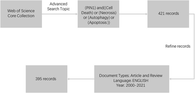
FIGURE 1. Flowchart of literature selection.
The content of bibliometric analysis includes publication year, country and region, institution, journal, author, keywords and key references. We imported all data extracted from the Web of Science Core Collection into VOSviewer (version 1.6.18; https://www.vosviewer.com/downloavosviewer) and CiteSpace (version 5.8. R3; https://sourceforge.net/project/citespace/files/latest/do) for bibliometric analysis and visualization.
CiteSpace is a citation visualization analysis software that focuses on analyzing the potential knowledge contained in scientific analysis. It has gradually developed under the background of scientometrics and data visualization. Since the structure, law and distribution of scientific knowledge are presented through visualization, the visualization graphs analyzed of such methods are called “Scientific knowledge graph”.
VOSviewer is a program for building and viewing bibliometric graphs. It can build a visual map of authors or journals, or a keyword map, based on the data. The main purpose of using VOSviewer is to analyze bibliometric networks and build visual network graphs, ultimately achieving a deep and comprehensive understanding of the structure and dynamic development of scientific research (van Eck and Waltman, 2010).
We used Microsoft Office Excel 2019 to analyze and visualize the relationship between the number of articles published and time. The parameters of VOSviewer are set as follows: Method (Linlog/Modular). The parameters of CiteSpace are set as follows: clustering method (LLR), time slice (2000–2021), number of years per slice (1), etymology (select all).
Of the 395 publications that were analyzed, 302 were articles (76.46%) and 85 (21.52%) were reviews. The developmental trend of Pin1 research in the field of cell death can be understood from the publication time in the literature. The distribution of publication times in the literature is shown in Figure 2, where the quantity of literature shows an unstable overall growth trend over time. From 2001 (n = 4, 1.01%) to 2011 (n = 15, 3.80%), the number of studies increased but stabilized at 20 or less every year. From 2012 (n = 21, 5.06%) to 2021 (n = 23, 5.82%), the growth was erratic although the number of studies exceeded 20. Data show that while the research in this field received a certain amount of attention, it did not present a research hotspot limited to a relatively lower number of published studies. This shows that study in this field is not in high demand. Improving the emphasis in this field and making greater research efforts may show good prospects for future research.
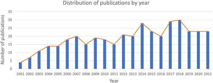
FIGURE 2. Trends of publications over the past 20 years. From 2001 to 2014, the number of documents showed an increasing trend, stabilized after 2014, and peaked in 2018.
Research teams from 43 countries and regions published 395 papers. In the distribution map of publishing countries, each circle represents a country. The size of the circle represents the publication outputs of the country, and the line between the circles represents cooperation between countries (Figure 3). Centrality describes the importance of a country or region represented by this node in this research field. The top ten countries and regions with the most literature are listed in Table 1. The country with the most literature is the United States (n = 147, 37.22%), followed by China (n = 91, 23.04%) and Canada (n = 54, 13.67%) (Figure 3). The country with the highest centrality in the literature is the United States (0.78), followed by Italy (0.43) and England (0.20) (Figure 4).
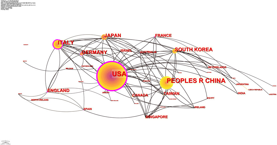
FIGURE 3. Distribution of publications from different countries/regions. The country with the largest number of publications is the United States, which also has the highest centrality of all countries. China has the second highest number of publications, but a lower centrality.
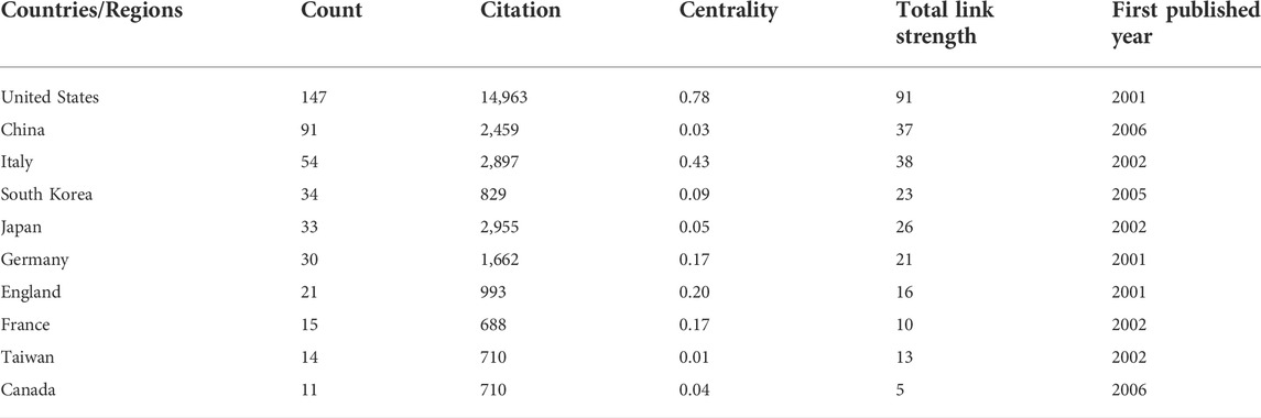
TABLE 1. Top 10 most productive countries and regions with publications.
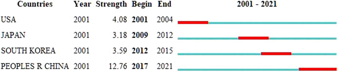
FIGURE 4. Top four countries/regions with the strongest citation bursts. A variable experiencing large changes in a short period of time indicates a strong burst of citations, and the red bar indicates the duration of the burst.
According to an analysis of CiteSpace and VOSviewer, 395 studies were published by 397 institutions. The top ten institutions with the most prolific publications are listed in Table 2. The most prolific institutions were University of Trieste (n = 22, 5.57%) and Harvard University (n = 22, 5.57%), followed by Tohoku University (n = 14, 3.54%). Of the top 10, two were American and two Italian. The most cited institutions include Harvard University (3,563), University of Trieste (2,234), Tohoku University (1,648), and Lab Nazl CIB (944); the co-occurrence relationship is shown in Figure 5. The evidence above indicates that institutions in the United States and Italy are leading research on Pin1 in the field of cell death.
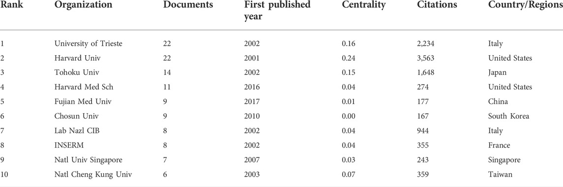
TABLE 2. Top 10 most productive organizations.
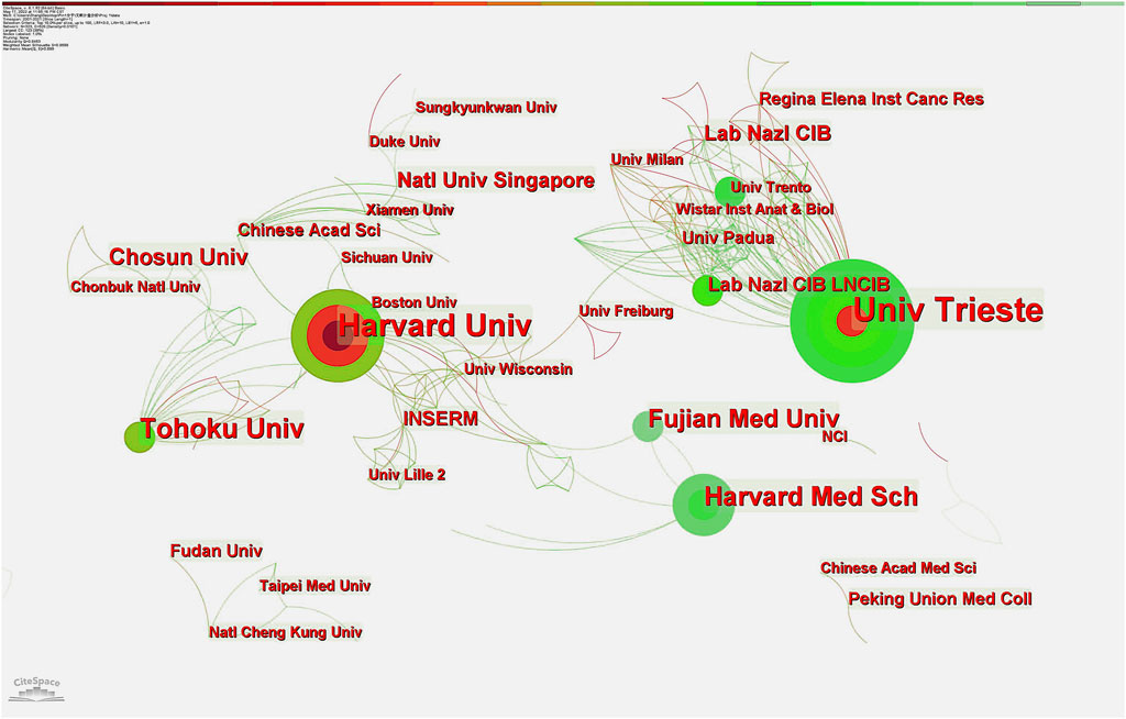
FIGURE 5. Distribution of publications from different institutions. Harvard University and the University of Trieste are tied for first place in the number of publications, with Harvard having the strongest centrality. These two core agencies have more cooperative links with most other institutions.
Based on the data visualization analysis using VOSviewer, the literature on Pin1 in the field of cell death from 2000 to 2021 was mainly distributed in 221 different journals, among which 64 published more than two articles. As shown in Table 3 and Figure 6, the journals with the highest citations are Nature and Journal of Biological Chemistry, which published 4 and 21 papers, respectively, and received 5,012 and 1,569 citations, respectively. This shows that the impact factors of the top 10 journals in 2021 range from 49.962 to 5.157. In 2021, Nature had the highest impact factor, while the Journal of Biological Chemistry had the lowest. Nature is the most influential journal in this research field based on the number of citations and journal impact factor data.
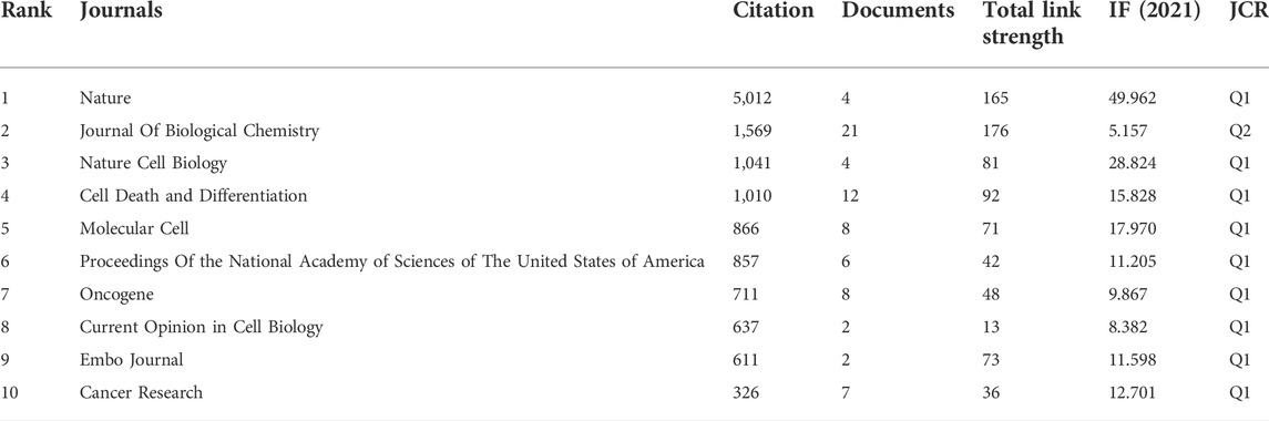
TABLE 3. Top 10 most cited journals.
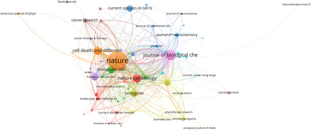
FIGURE 6. Journals with occurrence relations shown as an overlay graph plotted with VOSviewer 1.6.14. The analysis method is Linlog/modularity. The Weights was citations. The width of the line indicates the strength of the relationship.
A total of 510 authors participated in the writing of 395, studies. Collaborative clusters among different authors are shown by contact networks of distinct colors. The evaluation criteria for primary authors included the number of publications and H-index. The top 10 authors who published the most literature on Pin1 in the field of cell death from 2000 to 2021 are listed in Table 4. Giannino Del Sal of Lab Nazl CIB (LNCIB) ranks first among all authors, while Kun Ping Lu of Fujian Medical University ranks second with 15 publications. This shows the pattern of prolific authors from core institutions. The primary authors were from Italy, China, Japan, and Singapore. Figure 7 shows the patter of collaboration between authors. From the visualization diagram, we can observe that apart from a small number of author groups, author collaboration is centered on influential authors; the relationship between author groups is remarkably close. In terms of centrality, Giannino Del Sal tops the list with 0.12, and his citation number is also the highest among all the authors (n = 1,221), which shows that Giannino Del Sal has an important influence in this field of research; so does Kun Ping Lu.
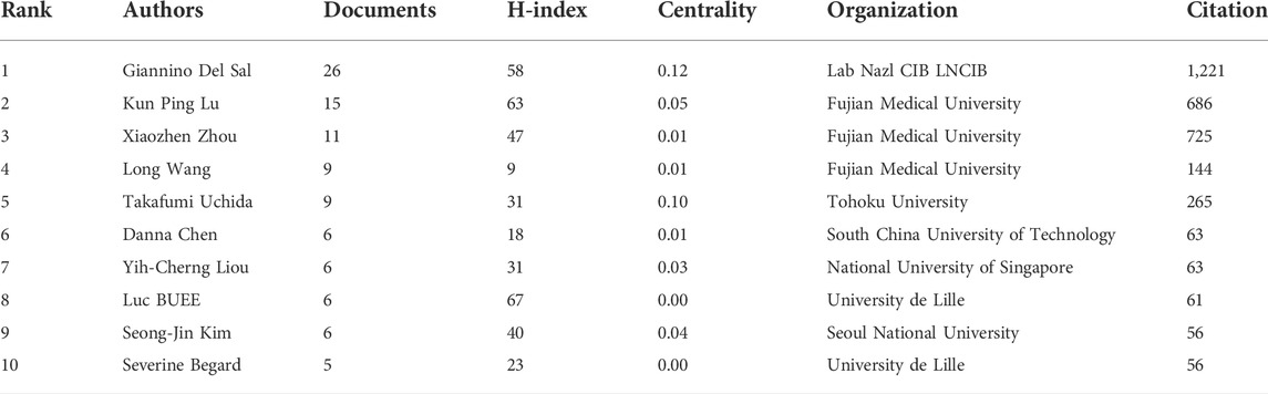
TABLE 4. Top 10 most productive authors.
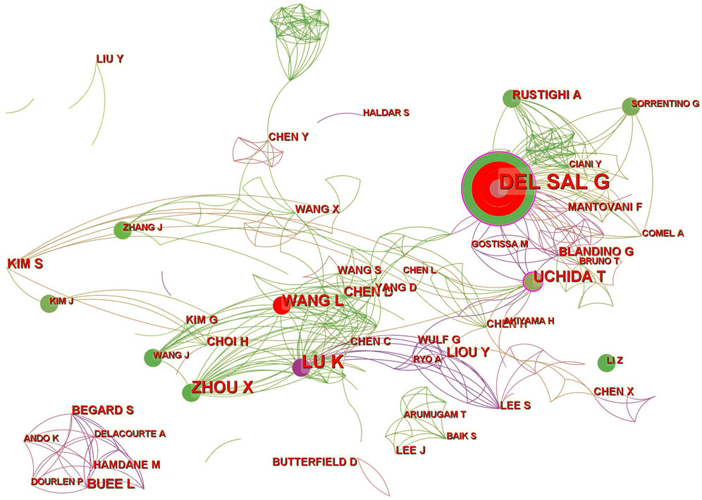
FIGURE 7. CiteSpace visualization map of authors involved. A circle represents an author, and the size of the circle represents the author’s number of articles. The purple part of the circle indicates the author’s centrality in the field.
Keywords can prompt or express the characteristics of the topic content of this study. By analyzing the frequency and distribution of keywords, we can understand the hot trends in research. A total of 903 keywords were retrieved from 395 articles. VOSviewer was used to draw the knowledge graph of the keywords (Figure 8). The frequencies and centralities of the keywords are listed in Table 5. As shown in Table 5, high-frequency keywords were Pin1 (206), apoptosis (80), phosphorylation (78), breast cancer (63), cell cycle (48), Alzheimer’s disease (37), and p53 (30). Figure 8 shows the frequency of keywords through the size of the circle and the clustering of keywords through the circle color. Owing to the difference in statistical methods between the two software tools, the keyword rankings in Figure 8 and Table 5 are somewhat different. It also should be noted that the “prolyl isomerase Pin1” and “Pin1” in Figure 8 and Table 5 are the same, which is due to the limitation of literature measurement tools.
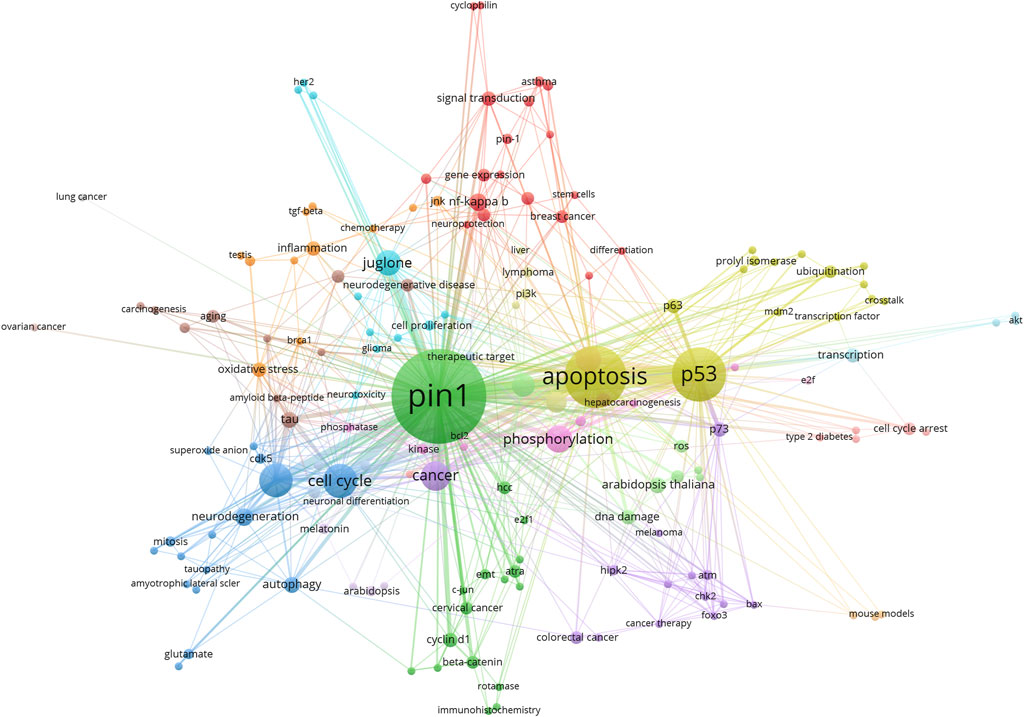
FIGURE 8. VOSviwer visualization map of keywords clustering analysis. The analysis method is Linlog/modularity. The size of the circle is the number of times the keyword appears. The colors of the circles in the figure represent the categories the keywords are in.
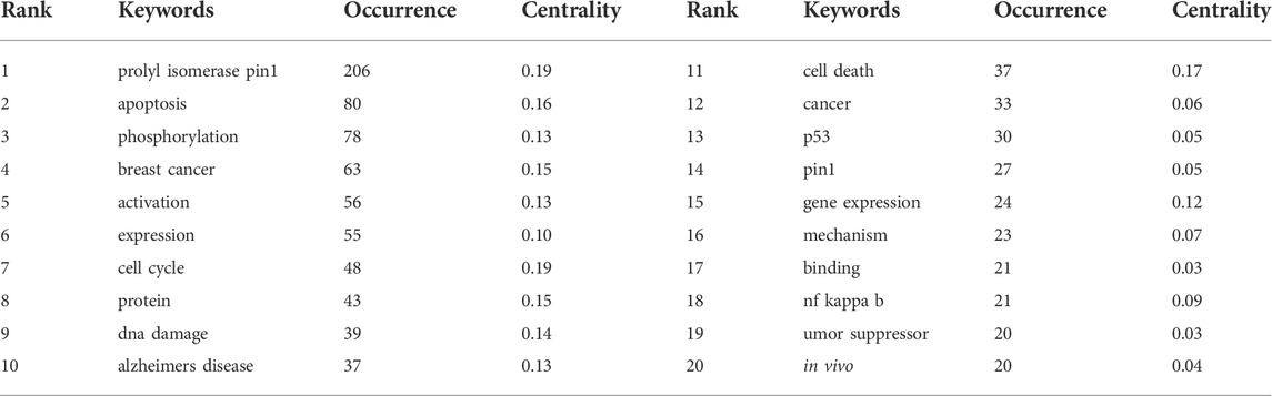
TABLE 5. Top 10 most productive keywords.
Co-citation refers to the fact that two (or more) papers are simultaneously cited by one or more subsequent papers, and the two papers are said to constitute co-citation relationships. The number of citations reflects the field’s influence. A total of 827 co-cited references were analyzed by co-clustering, and the top 10 highly cited references are listed in Table 6. As shown in Figure 9, the authors of the most frequently cited studies are Zacchi, Zheng, Zhou, Wei, and Lu.
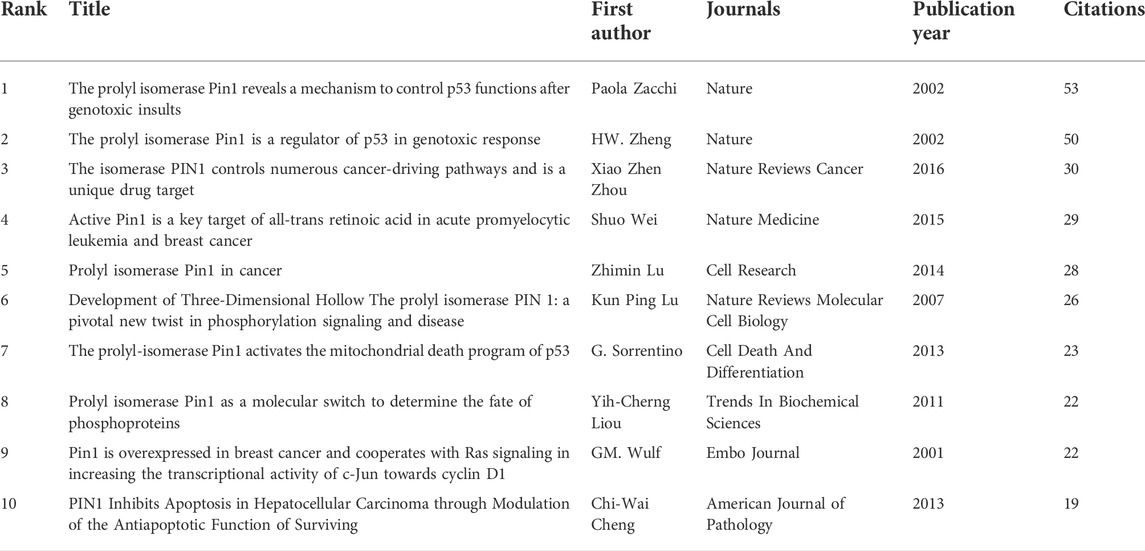
TABLE 6. Top 10 highly co-cited documents.
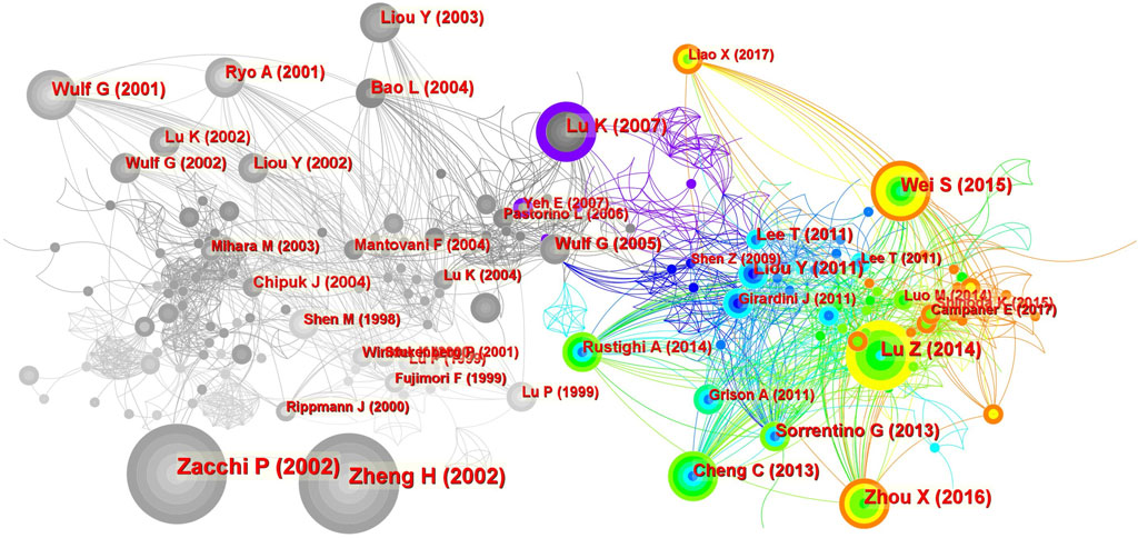
FIGURE 9. Co-citation analysis of references. The size of the circle represents the number of co-citations. The color of the circle represents the publication time of the co-cited literature, and the color of the outer circle represents the centrality of the literature.
A citation explosion refers to the fact that the number of citations of a paper increases rapidly in a brief period, indicating that the paper has received considerable attention from researchers in this field during this period. Figure 10 shows 40 articles with citation outbreaks. One of the two articles with high outbreak intensity cited in the last 2 years is Shuo Wei’s “Active Pin1 is a Key Target of All-trans Retinoic acid in acute Promyelocytic leukemia and Breast Cancer” in 2015. The outbreak intensity was 13.31, and the citation outbreak lasted from 2016 until present. Another study is “The Isomerase PIN1 Controls Numerous Cancer-Driving Pathways and is a Unique Drug Target,” published by Zhou and Xiao Zhen in 2016 with an outbreak intensity of 14.14. The citation outbreak lasted from 2017 to the present. At the same time, these two papers were also the fourth and third most cited papers, respectively, indicating that these two papers have had the greatest influence on Pin1 in the research field of cell death in recent years. After analyzing Figure 10, we found that almost all hot articles with high citation outbreaks after 2013 were related to cancer; representative cancers included breast and liver cancer. The high-citation burst articles before 2013 were more related to the molecular signaling mechanism of Pin1. This further verifies our thinking about the development trends in this field.
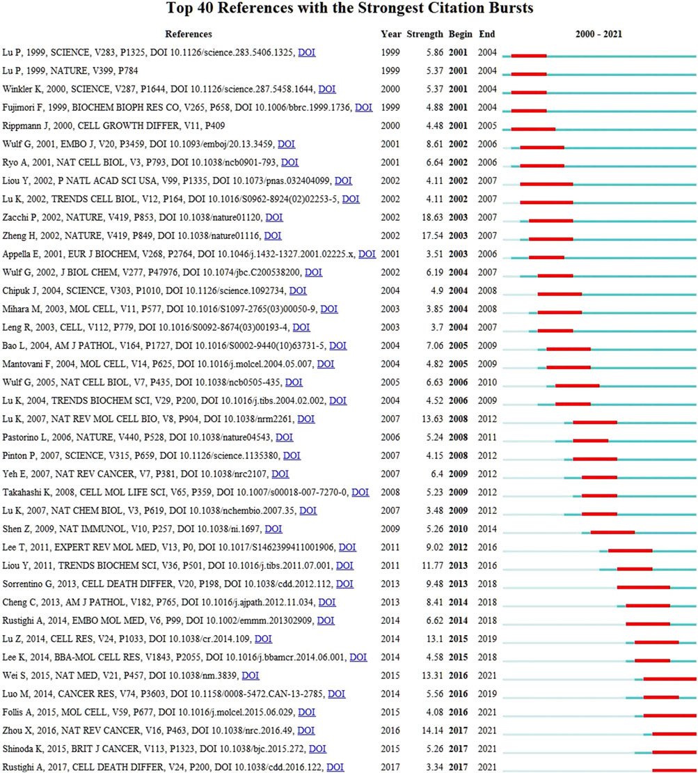
FIGURE 10. CiteSpace visualization map of top 40 references with the strongest citation bursts. A variable experiencing large changes in a short period of time indicates a strong burst of citations, and the red bar indicates the duration of the burst.
The 395 studies published in the past 20 years generally showed a trend of gradual increase over time and reached a peak in 2018 (Figure 2). From 2001 to 2014, research on Pin1 was in the stage of emerging increase, and the number of studies showed a growing trend. However, since 2014, research in this field has lost the trend of growth, which indicates that this field has not received much attention, or breakthrough research progress has not been made; thus, new research hotspots have not been formed. In conclusion, previous studies have improved the upstream and downstream molecular mechanisms and signaling pathways of Pin1 in cell death, and more studies on how to reduce or delay the occurrence of diseases by inhibiting Pin1 or blocking signaling pathways are needed.
According to the distribution of countries and regions in Table 1, the number of studies published (147) and cited (14,963) in the United States is the highest and far greater than that of other countries. Centrality is used to measure the importance of the nodes in the network. The relatively important countries with a threshold of over 0.1 are the United States (0.78), Italy (0.43), and England (0.20). In addition, the United States was the first country to publish literature in this field in 2001. All the above data show that the United States is the most influential country in this field. It is worth noting that China has the second-largest number of studies. Although centrality is low, the intensity of the literature explosion in China from 2017 to 2021 is as high as 12.76 (Figure 4). It is predicted that China will play a more significant role in this field in the future.
Harvard University in the United States has the highest number of literature publications and centrality. As can be seen from the co-occurrence figure (Figure 5), the cooperation within the cluster of regional research institutions is intricately linked and centrality is obvious. This means that Harvard University, the University of Trieste, and other research centers are taken as research centers, but there are few cooperative relationships between the clusters of research institutions. For example, there is little cooperation between Harvard University and the University of Trieste, which indicates that there is great space for development in cooperation between different countries, regions, and institutions in this field. According to the outbreak trend chart of research institutions, the outbreak intensity of Fujian Medical University in China has been as high as 4.17 since 2017, which indicates that Fujian Medical University has made many achievements in this field in recent years.
Giannino Del Sal (26, 6.6%) was the author of most publications, followed by Kun Ping Lu (15, 4.1%) and Xiaozhen Zhou (11, 2.78%). P Zacchi ranks first among the most co-cited authors, and HW Zheng ranks second. Both authors’ highly co-cited articles reveal the relationship between Pin1 and P53, and they are the founding authors of research in this field. The third most frequently cited article was published by Xiao Zhen Zhou and Kun Ping Lu in Nature Reviews on Cancer. In particular, Professor Kun Ping Lu found that over-activation of Pin1 in cancer interferes with the balance of oncogenes and tumor suppressors, thereby promoting the development of cancer and cancer stem cells. In addition, the role of Pin1 in cancer and the potential of Pin1 inhibitors to restore this balance have been discussed (Zhou and Lu, 2016). In 2018, Kozono et al. found that arsenic oxide inhibits Pin1 carcinogenesis by inhibiting and degrading Pin1. Additionally, they found that all-trans retinoic acid inhibits leukemia, breast cancer, and liver cancer by targeting isomerase Pin1 (Kozono et al., 2018). Among the top 10 co-cited articles, there are many studies on Pin1 and cancer. This indicates that research on Pin1 in the field of cell death has gradually shifted to the treatment of diseases represented by Pin1 inhibitors.
According to Table 3 and Figure 6, the most frequently cited journals in the field of Pin1 and cell death were Nature (4, 1.0%) and its subsidiary Nature Cell Biology (4, 1.0%), with a total of 6,053 citations. The Journal of Biological Chemistry (21, 5.3%) published the most articles with 1,569 citations, followed by Cell Death and Differentiation (12, 3.0%) with 1,010 citations. Of the top 10 journals, nine were classified in the Q1 region. According to the results, studies on Pin1 in cell death were mostly distributed in highlyinfluential journals, indicating that research achievements in this field have received significant attention.
Based on the analysis of keyword frequencies, popular research trends can be acknowledged. As shown in Table 6, in addition to PIN1, the most common keywords were apoptosis (80), phosphorylation (78), breast cancer (63), cell cycle (48), Alzheimer’s disease (37), and p53 (30). The keywords mainly focused on cell death, molecular mechanisms, and diseases. Pin1 is involved in three aspects of cell death: apoptosis, regulatory necrosis, and autophagy.
Apoptosis is controlled by several apoptotic genes and characterized by regulated cell death (Bai et al., 2020; Hu et al., 2021b; Nascimento et al., 2022). Pin1 has been reported to mediate neuronal apoptosis by promoting the expression of pro-apoptotic genes such as p53 (Grison et al., 2011; Balaganapathy et al., 2018). In Huntington’s disease (a neurodegenerative disease), the mutated Htt protein promotes the activation of Pin1, which subsequently interacts with p53 and triggers the dissociation of its inhibitors (inhibitors of p53’s apoptotic stimulator protein; IASPP). This ultimately promotes the expression of a series of apoptotic genes and leads to cell death (Grison et al., 2011). Recent studies have shown that DAPK1, which plays a key role in cell death, is a key modulator of Pin1 activity. The reduction in Pin1 activity is accompanied by dapK1-mediated increased expression of CIS PT231-Tau, which promotes neuronal cell death (Kim et al., 2021; Qiu et al., 2021). Knockout or functional inhibition of Pin1 almost completely prevented caspase-3 activation, DNA disruption, and cell death induced by 1-methyl-4-phenylpyridinium. This suggests that Pin1 promotes apoptosis (Ghosh et al., 2013). Other studies have shown that Pin1 binds to and stabilizes the NICD1 domain through its WW domain (Baik et al., 2015) and enhances its stability by inhibiting the F-box and WD repeat domains (containing polyubiquitination mediated by domain protein 7). This ultimately results in NICD1-induced apoptosis (Wang et al., 2014; Baik et al., 2015). The pro-apoptotic effect of Pin1 is also reflected in the activation of NICD1, which is involved in the regulation of p53 transactivation and the upregulation of pro-apoptotic genes (such as Bax) (Balaganapathy et al., 2018). Reactive oxygen species (ROS) production is a key effector of the PIN1-notch-p53 pathway (Li et al., 2019). The JNK pathway plays a complex role in many physiological and pathological processes such as apoptosis and differentiation and may be activated by various stress signals and inflammation (Zhang et al., 2021). The JNK pathway also promotes mitochondrial apoptosis by regulating the release of Bax under the control of Pin1 (Shen et al., 2009). As research on mitochondria, cell necrosis, and apoptosis is becoming increasingly important (Yan et al., 2021), it is necessary to determine the role of Pin1.
Regulatory necrosis (RN) includes necrotizing apoptosis induced by tumor necrosis factor, which is mediated by receptor-interacting protein three; scoria, which is dependent on caspase-1 activation and triggered by microbial infection; aerastin-mediated iron death, which requires excess iron; and RN, which is dependent on mitochondrial permeability transition (Yuan et al., 2019; Alu et al., 2020; Chen et al., 2021b; Liao et al., 2021; Tonnus et al., 2021). Pin1 acts as an important downregulatory factor in dapK1-induced excitatory toxic necrosis (Wang et al., 2018). In addition, activation of calpain-2 induces the cleavage of apoptosis-inducing factors, which are transferred to the nucleus and induce DNA degradation, resulting in neuronal RN (Wang et al., 2018; Wang et al., 2019b). Calpastatin (CAST) is an endogenous specific inhibitor of CAST (Wang et al., 2014; Cheng et al., 2018), and Pin1 promotes excitatory toxic glutamate injury. This results in a reduced inhibitory effect of CAST on calpain-2 and induction of cell necrosis (Wang et al., 2018; Wang et al., 2019a). In recent years, PANoptosis, which has been shown to exist in neuronal ischemia/reperfusion injury (Yan et al., 2022; Yan et al., 2023), and other new regulatory modes of necrosis (such as iron necrosis and copper necrosis) have been discovered successively. However, Pin1 has not received much attention in these fields, and the relevant regulatory mechanisms still need to be further studied.
Autophagy plays a critical role in cell homeostasis by degrading dysfunctional and degraded proteins and damaged organelles inside cells into autophagosomes, which fuse with lysosomes to form autolysosomes (Chao et al., 2022). Pin1 is thought to regulate the proteasome during autophagy (So and Sum, 2015). Pin1 inhibits GSK-3β expression by inhibiting proteasome degradation, and ultimately stimulates cell death through autophagy (So, 2015). In contrast, inhibition of GSK-3β induces Pin1 activation, suggesting that GSK-3β may be a mediator of Pin1 activity in autophagy (Chao et al., 2022).
Our literature retrieval work was completed on 7 May 2022, and the retrieval date ranged from 1 January 2000, to 31 December 2021. Therefore, it is possible that some articles that met the retrieval conditions could not be retrieved because their publication dates were too early or too late, making the number of studies in our database different from the actual number. Only articles and reviews were selected for this study because we wanted to control the quality of the included literature as much as possible. However, through this process, we might have ignored the research progress from other publications that have higher values. The fields of Pin1 and cell death cover many aspects of research, among which many primary keywords were not included in the retrieval formula. This made it impossible to retrieve more relevant research results, despite helping us avoid importing irrelevant articles. In addition, because of the limitation of tools, we can only search the literature on the Web of Science Core Collection; thus, the literature not included in the Web of Science Core Collection is omitted. Finally, bibliometric tools, such as CiteSpace and VOSviewer, still have limitations in their functions. They cannot replace systematic scientific retrieval and are still limited by the experience and knowledge level of researchers.
Research on the molecular mechanism between Pin1 and cell death, the difference between Pin1 functions in different signaling pathways, and the role of Pin1 in the prevention or delay of diseases is of great research value, especially has exciting application prospects in the field of cancer and nervous system diseases. We used bibliometrics to analyze and visualize the current situation of research in this field and found that research on Pin1 and cell death shows an increasing trend over time, and that there are opportunities for further research. Undoubtedly, the United States plays the most vital role in this field, and Harvard University is the most influential institution. China’s outstanding performance in recent years is sufficient to keep us hopeful that China will play a more key role in this field in the future. Professor Kun Ping Lu is an outstanding researcher in this field of study in that he and his team have published several valuable research results in high-influence journals. Another sign that research on Pin1 and cell death has attracted more attention is that many research results in this field have been included and cited by internationally influential journals. We speculate that pending the discovery of new molecular mechanisms or signaling pathways, future studies on Pin1 and cell death will focus more on the treatment of diseases.
The original contributions presented in the study are included in the article/supplementary material, further inquiries can be directed to the corresponding author.
S-CW designed the experiments, interpreted the data, and revised the manuscript. J-HZ performed the experiments, prepared the figures, and wrote the manuscript. S-YN and Y-TT helped to perform the experiments and prepared the figures. JL prepared the figures and gave helpful advice during the revision. All authors read, discussed, and approved the final manuscript.
The study was supported by National Natural Science Foundation of China, No. 82101126, Natural Science Foundation of Hunan Province, No. 2021JJ40873, the Hunan Provincial Natural Science Foundation of China (No.2019JJ50696), the Scientific Research Launch Project for new employees of the Second Xiangya Hospital of Central South University.
The authors declare that the research was conducted in the absence of any commercial or financial relationships that could be construed as a potential conflict of interest.
All claims expressed in this article are solely those of the authors and do not necessarily represent those of their affiliated organizations, or those of the publisher, the editors and the reviewers. Any product that may be evaluated in this article, or claim that may be made by its manufacturer, is not guaranteed or endorsed by the publisher.
Alu, A., Han, X. J., Ma, X. L., Wu, M., Wei, Y. Q., and Wei, X. W. (2020). The role of lysosome in regulated necrosis. Acta Pharm. Sin. B 10 (10), 1880–1903. doi:10.1016/j.apsb.2020.07.003
Avena, M. J., and Barbosa, D. A. (2017). Bibliometric indicators of the nursing journals according to the index databases. Rev. Esc. Enferm. Usp. 51, e03262. doi:10.1590/s1980-220x2017014603262
Bai, Y. J., Lam, H. C., and Lei, X. G. (2020). Dissecting programmed cell death with small molecules. Acc. Chem. Res. 53 (5), 1034–1045. doi:10.1021/acs.accounts.9b00600
Baik, S. H., Fane, M., Park, J. H., Cheng, Y. L., Fann, D. Y. W., Yun, U. J., et al. (2015). Pin1 promotes neuronal death in stroke by stabilizing notch intracellular domain. Ann. Neurol. 77 (3), 504–516. doi:10.1002/ana.24347
Balaganapathy, P., Baik, S. H., Mallilankaraman, K., Sobey, C. G., Jo, D. G., and Arumugam, T. V. (2018). Interplay between Notch and p53 promotes neuronal cell death in ischemic stroke. J. Cereb. Blood Flow. Metab. 38 (10), 1781–1795. doi:10.1177/0271678x17715956
Bianchi, M., D'Oria, V., Braghini, M. R., Petrini, S., and Manco, M. (2019). Liraglutide treatment ameliorates neurotoxicity induced by stable silencing of Pin1. Int. J. Mol. Sci. 20 (20), E5064. doi:10.3390/ijms20205064
Chao, X. J., Wang, S. G., Fulte, S., Ma, X. W., Ahamed, F., Cui, W., et al. (2022). Hepatocytic p62 suppresses ductular reaction and tumorigenesis in mouse livers with mTORC1 activation and defective autophagy. J. Hepatol. 76 (3), 639–651. doi:10.1016/j.jhep.2021.10.014
Chen, C. M. (2006). CiteSpace II: Detecting and visualizing emerging trends and transient patterns in scientific literature. J. Am. Soc. Inf. Sci. Technol. 57 (3), 359–377. doi:10.1002/asi.20317
Chen, W., Jiang, L. F., Hu, Y. Q., Fang, G., Yang, B. L., Li, J. H., et al. (2021a). Nanomedicines, an emerging therapeutic regimen for treatment of ischemic cerebral stroke: A review. J. Control. Release 340, 342–360. doi:10.1016/j.jconrel.2021.10.020
Chen, Y. H., Li, Y., Guo, L. M., Hong, J., Zhao, W. J., Hu, X. M., et al. (2021b). Bibliometric analysis of the inflammasome and pyroptosis in brain. Front. Pharmacol. 11, 626502. doi:10.3389/fphar.2020.626502
Cheng, S. Y., Wang, S. C., Lei, M., Wang, Z., and Xiong, K. (2018). Regulatory role of calpain in neuronal death. Neural Regen. Res. 13 (3), 556–562. doi:10.4103/1673-5374.228762
Ghosh, A., Saminathan, H., Kanthasamy, A., Anantharam, V., Jin, H. J., Sondarva, G., et al. (2013). The peptidyl-prolyl isomerase Pin1 up-regulation and proapoptotic function in dopaminergic neurons relevance to the pathogenesis of Parkinson disease. J. Biol. Chem. 288 (30), 21955–21971. doi:10.1074/jbc.M112.444224
Grison, A., Mantovani, F., Comel, A., Agostoni, E., Gustincich, S., Persichetti, F., et al. (2011). Ser46 phosphorylation and prolyl-isomerase Pin1-mediated isomerization of p53 are key events in p53-dependent apoptosis induced by mutant huntingtin. Proc. Natl. Acad. Sci. U. S. A. 108 (44), 17979–17984. doi:10.1073/pnas.1106198108
Hleihel, R., El Hajj, H., Wu, H. C., Berthier, C., Zhu, H. H., Massoud, R., et al. (2021). A Pin1/PML/P53 axis activated by retinoic acid in NPM-1c acute myeloid leukemia. Haematologica 106 (12), 3090–3099. doi:10.3324/haematol.2020.274878
Hu, X. M., Li, Z. X., Lin, R. H., Shan, J. Q., Yu, Q. W., Wang, R. X., et al. (2021a). Guidelines for regulated cell death assays: A systematic summary, A categorical comparison, A prospective. Front. Cell. Dev. Biol. 9, 634690. doi:10.3389/fcell.2021.634690
Hu, X. M., Zhang, Q., Zhou, R. X., Wu, Y. L., Li, Z. X., Zhang, D. Y., et al. (2021b). Programmed cell death in stem cell-based therapy: Mechanisms and clinical applications. World J. Stem Cells 13 (5), 386–415. doi:10.4252/wjsc.v13.i5.386
Kim, N., Wang, B., Koikawa, K., Nezu, Y., Qiu, C. X., Lee, T. H., et al. (2021). Inhibition of death-associated protein kinase 1 attenuates cis P-tau and neurodegeneration in traumatic brain injury. Prog. Neurobiol. 203, 102072. doi:10.1016/j.pneurobio.2021.102072
Koikawa, K., Kibe, S., Suizu, F., Sekino, N., Kim, N., Manz, T. D., et al. (2021). Targeting Pin1 renders pancreatic cancer eradicable by synergizing with immunochemotherapy. Cell. 184 (18), 4753–4771. e27. doi:10.1016/j.cell.2021.07.020
Kozono, S., Lin, Y. M., Seo, H. S., Pinch, B., Lian, X. L., Qiu, C. X., et al. (2018). Arsenic targets Pin1 and cooperates with retinoic acid to inhibit cancer-driving pathways and tumor-initiating cells. Nat. Commun. 9, 3069. doi:10.1038/s41467-018-05402-2
Kumari, A., Kumar, C., Pergu, R., Kumar, M., Mahale, S. P., Wasnik, N., et al. (2021). Phosphorylation and Pin1 binding to the LIC1 subunit selectively regulate mitotic dynein functions. J. Cell. Biol. 220 (12), e202005184. doi:10.1083/jcb.202005184
Lammert, C. R., Frost, E. L., Bellinger, C. E., Bolte, A. C., McKee, C. A., Hurt, M. E., et al. (2020). AIM2 inflammasome surveillance of DNA damage shapes neurodevelopment. Nature 580 (7805), 647–652. doi:10.1038/s41586-020-2174-3
Li, L., Su, Z. J., Zou, Z. M., Tan, H. P., Cai, D. Z., Su, L., et al. (2019). Ser46 phosphorylation of p53 is an essential event in prolyl-isomerase Pin1-mediated p53-independent apoptosis in response to heat stress. Cell. Death Dis. 10, 96. doi:10.1038/s41419-019-1316-8
Liao, L. S., Lu, S., Yan, W. T., Wang, S. C., Guo, L. M., Yang, Y. D., et al. (2021). The role of HSP90 alpha in methamphetamine/hyperthermia-induced necroptosis in rat striatal neurons. Front. Pharmacol. 12, 716394. doi:10.3389/fphar.2021.716394
Lu, K. P., Finn, G., Lee, T. H., and Nicholson, L. K. (2007). Prolyl cis-trans isomerization as a molecular timer. Nat. Chem. Biol. 3 (10), 619–629. doi:10.1038/nchembio.2007.35
Makinwa, Y., Musich, P. R., and Zou, Y. (2020). Phosphorylation-dependent Pin1 isomerization of ATR: Its role in regulating ATR's anti-apoptotic function at mitochondria, and the implications in cancer. Front. Cell. Dev. Biol. 8, 281. doi:10.3389/fcell.2020.00281
Nascimento, F. R., Baeta, J., de Franca, A. A. P., Oliveira, M., Pizziolo, V. R., dos Santos, A. A., et al. (2022). Dibenzoylmethane derivative inhibits melanoma cancer in vitro and in vivo through induction of intrinsic and extrinsic apoptotic pathways. Chem. Biol. Interact. 351, 109734. doi:10.1016/j.cbi.2021.109734
Qiu, C. X., Albayram, O., Kondo, A., Wang, B., Kim, N., Arai, K., et al. (2021). Cis P-tau underlies vascular contribution to cognitive impairment and dementia and can be effectively targeted by immunotherapy in mice. Sci. Transl. Med. 13 (596), 15. doi:10.1126/scitranslmed.aaz7615
Shen, Z. J., Esnault, S., Schinzel, A., Borner, C., and Malter, J. S. (2009). The peptidyl-prolyl isomerase Pin1 facilitates cytokine-induced survival of eosinophils by suppressing Bax activation. Nat. Immunol. 10 (3), 257–265. doi:10.1038/ni.1697
Smith, D. R. (2008). Bibliometrics, dermatology and contact dermatitis. Contact Dermat. 59 (3), 133–136. doi:10.1111/j.1600-0536.2008.01405.x
Sung, P.-S., and Hsieh, S.-L. (2021). C-type lectins and extracellular vesicles in virus-induced NETosis. J. Biomed. Sci. 28 (1), 46. doi:10.1186/s12929-021-00741-7
Theobald, S. J., Graeb, J., Fritsch, M., Suarez, I., Eisfeld, H. S., Winter, S., et al. (2021). Gasdermin D mediates host cell death but not interleukin-1 beta secretion in Mycobacterium tuberculosis-infected macrophages. Cell. Death Discov. 7 (1), 327. doi:10.1038/s41420-021-00716-5
Tonnus, W., Belavgeni, A., Beuschlein, F., Eisenhofer, G., Fassnacht, M., Kroiss, M., et al. (2021). The role of regulated necrosis in endocrine diseases. Nat. Rev. Endocrinol. 17 (8), 497–510. doi:10.1038/s41574-021-00499-w
van Eck, N. J., and Waltman, L. (2010). Software survey: VOSviewer, a computer program for bibliometric mapping. Scientometrics 84 (2), 523–538. doi:10.1007/s11192-009-0146-3
Wang, L. X., Ye, X. T., Liu, Y. Y., Wei, W. Y., and Wang, Z. W. (2014). Aberrant regulation of FBW7 in cancer. Oncotarget 5 (8), 2000–2015. doi:10.18632/oncotarget.1859
Wang, S. C., Hu, X. M., and Xiong, K. (2023). The regulatory role of Pin1 in neuronal death. Neural Regen. Res. 18 (1), 74–80. doi:10.4103/1673-5374.341043
Wang, S. C., Huang, Y. X., Yan, Y. H., Zhou, H. K., Wang, M., Liao, L. S., et al. (2019a). Calpain2 but not calpain1 mediated by calpastatin following glutamate-induced regulated necrosis in rat retinal neurons. Ann. Anatomy-Anatomischer Anzeiger 221, 57–67. doi:10.1016/j.aanat.2018.08.005
Wang, S. C., Liao, L. S., Huang, Y. X., Wang, M., Zhou, H. K., Chen, D., et al. (2019b). Pin1 is regulated by CaMKII activation in glutamate-induced retinal neuronal regulated necrosis. Front. Cell. Neurosci. 13, 276. doi:10.3389/fncel.2019.00276
Wang, S. C., Liao, L. S., Wang, M., Zhou, H. K., Huang, Y. X., Wang, Z., et al. (2018). Pin1 promotes regulated necrosis induced by glutamate in rat retinal neurons via CAST/Calpain2 pathway. Front. Cell. Neurosci. 11, 425. doi:10.3389/fncel.2017.00425
Yan, W.-T., Zhao, W.-J., Hu, X.-M., Ban, X.-X., Ning, W.-Y., Wan, H., et al. (2023). PANoptosis-like cell death in ischemia/reperfusion injury of retinal neurons. Neural Regen. Res. 18 (2), 357–363. doi:10.4103/1673-5374.346545
Yan, W. T., Lu, S., Yang, Y. D., Ning, W. Y., Cai, Y., Hu, X. M., et al. (2021). Research trends, hot spots and prospects for necroptosis in the field of neuroscience. Neural Regen. Res. 16 (8), 1628–1637. doi:10.4103/1673-5374.303032
Yan, W. T., Yang, Y. D., Hu, X. M., Ning, W. Y., Liao, L. S., Lu, S., et al. (2022). Do pyroptosis, apoptosis, and necroptosis (PANoptosis) exist in cerebral ischemia? Evidence from cell and rodent studies. Neural Regen. Res. 17 (8), 1761–1768. doi:10.4103/1673-5374.331539
Yuan, J. Y., Amin, P., and Ofengeim, D. (2019). Necroptosis and RIPK1-mediated neuroinflammation in CNS diseases. Nat. Rev. Neurosci. 20 (1), 19–33. doi:10.1038/s41583-018-0093-1
Zhang, H. Y., Jiao, W. J., Cui, H. L., Sun, Q. H., and Fan, H. G. (2021). Combined exposure of alumina nanoparticles and chronic stress exacerbates hippocampal neuronal ferroptosis via activating IFN-γ/ASK1/JNK signaling pathway in rats. J. Hazard. Mat. 411, 125179. doi:10.1016/j.jhazmat.2021.125179
Zheng, F. L., Li, Y. Q., Zhang, F. S., Sun, Y., Zheng, C. Y., Luo, Z. S., et al. (2021). Cobalt induces neurodegenerative damages through Pin1 inactivation in mice and human neuroglioma cells. J. Hazard. Mat. 419, 126378. doi:10.1016/j.jhazmat.2021.126378
Keywords: bibliometric analysis, Pin1, cell death, apoptosis, autophagy, necrosis, CiteSpace, VOSviewer
Citation: Zhang J-H, Ni S-Y, Tan Y-T, Luo J and Wang S-C (2022) A bibliometric analysis of PIN1 and cell death. Front. Cell Dev. Biol. 10:1043725. doi: 10.3389/fcell.2022.1043725
Received: 13 September 2022; Accepted: 17 October 2022;
Published: 31 October 2022.
Edited by:
Degui Wang, Lanzhou University, ChinaReviewed by:
Yan Xiong, Wuhan University, ChinaCopyright © 2022 Zhang, Ni, Tan, Luo and Wang. This is an open-access article distributed under the terms of the Creative Commons Attribution License (CC BY). The use, distribution or reproduction in other forums is permitted, provided the original author(s) and the copyright owner(s) are credited and that the original publication in this journal is cited, in accordance with accepted academic practice. No use, distribution or reproduction is permitted which does not comply with these terms.
*Correspondence: Shu-Chao Wang, d2FuZ3NodWNoYW9AY3N1LmVkdS5jbg==
Disclaimer: All claims expressed in this article are solely those of the authors and do not necessarily represent those of their affiliated organizations, or those of the publisher, the editors and the reviewers. Any product that may be evaluated in this article or claim that may be made by its manufacturer is not guaranteed or endorsed by the publisher.
Research integrity at Frontiers
Learn more about the work of our research integrity team to safeguard the quality of each article we publish.