 Feng Han
 Qingchen Meng1†
 Jie Hu
 Jiaying Li
 Fengxuan Han
94% of researchers rate our articles as excellent or good
Learn more about the work of our research integrity team to safeguard the quality of each article we publish.
Find out more
REVIEW article
Front. Bioeng. Biotechnol., 04 July 2023
Sec. Biomaterials
Volume 11 - 2023 | https://doi.org/10.3389/fbioe.2023.1205792
This article is part of the Research TopicEngineered Biomimetic Micro/Nano-Materials for Tissue RegenerationView all 11 articles
The incidence of tissue and organ damage caused by various diseases is increasing worldwide. Tissue engineering is a promising strategy of tackling this problem because of its potential to regenerate or replace damaged tissues and organs. The biochemical and biophysical cues of biomaterials can stimulate and induce biological activities such as cell adhesion, proliferation and differentiation, and ultimately achieve tissue repair and regeneration. Micro/nano materials are a special type of biomaterial that can mimic the microstructure of tissues on a microscopic scale due to its precise construction, further providing scaffolds with specific three-dimensional structures to guide the activities of cells. The study and application of biomimetic micro/nano-materials have greatly promoted the development of tissue engineering. This review aims to provide an overview of the different types of micro/nanomaterials, their preparation methods and their application in tissue regeneration.
Due to lifestyle changes and population aging, the incidence of various diseases, such as degenerative diseases, cancer and trauma, has been increasing. Therefore, there is an increasing demand for tissue and organ transplantation in clinical practice (Bakhshandeh et al., 2017; Nii and Katayama, 2021). However, the repair or replacement of organs and tissues remains a complex and unsolved problem. Current graft materials cannot be widely used due to limitations such as material mismatch and immune rejection (Chung et al., 2017). Tissue engineering which integrates different disciplines, such as life sciences and materials sciences, has a potential for the development of alternative therapeutic strategies to repair damaged tissues and organs.
The basic concepts of tissue engineering include seeding cells on scaffolds of a certain shape, expanding the cells continuously to form a composite system of cells and biomaterials, and finally implanting them into the patient. After in vivo implantation, the polymer system will gradually degrade, allowing regeneration of new tissues. In this functional and biomimetic system, the three major elements are the selection of seed cells, the construction of scaffolds, and the regulation of cell-biomaterial interactions (Li et al., 2020). Biomaterials used for tissue engineering must have the following properties: (1) It has good biocompatibility, non-toxic side effects on cells and tissues; (2) The surface of the materials is conducive to cell adhesion and proliferation, and can induce cell growth according to the predetermined morphology; (3) The degradation rate should match the new tissue formation rate; (4) Three-dimensional (3D) structure. Studies have shown that the surface microstructure of biomaterials has a great influence on cell morphology, adhesion, directional growth and bioactivity (Yu et al., 2020; Han et al., 2022).
Biomaterials are natural or artificial materials used to replace or repair tissue. For instance, the annulus fibrosus (AF) has a collagen-rich fibrous lamellar with highly aligned collagen fibers tilted 30° from the horizontal axis. The microstructure allows the spine to resist complex loading. Tendon has a hierarchical organization, and the subunits of the tendon are the fascicles, which consist of highly aligned collagen fibers. With the continuous improvement of tissue engineering and regeneration, plenty of biomaterials have been developed. With the in-depth understanding of the microstructure of different tissues and organs, researchers have noted that it is important to simulate the microstructure of tissues and organs by biomaterials at a more microscopic scale to achieve better tissue repair (Craciunescu et al., 2021). Tissue engineering generally controls the growth and development of cells in three scales. The millimeter scale determines the overall shape of the engineering tissue. The micron scale determines the pore size of the material and regulates cell migration and growth. The nanometer scale determines the physical and chemical properties of material surface, which regulates cell adhesion and gene expression. The properties of extracellular matrix (ECM), such as matrix stiffness, surface topography, and chemical composition, can affect cell behavior spatially and temporally, which is attributed to its fusion with mechanical transduction and molecular recognition (Wang et al., 2018). Those will affect cell phenotype, gene expression, and ultimately cell fate. Moreover, the geometry of ECM also has a great influence on their mechanical properties. It can be seen that the micro/nano structure of ECM has a great impact on regulating cell behavior, which in turn affects tissue organization. Therefore, the preparation of biomaterials with certain micro and nano structures is of great significance for tissue repair and regeneration.
As one of the three elements of tissue engineering, the scaffold is the foundation of constructing engineered tissues. Suitable 3D scaffolds can simulate the microstructure of tissues and provide a suitable microenvironment for cell and tissue growth. Micro/nano-materials have good properties of general biomaterials. The special pores of micro/nano-materials can support nutrient transport, absorption, and tissue growth (Carotenuto et al., 2022). For specific tissues, such as bone and cartilage, higher requirements are also put forward for the mechanical properties of micro/nano-materials. Therefore, more and more attention has also been paid to the mechanical properties. In recent years, a variety of multi-functional micro/nanomaterials have been applied in tissue engineering, exhibiting a broad application prospect. However, the application of micro/nano-materials in tissue engineering is still in the early stages, and there are still many problems regarding the clinical application that need to be solved. The problems include developing suitable micro/nanomaterials to simulate the microstructure of tissue, constructing an ideal cell-material interface to induce directional differentiation of cells, making allogeneic biological tissues and cells cultured on micro/nano scaffolds from immune system recognition and rejection, maintaining the viability and function of cultured cells for a long time, and further improving the biocompatibility of micro/nano-materials.
Herein, we provide a comprehensive overview of the current research progress on micro/nanomaterials and introduce the types of micro/nanomaterials and their applications (Figure 1). First, the importance of scaffold micro/nanomaterials on cell behavior and biomimetic microstructure is highlighted. Next, several preparation methods of scaffolds are introduced. Then, examples of the application of micro/nanomaterials in bone, cartilage, blood vessels and other tissues are introduced. Finally, some limitations and challenges are briefly discussed.
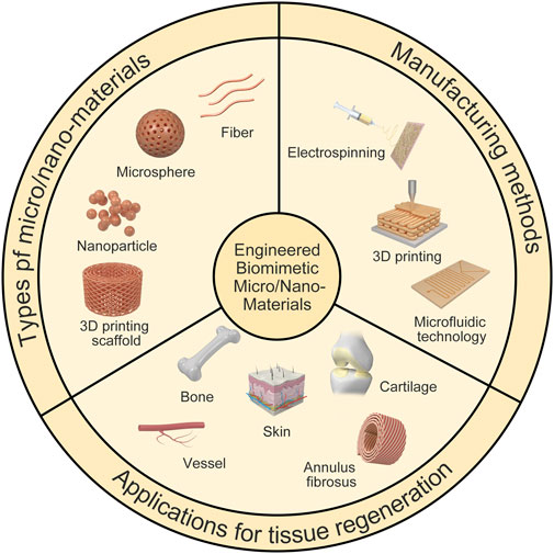
FIGURE 1. Illustration of engineered biomimetic micro/nano-materials for tissue regeneration. The micro/nano structures including micro/nano fiber, nanoparticle, microsphere and 3D printing scaffold, and the biomedical applications referring to bone repair, AF repair and vascular grafts are shown as examples. Manufacturing methods for different micro/nano-materials are also shown.
Electrospinning technology can continuously produce micro/nano-sized ultrafine fibers (Zamani et al., 2018). The scaffold prepared using the electrospinning technology has a unique micro/nano structure and proper mechanical properties (Figure 2). It can simulate the micro/nano-network structure of natural ECM and has unique advantages in the preparation of tissue engineered scaffolds (Recum et al., 1996; Flemming et al., 1999; Mitragotri et al., 2015). Generally, the electrospinning device consists of a high-voltage source, a solution reservoir, and spraying and receiving devices. The electrospinning process can be divided into five processes, including fluid charging, Taylor cone formation, jet stretching and whipping, jet solidification, and fiber reception (Liu et al., 2012; Zamani et al., 2018). The most important process is the formation of the Taylor cone. The electrospinning process can be influenced by many factors such as the solvent properties (e.g., polymer type, concentration, viscosity, conductivity), the processing factors (e.g., electric field strength, flow rate, spray distance), and the ambient parameters (e.g., temperature, humidity) (Bhardwaj and Kundu, 2010; Pelipenko et al., 2015; Ye et al., 2019).
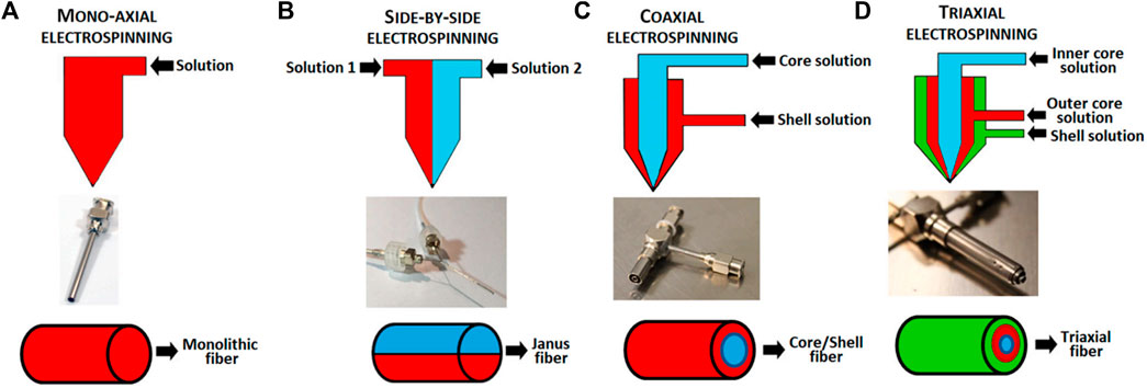
FIGURE 2. Schematic of electrospinning setup and the fibers output. (A) mono-axial electrospinning; (B) side-by-side electrospinning; (C) coaxial electrospinning and (D) triaxial electrospinning (Luraghi et al., 2021).
Core-shell nanofiber scaffold is a special type of nanofibers. Core-shell nanofiber can combine the excellent properties of two or more polymers. It uses the core-shell structure to encapsulate specific substances, such as drugs and growth factors, into nanofibers (Kim et al., 2004; Bölgen et al., 2007; Nie and Wang, 2007). In the fiber material system, nanofibers have the characteristics of high porosity, high specific surface area, and high surface activity. This structure is suitable for processes in which the fiber needs to fully contact and react with the surrounding environment, such as drug release. Mixing the drug directly into the nanofibers often results in burst release of the drug, while the core-shell nanofiber can encapsulate drugs in the core layer and prolong their release (Mohiti-Asli et al., 2017; da Silva et al., 2019). Some drugs or active substances should not be exposed to organic solvents. The core structural material can enclose the drugs or active substances inside to prevent them from being exposed to organic solvents. Because the drug or active substance is dissolved in the core solution, the surface modification of the fiber will not affect the activity of the internal drug. The main manufacturing methods of core-shell nanofibers reported in the literature are coaxial electrospinning and emulsion electrospinning.
The difference between coaxial electrospinning and ordinary electrospinning lies in the design of the spinneret. Ordinary electrospinning uses a single-layer capillary while coaxial electrospinning uses a coaxial nozzle (Zupančič et al., 2016). A coaxial nozzle is formed by nesting two or more coaxial capillaries with each other, and a certain gap is left between the inner and outer capillaries to ensure the smooth flow of the shell liquid. In coaxial electrospinning, the solution of the core and shell material is divided into two different syringes, and the spinning system is composed of two coaxial but different inner diameter capillary tubes. Under the action of a high-voltage electric field, the outer shell liquid flows out (Huang et al., 2006). Coaxial electrospinning relies on complex electrospinning equipment such as coaxial nozzles.
Emulsion electrospinning is a technical method capable of preparing nanofibers with core-shell structure in one step (Bazilevsky et al., 2007; Moydeen et al., 2018). In general, emulsion electrospinning solution mainly uses a water-in-oil or oil-in-water two-phase dispersion system to form an external phase by continuous polymer organic solution and an internal phase with drug aqueous solution in the emulsion. Amphiphilic emulsifiers are used to stabilize the interaction between the external phase and the internal phase to form an emulsion (Qi et al., 2006). The drug-loaded nanofibers prepared by this method can effectively solve the problem of burst drug release and can slow and control the release of the loaded substance (Liu et al., 2018). Compared with coaxial electrospinning, the advantage of emulsion electrospinning is that the device is simple without the requirement of coaxial nozzles. However, it has higher requirements for spinning solution, and the instability of the emulsion limits its wide application.
3D printing technology, a type of rapid prototyping technology, is a digital model file based on the use of adhesive materials through layer-by-layer printing to construct an object. As a new technology, 3D printing is developing rapidly, as there are many established manufacturing techniques and a large number of experimental techniques (Li et al., 2020). The most popular 3D bioprinting technologies include laser-assisted bioprinting, inkjet printing, and extrusion and robotic dispensing bioprinters (Figure 3). In recent years, 3D printing technology has been applied to tissue engineering with the aim of constructing biomaterials that simulate the microstructure of tissues and organs. The 3D bio-printing process comprises three steps. (1) Data collection and software modeling. This mainly includes 3D image collection and digital 3D design; (2) choice of printing ink (materials and cells); and (3) improvement of the biomimetic structure, mechanical properties, and biological activity of the scaffolds (Zhu et al., 2016).
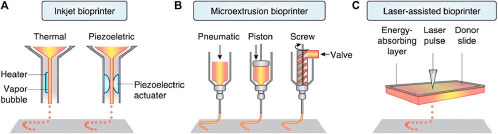
FIGURE 3. Three main 3D bioprinting technologies. (A) Inkjet printing; (B) Extrusion or robotic dispensing bioprinters; (C) Laser-assisted bioprinting (Bartolo et al., 2022).
At present, many materials such as synthetic polymer materials (polyethylene glycol (PEG), polylactic acid (PLA), polycaprolactone (PCL), polyetheretherketone (PEEK)) and natural polymer materials (collagen, gelatin, hyaluronic acid) have been used in 3D printing. However, the accuracy of 3D printing is limited. Generally, it is used to print micron-sized materials; therefore, it is often necessary to combine it with other technologies to obtain tissue engineering scaffolds with higher accuracy.
In recent years, the technological advances in materials science have contributed to the production of various micro/nano-materials such as microspheres for biomedical applications (Tien and Dance, 2021). Interestingly, the characteristics of microfluidic technology are helpful for preparing these micro-nano materials with precise control of reaction parameters in a short time and minimal reagent consumption. Generally, the synthesis of micro-nano materials in microfluidic platform can be achieved by mixing single or multiple miscible solvents (single-phase microfluidic) or mixing multiple immiscible solvents (multiphase microfluidic). The microfluidic reaction system has become a high-throughput and controlled tool, which can be used for materials with diverse biological and chemical applications. Due to the typical laminar flow environment of microfluidic channels, the reaction can be carried out in a highly controlled and reproducible manner (Domachuk et al., 2010). In addition, the size and composition of the product can be flexibly designed by adjusting the flow rate, characteristic geometry and the properties of the input reagent. Different from conventional methods, microfluidic technology allows the continuous production of new materials in a single workflow. Most importantly, many reactions in the microfluidic platform can be carried out under mild conditions, which is very beneficial to many bioactive reaction systems. Therefore, researchers use microfluidics to prepare a large number of materials for biological applications, including microspheres for biomedical applications (Figure 4), microcapsules for medical diagnosis applications, and micron (or nano) fibers for tissue engineering.
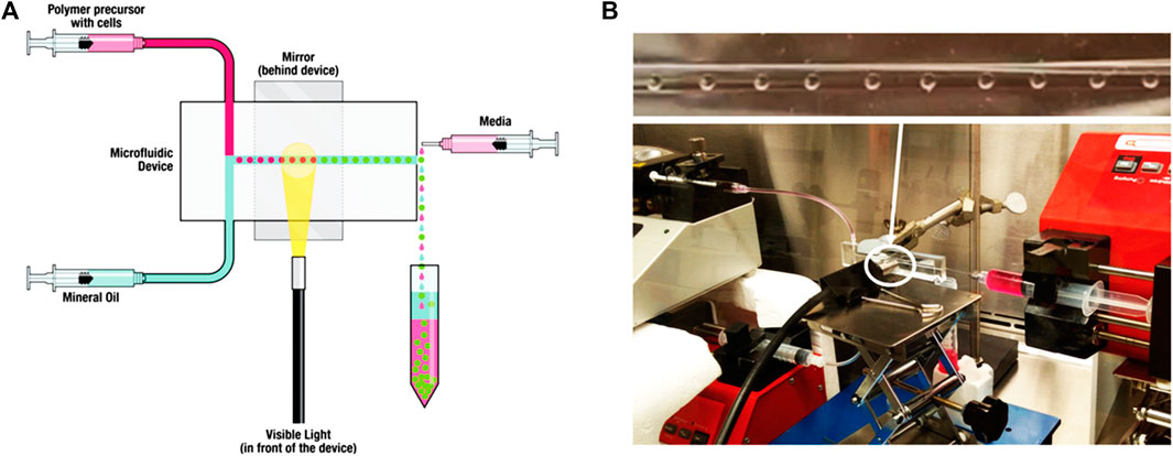
FIGURE 4. Microfluidic encapsulation platform using a novel custom design and device molding technique enables production of uniform hydrogel microspheres with a wide range of diameters. (A) Schematic of the microfluidic encapsulation platform. (B) Setup of the microfluidic encapsulation platform in a biosafety cabinet (Seeto et al., 2019).
Electrospinning is a feasible technology to produce nanofibers, which can be prepared from various types of biomaterials (Xue et al., 2019). The most commonly used materials are synthetic polymers, but natural polymers such as alginate are also used. In addition, synthetic materials offer many advantages over natural polymers, such as reliable sources, low immunogenicity and cost-effectiveness. Biodegradability and structural modifiability are additional advantages of synthetic polymers. The most commonly used polymers include PLA, polyglycolic acid (PGA), PCL, polyurethane (PU), poly (vinyl alcohol) (PVA) and poly- (ethylene oxide) (PEO). The microstructure of nanofibers is also very important for tissue repair and regeneration. Compared with random PCL nanofibers, aligned nanofibers have stronger AF repair effect (Gluais et al., 2019). Besides the arrangement of nanofibers, the stiffness and fiber size of nanofibers will also affect AF stem cell differentiation. According to different needs, specific stiffness and fiber size can be designed to guide cells to differentiate in a specific direction, regulate cell morphology and promote the expression of related ECM (Chu et al., 2019). To further improve the mechanical properties of nanofibers and enhance the interaction with cells, synthetic polymers are also often functionalized through the combination of two or more materials. Nanofibers can also be modified with bioactive cues such as ECM proteins, growth factors, peptides, and nucleic acids to control the cell attachment, migration, spreading, proliferation, and differentiation (Figure 5) (Taskin et al., 2021).
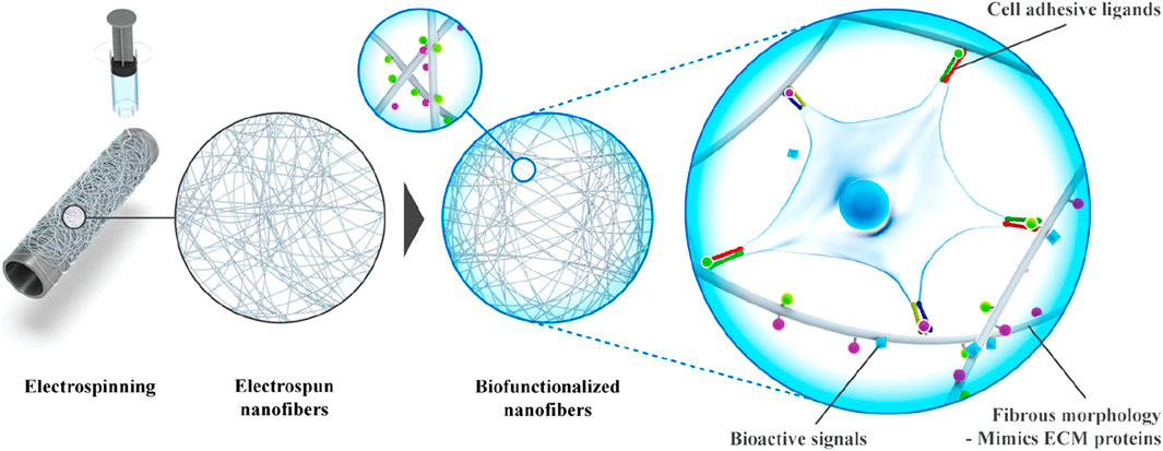
FIGURE 5. Fabrication of bioactive electrospun fibers mimicking the native ECM (Taskin et al., 2021).
When hydrophobic materials such as PCL are delivered to the body, immunological rejection may occur, resulting in unspecific protein adsorption and an inflammatory response (Ratner and Bryant, 2004). The incorporation of hydrophilic bioinert materials such as PEG, can effectively shield hydrophobic fibers, thus making the biomaterial resistant in the biological environment (Dash and Konkimalla, 2012). In addition, the surface of poly (L-lactic acid) (PLLA) scaffolds treated with plasma is more hydrophilic (Jin Seo et al., 2013). It was shown that PCL-α-CD fibers obtained from PCL co-electrospinning with cyclodextrins (CDs) promoted osteogenic differentiation of human adipose-derived stem cells compared with pristine PCL fibers (Zhan et al., 2012). The plasma-treated polyester fiber further improves the adhesion and proliferation of human mesenchymal stem cells (hMSCs), fibroblasts, osteoblasts and Schwann cells (Jia et al., 2008; Martins et al., 2009; Nandakumar et al., 2013). Moreover, studies have confirmed that polydopamine-coated nanofibers (e.g., PCL, PLLA and PLGA) support the deposition of serum proteins, accelerate cell attachment and are widely used for bone, cardiac, skin, nerve and vascular tissue engineering (Chen et al., 2007; Taskin et al., 2016). Furthermore, a variety of active proteins can be modified to nanofibers to enhance the biological activity of fibers, such as vascular endothelial growth factor (VEGF) and epidermal growth factor (EGF) (Choi et al., 2008; Li et al., 2008; Guex et al., 2014; Heo et al., 2015). For instance, VEGF was immobilized on a nanofibrous scaffold and effectively stimulated endothelial cell proliferation (Guex et al., 2014). Shin et al. demonstrated that gelatin and EGF immobilized together on acrylic acid-grafted poly (lactide-co-ε-caprolactone) (PLCL) significantly enhanced the ability of hMSCs to differentiate into keratinocytes (Shin et al., 2015). Stable and self-assembled coatings on the surface of nanofibers play an important role in the delivery system, enabling precisely temporal release of encapsulated molecules (Yoo et al., 2009; Shah et al., 2011). For instance, polylaminate coating on PCL nanofibers can be used for the quick release of connective tissue growth factor (CTGF) and to regulate the release of bone morphogenetic protein-2 (BMP-2), with the aim of mimicking the natural bone healing process (Cheng et al., 2019). Functionalization of nanofibers can also be achieved through click chemistry (Callahan et al., 2013). The introduction of RGD peptide and osteogenic growth protein (OGP) on PCL nanofibers promoted the differentiation of preosteoblast cells into osteoblasts (Kim et al., 2015).
In addition to electrospinning, microfibers can be prepared by microfluidics (Costantini et al., 2016; Zuo et al., 2016; Wang et al., 2019). Zuo et al. designed a dual coaxial capillary microfluidic device that mimics the unique structure of the osteon (Zuo et al., 2016). The middle and outer layers of the fabricated bilayer hollow microfibers are composed of human umbilical vein epithelial cells (HUVECs) and human osteoblast-like cells (MG63), respectively, imitating the vascular layer and bone tissue. Furthermore, the microfibers consist of an alginate-GelMA composite hydrogel in the middle and outer layers, and hyaluronic acid in the inner layer. The incorporation of GelMA reduces the concentration of alginate and improves the biocompatibility of the composite hydrogel, without altering the mechanical properties of the composite hydrogel. Topological hydrogel microfibers have been successfully used to mimic muscle-like bundles (Costantini et al., 2017; Miri et al., 2018; Bansai et al., 2019). Bansai et al. demonstrated that C2C12 muscle precursor cells reorganized their cytoskeleton and formed longitudinal myofibrillar-like structure when wrapped in the collagen-rich core of hydrogel core-shell microfibers (Bansai et al., 2019). Moreover, the alginate shell enhances the mechanical stability of the microfibers and promotes C2C12 cell elongation and myogenesis.
The structure, physical and chemical properties and biological effects of nanofiber scaffolds are of great significance to the development of tissue engineering scaffolds. Although some achievements have been made in the preparation and research of nanofiber materials, how to improve the preparation process, shorten the preparation period of materials, reduce the processing cost of materials, increase the porosity of scaffold materials and control the size and distribution of pore size needs further study. In addition, ECM contains both micron pores and nano-space. There is a synergistic effect between them, which provides the necessary space for cell planting, growth and ECM formation, and provides channels for oxygen and nutrition transmission, information transmission, gene expression and metabolite excretion. Therefore, the ideal scaffold not only needs to be composed of continuous nanofibers with sufficient mechanical properties, but also needs to contain both micro and nano spaces. But up to now, we still lack sufficient understanding of the optimal fiber diameter and its corresponding pore size and distribution, the ideal ratio of nano-space to micro-space, and the interaction between nano-space and body tissue. It is believed that in the future, with the development of nanotechnology and its continuous penetration in the field of tissue engineering, there will be a breakthrough in the design, preparation and performance regulation of nanofiber scaffolds.
Microspheres have become advanced functional materials used in a wide range of biomedical applications, such as researchers prepared microcapsules and microcarriers as a vehicle for drug delivery, summarized the recent progress of engineering particles and their emerging applications in biomedical delivery and diagnosis, and reviewed a comprehensive and in-depth insight into droplet microfluidics, covering fundamental research from microfluidic chip fabrication and droplet generation to the applications of droplets in bio (chemical) analysis and materials generation (Hernandez et al., 2010; Choi et al., 2017; Shang et al., 2017). It is suggested that the distribution of microsphere-sized particles is thought to be an important factor affecting the choice of drug delivery route and the rate of drug release. The properties of microspheres are closely related to their size, structure, and composition; therefore, it is essential to manufacture microparticles in a controlled manner to improve the reliability of their application (Tran et al., 2011; Duncanson et al., 2012; Thiele, 2017). Currently, most of the custom microspheres are manufactured by microfluidics and electrohydrodynamic co-jetting (Yeh et al., 2006; Dendukuri and Doyle, 2009; Seo et al., 2015). Moreover, microspheres can be prepared from a host of materials, including nature polymer (collagen, gelatin, hyaluronic acid) and synthetic polymer (PEG, PLGA, polyglycerol, poly (acrylic acid) and poly (acrylamide)) (Velasco et al., 2012; Rossow et al., 2017).
Microspheres produced by droplet microfluidics can be used as excellent drug delivery microcarriers. To ensure the effectiveness of drug encapsulation, it is necessary to select the material with great compatibility with the drug. For instance, chitosan-based microspheres enable ampicillin or bovine serum albumin (BSA) encapsulation (Yang et al., 2007; Xu et al., 2012). Microspheres with core-shell structure allows the drug to be encapsulated in the core, while the shell acts as a diffusion barrier and controls the release curve (Lee et al., 2017). In addition, microspheres with pH-responsive properties show great potential for targeted drug delivery. Core-shell alginate microspheres tolerate acidic environments and degrade in alkaline environments while releasing encapsulated substance for intestine-targeted drug delivery (Huang et al., 2014). Hydrogel microspheres are exciting platforms for cell culture, because they act as 3D matrices that mimic ECM (Shum et al., 2008; Velasco et al., 2012; Zheng et al., 2023). In addition, hydrogel microspheres with porous structures and the high surface-to-volume ratios are conducive to the exchange of oxygen and nutrients between cells and the external environment, while maintaining cell activity. These advantages allow for the potential use of cell-loaded hydrogel microspheres in the field of cell life research (Huang et al., 2017; Rossow et al., 2017), drug delivery (Shembekar et al., 2016; Agarwal et al., 2017; Zhao et al., 2021), and tissue engineering (Chung et al., 2012; Jiang et al., 2016). For instance, a 3D liver model made of heterotypic cells and core-shell microspheres was assembled (Chen et al., 2016). The layered assembly of hepatocytes in the core and fibroblasts in the outer shell leads to the formation of a spheroid of allotypic cells. The alginate shell allows the spheroid to be cultured for long periods, and the hepatocytes and fibroblasts to be spatially separated and co-cultured, which facilitates the expression of liver-specific functions. Furthermore, microspheres containing sensing components prepared by microfluidics have been used in sensing applications (Le Goff et al., 2015). Fluorescent polyacrylamide hydrogel microspheres are sensors with high sensitivity, biostability, durability, and injectability for continuous monitoring of in vivo blood glucose levels (Shibata et al., 2010).
However, there are still many problems in the preparation of microspheres, such as (1) the size of microspheres is difficult to be unified and the size distribution is uneven; (2) microspheres are easy to aggregate; (3) the preparation cost of microspheres is high. Therefore, it can be predicted that the future research trends of microspheres may be: (1) exploring how to unify the size of microspheres to improve the preparation efficiency; (2) choosing suitable methods to disperse the microspheres to prevent them from aggregation; (3) reducing the preparation cost of microspheres; (4) Further broadening the clinical application of microspheres.
Nanoparticles with high specific surface area and excellent encapsulation capabilities have a wide range of applications, including drug delivery, sensors, bioimaging, and catalytic and diagnostic systems (Chen et al., 2016; Li et al., 2017). Nanoparticle-based drug delivery systems offer significant advantages over traditional drugs, such as increasing drug solubility and stability, achieving controlled drug release, and overcoming biological barriers (Cui et al., 2016). The major classes of materials used to construct nanoparticles include lipid, polymers and inorganic materials (Ju et al., 2022).
Lipid nanoparticles (LNanoparticles), with a liposome-like structure, are widely used for the delivery of drugs (Mitchell et al., 2021; Eygeris et al., 2022). Nanoparticles can achieve drug loading in different ways including post-loading, co-loading, and pre-loading (Figure 6) (Liu et al., 2020). The efficacy of LNanoparticles for drug delivery, together with their simple synthesis, small size and serum stability, allow LNanoparticles to play an important role in personalized gene therapy, and in the penetration of the blood-brain barrier (Berraondo et al., 2019; Cheng et al., 2020; Hou et al., 2021).
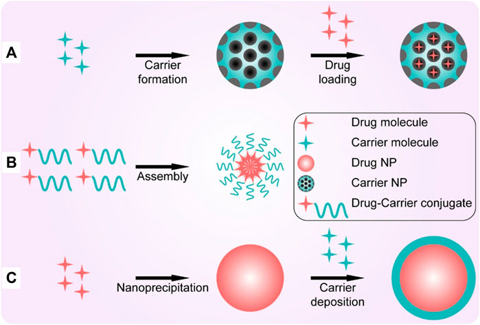
FIGURE 6. Three representative strategies for making high drug-loading nanoparticles. (A) Post-loading; (B) co-loading; (C) pre-loading (Liu et al., 2020).
Polymeric nanoparticles can be formed from natural or synthetic materials and monomeric or pre-polymeric polymers to effectively deliver a wide range of drugs, including hydrophobic and hydrophilic compounds, and carriers of varying molecular weights, allowing the precise control of the loading efficiency and release kinetics of these therapeutic agents by modulating properties such as composition, stability, reactivity, and surface charge (Caldorera-Moore et al., 2019; Knight et al., 2019; Zhang et al., 2020). PLGA has been approved by the FDA for its excellent biosafety, biodegradability and versatility, and is considered to be one of the most successful polymers in the biomedical field (Ulery et al., 2011; Martins et al., 2018; Niza et al., 2021). PLGA-based micro/nanoparticles have been used as carriers for small molecule and macromolecular drugs, with therapeutic benefits in diverse conditions such as diabetes or CNS related diseases (Bala et al., 2004; Rezvantalab et al., 2018; Essa et al., 2020; Ghitman et al., 2020). Furthermore, recent studies have proposed that PLGA itself can act as an active molecule. For instance, impaired lysosomal acidification occurs in neurodegenerative diseases, leading to lysosomal dysfunction (Udayar et al., 2022). This negative effect can be offset by the products of PLGA cleavage, such as lactic acid and glycolic acid (Bourdenx et al., 2016; Zeng et al., 2019). Bourdenx et al. demonstrated that restoration of lysosomal function significantly reduced substantia nigra dopaminergic neurodegeneration (Bourdenx et al., 2016). Moreover, Paul et al. verified that PLGA nanoparticles could inhibit Aβ aggregation and improved the cell survival rate of cortical neurons in Aβ1-42 treated mice (Paul et al., 2022).
With the increase in research on silk sericin, it was found that silk sericin has remarkable biological functions and can be used as a potential biomaterial (Liu et al., 2022). Numerous studies have indicated that silk sericin has low immunogenicity and does not cause significant immunoreaction (Chouhan and Mandal, 2020; Siavashani et al., 2020; Ode Boni et al., 2022). In addition, silk sericin with antioxidant properties has been shown to regulate glycolipid metabolism and promote the proliferation of various cell types while maintaining their functions (Kato et al., 1998; Ersel et al., 2016; Jena et al., 2018). Due to their good bioactivity and biocompatibility, sericin-based nanomaterials are currently being widely used in tissue engineering. Sericin-based nanoparticles act as nanocarriers for insoluble molecules, providing curative effects and reducing side effects (Kanoujia et al., 2016; Suktham et al., 2018). In addition to being a drug carrier, silk sericin serves as a biomineralization matrix (Wang et al., 2017; He et al., 2017; Chaisabai et al., 2018; Tian et al., 2020). For instance, nucleation of hydroxyapatite (HA) crystals can be mediated by using sericin as a template (Yang et al., 2014).
Inorganic materials can be designed in a wide range of sizes, structures, and geometries, and for various drug delivery and imaging applications (Mitchell et al., 2021). CaCO3 nanoparticles have the characteristics of biocompatibility and biodegradability, and controlled synthesis and easy functionalization (Trofimov et al., 2018). In addition, as a porous microcarrier, CaCO3 nanoparticles can be used for drug delivery. Animal and clinical studies have shown that nasal delivery of porous CaCO3 microcarrier can rapidly and effectively control blood glucose levels in diabetic patients (Haruta et al., 2003). No drug loss was observed when nifedipine, ibuprofen and other drugs with poor water solubility loaded onto CaCO3 nanoparticles (Preisig et al., 2014). In addition, Ca2+ produced by the decomposition of CaCO3 may serve as exogenous calcium sources to promote tissue repair, such as bone formation (Combes et al., 2006; Zhong et al., 2016).
Iron oxide is a commonly studied inorganic material and nanomedicines made from iron oxide nanoparticles are approved by the FDA (Bobo et al., 2016). Magnetic iron oxide nanoparticles consisting of magnetite (Fe3O4) or maghemite (Fe2O3) have superparamagnetic properties at specific sizes and have shown excellent promise as drug delivery carriers, contrast agents and thermotherapy agents (Arias et al., 2018). As common inorganic nanoparticles, silicon quantum dots are mainly used as unique nanoparticles for imaging applications in vitro, but they show prospect for in vivo diagnostics (Xu et al., 2019; Huang et al., 2020). Due to the different oxidation states of cerium (Ce3+/Ce4+), cerium oxide (CeO2) nanoparticles have excellent resistance to oxidation, which has been reported in the treatment of diseases, such as spinal cord injury, inflammation, sepsis and Alzheimer disease (AD) (Selvaraj et al., 2015; Kwon et al., 2016; Kim et al., 2017). For instance, in a mouse model of AD, small and positively charged CeO2 nanoparticles conjugated with triphenylphosphonium attenuated neuronal death, alleviated mitochondrial damage, and reduced reactive gliosis by scavenging reactive oxygen species (ROS) (Kwon et al., 2016).
Modulation of macro, micro and nano structures can be achieved by bioprinting strategies (Tarassoli et al., 2018; Chakraborty et al., 2022). 3D bioprinting enables the rapid and precise spatial patterning of cells, growth factors and biomaterials to create complex 3D tissue structures (Matai et al., 2020; Kim et al., 2021). The printed structures should have excellent biocompatibility, great mechanical and biomimetic properties. To achieve this, a variety of bioinks are used for bioprinting, such as fibrinogen, agarose, alginate, collagen, gelatin, hyaluronic acid, pluronic or poly (ethylene glycol) (Shao and Vollrath, 2002; Muller et al., 2015; Wang et al., 2015; Axpe and Oyen, 2016; Mandrycky et al., 2016).
3D porous polymer scaffolds have been used to generate substitutes for skin tissue. A 3D bioprinted skin tissue model was developed using silk fibroin-gelatin (SF-G) bioink and human dermal fibroblasts, keratinocytes and melanocytes (Admane et al., 2019). The 3D bioprinted structure mimics the morphology of the dermal-epidermal junction and replicates the mechanical properties and biochemical characteristics of human skin, ultimately facilitating full-thickness wound healing (Xiong et al., 2017; Admane et al., 2019). In addition, 3D bioprinted skin tissue equivalents serve as in vitro models to test the biosafety of pharmaceuticals and cosmetics, contributing to the study of cell signaling and physiological response mechanisms (Chakraborty et al., 2022).
Multiple causes, such as trauma, can lead to bone defects. However, no consistent results have been achieved in terms of bone replacement in current bone tissue engineering strategies. Although it is challenging to simulate the complex anatomical structure and functional dynamics of native bone, 3D bioprinting can be a potential method for bone tissue replacement (Kacarevic et al., 2018). For instance, 3D printed scaffolds through combining SF, gelatin, hyaluronic acid and tricalcium phosphate (TCP) can support osteogenic differentiation of human adipose-derived mesenchymal stem cells (Du et al., 2019). It has also been shown that bioprinted BMSC-loaded GelMA/SFMA scaffolds can promote bone repair through vascularized osteogenesis (Yang et al., 2022). The microstructure of intervertebral disc is complex, which can be simulated by 3D printing technology. 3D printing of flexible polylactic acid (FPLA) can be used to fabricate a viscoelastic scaffold with tunable biomimetic mechanics for whole spine motion segment applications (Marshall et al., 2021).
Although 3D printing technology has many advantages that other materials cannot achieve, the types of printing materials are still limited, which is the main bottleneck that hinders the development of 3D printing technology. At present, the main way of 3D printing is to print materials in the form of “ink” and quickly solidify them. Therefore, while “ink” has good biocompatibility and biodegradability of general biomaterials, it should also be considered that after printing, it still needs to maintain its original biological activity and mechanical strength, which greatly limits the application scope of 3D printing. The existing 3D printing equipment also has low printing precision and cannot meet the bionic requirements of tissues and organs.
Smart hydrogels, a cross-linked polymer, can respond to small changes in environmental stimuli by significantly altering their volumes and other physico-chemical properties. The uniqueness of these hydrogels lies in their non-linear feedback. They respond to external stimuli primarily through reversible, intensity-scalable, repeatable and predictable phase volume shifts and are able to return to their original state after the stimulus has been removed. These transformations include changes in physical state and hydrophilicity, etc (Bordbar-Khiabani and Gasik, 2022). In addition, the flexibility of different response behaviors in response to different stimuli is important for broadening the application of smart hydrogels in various conditions. It is worth noting that micro-/nano-structured smart hydrogels show higher elasticity and respond faster and stronger to external stimuli than smart hydrogels without micro-/nano-structures (Wang et al., 2021).
Poly (N-isopropyl acrylamide) (PNIPAM) hydrogels are commonly used thermal responsive hydrogels with reversible swelling/contraction changes when the temperature is varied within the volume phase transition temperature (VPTT) range. In general, PNIPAM hydrogels formed by chemical crosslinking of N, N′-methylenebisacrylamide (BIS) and monomer N-isopropylacrylamide (NIPAM) have a slow response and poor elasticity. However, when PNIPAM nanogels with unsaturated C=C bonds are used to cross-link PNIPAM chains, micro/nanostructured PNIPAM hydrogels with remarkable elastic and responsive properties can be obtained (Xia et al., 2013). The transition from the fully swollen to the fully collapsed state of the nanogel cross-linked hydrogel takes only 6 min, much faster than the normal PNIPAM hydrogels. Moreover, the swelling ratio of nanogel cross-linked hydrogel is 10 times that of the normal PNIPAM hydrogel. In addition to responsive volume changes, smart hydrogels can achieve various stimuli responses (Isapour and Lattuada, 2018; Wen et al., 2019; Jia et al., 2021). For instance, it is often possible to obtain smart hydrogels with responsive color changes by designing colloidal nanocrystal structures in their polymer networks (Jia et al., 2021). BIS and graphene oxide nanosheets with photothermal properties can be used as double crosslinking agents to produce PNIPAM hydrogels with micron-scale network. This double cross-linked hydrogel allows ultra-high stretchability and rapid volume phase transition in response to NIR light (Wang et al., 2022).
As a new biological material, liquid metal (LM) has shown promising prospects in the biomedical field, such as tissue therapeutics, repair, bioimaging, and biosensors (Yan et al., 2018; Zeng and Fu, 2018; Miyako, 2021). LM with preeminent deformability can be molded into various shapes with different properties to cope with diverse application scenarios (Wang et al., 2018; Sun et al., 2019). In addition, the morphology of LM changes significantly in response to external stimuli such as pH, light, temperature, and magnetic fields. Additionally, the morphological changes induced by stimuli can help to expand the application of LM in drug delivery, thermotherapy and antimicrobial therapy (Wang et al., 2022) (Figure 7). For instance, LM nanoparticles generally have an oxide layer on their surface, which is due to self-limiting oxidation. The oxide layer can react with protons and dissolve in an acidic environment. Therefore, drug release from LM can be facilitated in an acidic microenvironment due to the removal of the oxide layer (Lu et al., 2015). Furthermore, under NIR irradiation, the shape of polydopamine (PDA)-coated LM nanoparticle changes from spheres to ellipsoids (Gan et al., 2019). Based on this transformation, the prepared LM nanorobots exhibit excellent photothermal properties and can be used for photothermal antibacterial therapy (Xu et al., 2021). LM with good biocompatibility has emerged as a potential material for tissue repair, including bone defect repair and nerve connectors (Yi et al., 2014; Liu et al., 2016; He et al., 2021). LM used in biomedicine generally include gallium (Ga) and its alloys, which can achieve solid-liquid transition at relatively low temperatures to satisfy the needs of different application environments via the component modulation. Kinds of Ga-based biomaterials such as Ga droplets and Ga-based hybrids show low toxicity in aqueous environments and mice bodies. At the same time, Ga-based LM possess good degradability in acidic biological environments, thus helping to reduce their potential systemic toxicity. In particular, Ga ions also exhibit antibacterial abilities and can be used to treat bacterial infections. In addition, an actuator using LM based on Ga can circumvent the problems of conventional actuators such as high drive potential, low strain and their time responsiveness. Thus, Ga and its alloys have great potential for tissue engineering applications (Wang et al., 2022).
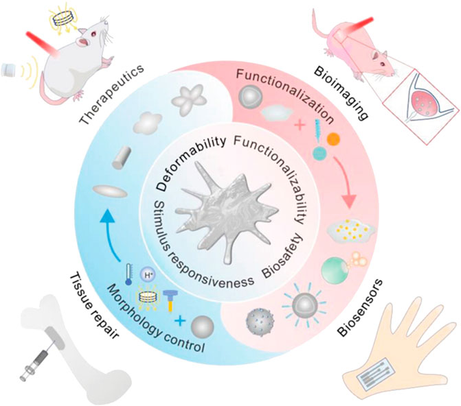
FIGURE 7. Schematic illustration of advantages and design of LM and their applications in biomedicine (Wang et al., 2022).
Micro/nanomaterials can simulate the microstructure of tissues and can also be used as carriers of drugs and growth factors. Research on micro/nanomaterials in tissue repair and regeneration is gaining increasing attention. Micro/nanomaterials play a very important role in the treatment of bone defects, cartilage injuries, cardiovascular diseases, skin injuries, intervertebral disc degeneration, and other diseases.
Bone tissue defect is caused by trauma, tumor and other diseases, and has become a common clinical disease. Currently, the use of orthopedic implants is an available option for treating these diseases. The use of bone grafts is on the rise, with more than 2.2 million cases of bone grafting worldwide each year (Wang and Yeung, 2017). In recent years, the development of nanotechnology, biomaterial science and tissue engineering has provided a broad prospect for the preparation of bone graft materials (Liu et al., 2022). Natural bone tissue has a unique structure, mainly composed of collagen, nanofibrils and nano-hydroxyapatite (Laubach et al., 2022). The use of micro-nanomaterials can play a key role in accelerating cell responses and guiding tissue regeneration. Therefore, it is necessary to construct bone tissue engineering scaffolds with consideration of aspects such as surface morphology, mechanical strength, and regulation of bioactive molecules. As a 3D scaffold for cells, bone tissue engineering scaffolds provide cells with the unique microstructure and microenvironment of bone tissue to maintain the survival and differentiation of cells (Park et al., 2016).
Nanofiber scaffold is a commonly used scaffold for bone tissue engineering (Shalumon et al., 2015). Biomimetic composite scaffold of HA/gelatin-chitosan core-shell nanofibers has been used for bone regeneration, as it can mimic both the specific structure and the chemical composition of natural bone. MG-63 was seeded on the nanofibers and it was proved that the nanofibers could enhance osteoblast cell proliferation (Chen et al., 2019). Incorporation of dual factors, HA, and laminin within the shell and core of nanofibers respectively via emulsion electrospinning is another method of fabricating core-shell nanofibers. Nanofibers play important roles in osteoblast proliferation and maturation (Tian et al., 2013). Electrospun core-shell nanofibers loaded with metronidazole (MNA) and nano-hydroxyapatites (nHA) have both anti-infection and osteogenesis capabilities (Wang et al., 2019). Another type of core-shell nanofiber can incorporate an osteogenic inductive peptide H1, derived from the cysteine knot (CT) domain of CTGF, in the core of SF and co-deliver HA from the shell of poly (L-lactic acid-co-ε-caprolactone) (PLCL). It can promote proliferation and osteoblastic differentiation of human induced pluripotent stem cell-derived mesenchymal stem cells (hiPS-MSCs). In vivo experiments further verified that SF-H1/PLCL-HA core-shell nanofibers can promote the repair of bone defects (Xu et al., 2019). Platelet-rich plasma (PRP) can be incorporated into SF/PCL/PVA nanofibers by coaxial electrospinning to avoid the rapid degeneration of the growth factors, and this core-shell nanofiber can release multiple growth factors to promote bone regeneration (Cheng et al., 2018). BMP-2 is a growth factor commonly used in bone tissue engineering to promote bone regeneration. The core-shell nanofiber scaffolds can work as a sustained delivery vehicle for BMP-2 protein. The controlled release of BMP-2 can effectively promote the regeneration of bone tissue (da Silva et al., 2019). BMP-2 and other drugs or growth factors are often co-encapsulated in the core-shell nanofiber scaffold to promote bone regeneration. BMP-2 and dexamethasone (DEX) were successfully incorporated into PLLACL/collagen nanofibers by means of coaxial electrospinning. The controlled release of the two growth factors from PLLACL/collagen nanofibrous mats can induce hMSC to differentiate into osteoblasts for bone tissue engineering (Su et al., 2012). Zein/PLLA nanofibers can also be fabricated to load BMP-2 and DEX. It can achieve the burst release of DEX and sustained release of BMP-2. It can enhance the osteogenic differentiation of MSCs and has great potential in bone tissue engineering applications (Li et al., 2018). Core-shell SF/PCL/PVA nanofibrous mats using coaxial electrospinning and layer-by-layer (LBL) techniques, where BMP-2 was incorporated into the core of the nanofibers and CTGF was attached onto the surface were also fabricated. According to the physiological needs of bone regeneration, this core-shell nanofiber scaffold induced sustained release of BMP-2 and sudden release of CTGF. In vivo experiments found that this scaffold has the largest bone repair area, and the new bone formed has the same tissue structure as normal bone. Therefore, it can provide a promising strategy to facilitate bone healing (Figure 8) (Cheng et al., 2019). The mechanical properties of bone repair materials are required. Studies have shown that nHA and Fe3O4 nanoparticles can improve the mechanical properties of materials and further promote the repair and regeneration of bone tissue (Zhao et al., 2019).
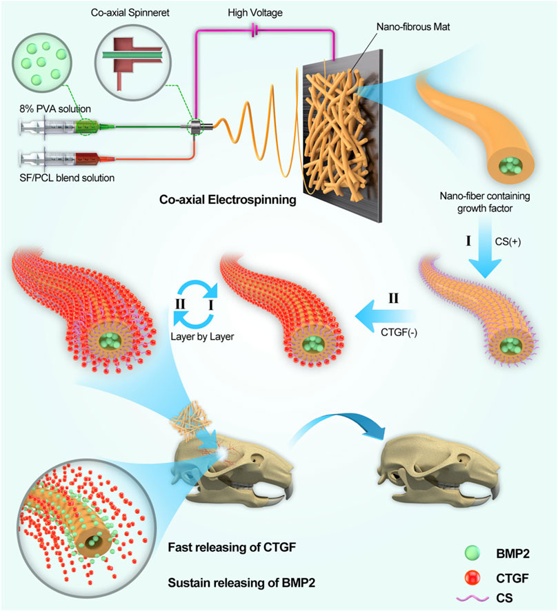
FIGURE 8. Schematic illustration of the (SF/PCL)1:5/PVA-LBL20 coaxial fibers loaded with BMP2 and CTGF for bone tissue engineering (Cheng et al., 2019).
3D printing technology is considered to be an advantageous technology, which can also be used to construct simulated bone tissue microstructure. In addition, scaffolds produced through 3D printing can provide excellent delivery vehicles for topical, continuous delivery of drugs and/or biomolecules. PLA/PCL/HA composite bone scaffold prepared by 3D printing can effectively simulate the microstructure of bone and has good biocompatibility (Hassanajili et al., 2019). Hierarchical porous and recombinant human bone morphogenetic protein-2 (rhBMP-2)-loaded calcium phosphate (Ca-P) nanoparticle/poly (L-lactic acid) nanocomposite scaffolds can also be fabricated by 3D printing, in which sustained releases of Ca2+ ions and rhBMP-2 can be achieved (Wang et al., 2017).
Microspheres and nanoparticles are also widely used in bone tissue engineering because of their good injectability and drug-controlled release ability and etc. nHA microspheres prepared via a hydrothermal transformation method significantly improved the ability of the microspheres to adsorb the bioactive protein (BMP-2) and realize bone regeneration (Zhou et al., 2018). rhBMP-2 can be grafted on the surface of mesoporous bioglass nanoparticles (MBGNs) with an amide bond. It can realize the rhBMP-2 release in a controllable program during the early bone regeneration period and then sustained release of calcium and silicon ions to keep promoting osteogenesis in a long term (Xin et al., 2020).
LM have good biocompatibility, deformability, good electrical and thermal conductivity and have become a potential material in the field of tissue repair. Severe bone defects caused by some diseases, such as bone tumors and external trauma, often require bone repair materials to replace the missing bone. Bismuth (Bi) alloys can be introduced as a bone defect repair material (bone cement) to fill the defect locations. Bi alloys have a low melting point and can be solidified at low temperatures to avoid damage to normal tissue. At the same time, the Bi alloys with its strong magneto-thermal effect can be utilized to alleviate mechanical and thermal pain sensitivity, so pain is relieved. This bone cement has a high affinity to bone and does not move significantly after implantation. Furthermore, the Bi alloys exhibited excellent imaging ability (Wang et al., 2022). In summary, LM have potential application in the field of tissue repair due to their unique advantages.
Cartilage lesions and defects caused by trauma, tumors, and inflammation are more common in the clinic. Cartilage is a tissue without blood vessels and nerves (Mow et al., 1992; Ahmed and Hincke, 2010; Gu et al., 2023), and it is extremely difficult to repair itself after injury, which seriously affects the health of patients. At present, cartilage defects can be repaired and reconstructed by cartilage tissue transplantation, chondrocyte or mesenchymal stem cell transplantation, and biomaterial filling. However, these methods have certain limitations, such as limited sources of autologous cartilage and allogeneic cartilage rejection (Haq-Siddiqi et al., 2023). Therefore, repair of cartilage defects and functional reconstruction are still difficult problems in surgical treatment. In recent years, the rapid development of tissue engineering technology has provided new directions for solving the above problems (Ma et al., 2005). Nanofibers loaded with growth factors can combine with seed cells to repair and regenerate cartilage tissue. Kartogenin (KGN) is a newly discovered small molecule compound that has a strong ability to promote cartilage differentiation and can effectively promote the differentiation of mesenchymal stem cells into chondrocytes (Xu et al., 2015). Coaxial electrospun fibers using poly (L-lactic acid-co-caprolactone) and collagen solution as shell fluid and KGN solution as core fluid was fabricated via coaxial electrospinning. KGN encapsulated in the core-shell nanofibers can be sustainedly released for about 2 months. The chondrogenic differentiation of bone marrow mesenchymal stem cells cultured on core-shell nanofibrous scaffold was promoted obviously (Yin et al., 2017). KGN can also be loaded into microspheres via microfluidics to realize the sustained release and eventually repair the cartilage (Wu et al., 2020). An injectable double positively charged functional hydrogel microsphere with “targeting cartilage extracellular matrix”, “cartilage penetration”, and “cellular phagocytosis” can also be designed for matching the structural characteristics of joints and addressing the difficulties of drug delivery in cartilage (Lin et al., 2022). Cartilage repair requires a large supply of cells, therefore microspheres can also be used as injectable cell carriers for cartilage tissue engineering (Li et al., 2023) (Figure 9).
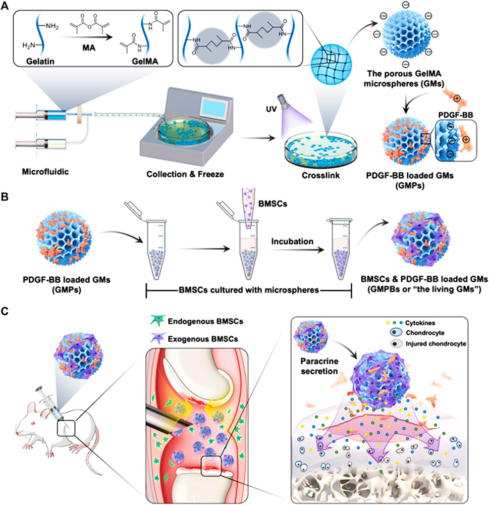
FIGURE 9. Schematic illustrations of the living GMs for cartilage regeneration. (A) The fabrication of GMs using microfluidic technology, and incorporation of PDGF-BB via the electrostatic force to engineer GMPs. (B) The BMSCs and PDGF-BB loaded GMs, or “the living GMs” were further developed through incubation with exogenous BMSCs. (C) The living GMs were injected into the joint cavity of the DMM rat. The enhanced paracrine activity was achieved by integrating the endogenous and exogenous regeneration mechanisms (Li et al., 2023).
In recent years, cardiovascular diseases have become one of the most serious diseases that threaten human health (Tan et al., 2013). When vascular disease reaches the end stage, it is often necessary to use biologically active blood vessels for transplantation or bypass (Ezhilarasu et al., 2019). Due to the limited sources of autologous blood vessel transplantation and the limitations of their own vascular conditions, many of artificial blood vessel replacements are clinically required (Weinberg and Bell, 1986; Jia et al., 2013). In the past, various methods of allogeneic vascular transplantation have been tried, but their long-term effects were poor, including increased thrombosis, vascular degeneration and hyperplasia, and vascular stenosis. The introduction of tissue engineering in recent years have provided new options for this clinical problem. Based on cell biology, material science, and engineering as the basic theory, it can culture and expand seed cells in vitro and plant them in biocompatible organisms. A cell–biomaterial complex is formed and implanted in the body to replace tissue defects caused by diseases and trauma, in order to promote the repair and functional reconstruction of the host tissue structure (Wu et al., 2010; Fu et al., 2014). Micro/nano-materials can mimic the microstructure of blood vessels, regulate the microenvironment, and provide biological signals for blood vessel regeneration.
Nanofiber scaffolds are commonly used in vascular tissue engineering. Many studies have shown that it plays an important role in the repair and regeneration of blood vessels (Merkle et al., 2015a). Nanofiber scaffold can simulate the structure of blood vessels. PCL/collagen scaffold prepared via coaxial electrospinning was a suitable scaffold for vascular regeneration. PCL as the core can provide the mechanical property and integrity while collagen as the shell can improve the attachment and proliferation of vascular cells due to its excellent biocompatibility. The collagen shell was crosslinked by genipin and further bound with heparin. The scaffold can support the attached vascular cells to grow and proliferate on its surface, and also allows the infiltration of SMCs into its interior. The heparinized PCL/collagen scaffold with core/shell fiber structure has a promising application in vascular tissue engineering (Duan et al., 2016). Coaxial electrospun PCL/Gelatin-MA fibers which are composed of a PCL core and a functionalized gelatin (GelMA) shell were also proved to be suitable for vascular regeneration (Coimbra et al., 2017). Core-shell PVA/Gelatin electrospun nanofibers promote vascular tissue regeneration due to the potential of altering the proliferation and migration of HUVECs and rat smooth muscle cells (rSMC) (Merkle et al., 2015b). Highly aligned hyaluronan/PLLA nanofibers in core-shell structure were also reported to be an optional vascular graft (Yuan et al., 2016). The hollow microfibers created by microfluidic technology may simulate the structural characteristics of blood vessels. The scaffold is also biocompatible, allowing cells to proliferate and spread within the tube (Jia et al., 2019) (Figure 10).
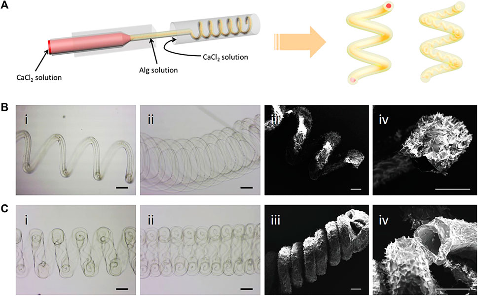
FIGURE 10. Generation of helical hollow alginate hydrogel microfibers with different channels. (A) Schematic illustration of the fabrication of the helical hollow microfibers. (B, C-i, ii) Optical microscopy and iii, iv) SEM images of helical hollow microfibers with (B) straight and (C) helical channels. Scale bars, 200 μm (Jia et al., 2019).
Skin trauma, especially large-area full-cortex trauma is still one of the main diseases. The methods of accelerating the healing of the wound surface and reducing the occurrence of scars remain a clinical problem (Chaudhari et al., 2016; Yao et al., 2017). Skin transplantation is the primary method of treating such skin trauma, but patients with large-scale skin defects or skin burns who receive their own skin transplantation will have new wounds on the skin from other areas. The shortage of autologous skin supply is another major factor that limits its application (Wu et al., 2021). Tissue-engineered skin organically combines engineering and life science principles to build skin replacements that improve, maintain, and restore function of the skin, and is expected to solve problems such as insufficient donor skin in repairing skin defects (Dai et al., 2004; Groeber et al., 2011; Li et al., 2023).
The nanofiber scaffold as an important part of skin tissue engineering provides a feasible method for promoting wound healing (Movahedi et al., 2020). Compared with traditional wound dressings, nanofibers have remarkable properties as dermal substitutes, because their structures are very similar to the skin’s primary ECM, and cells can adhere, proliferate, and permeate scaffolds, inducing the regeneration of skin. Lawsone (2-hydroxy-1,4-naphthoquinone) was electrospun in polycaprolactone-gelatin (PCL/Gel) polymers in core-shell architecture to detect the effect of new core-shell nanofibers on wound regeneration. The core-shell nanofibers-encapsulated lawsone prolonged the release of lawsone over a period of 20 days. Moreover, it can promote cell adhesion and proliferation and promote the expression of healing-related genes. In vivo experiments further demonstrated that it has a strong promotion effect on wound healing (Adeli-Sardou et al., 2019). Hyaluronic acid-silk fibroin/zinc oxide (ZO) nanofibers can be used in the treatment of burn injuries. ZO as an antibacterial agent can be released sustainedly from the nanofibers. Nanofibers with appropriate ZO loading can effectively inhibit the inflammatory response at the injury site and promote wound healing (Hadisi et al., 2020). Single or multiple growth factors or drugs can be loaded into the core-shell nanofibers in order to achieve the slow release of growth factors and promote wound healing. CTGF is a signaling molecule that has multiple roles in tissue repair and regeneration. CTGF encapsulated electrospun dual porous PLA-PVA core-shell fiber based membranes can be used as excellent wound dressing mats for the treatment of diabetic wounds and other chronic ulcers due to the promotion of cell proliferation, migration and angiogenic potential (Augustine et al., 2019). Biodegradable and biocompatible scaffolds incorporated with multiple epidermal induction factors might serve as the most promising medical devices for skin tissue regeneration. Other studies have shown that aligned nanofiber scaffolds can mimic skin microstructure and induce macrophage M2 polarization (Xie et al., 2022).
The rise of 3D printing technology also provides a new method for the construction of skin tissue engineering scaffolds. Different 3D printing technologies can be used to construct stents with different diameters. 3D printing scaffolds have good biocompatibility and are suitable for cell proliferation and differentiation (Gao et al., 2021). According to the shape and depth of the wound, the 3D printing technology can print out biomaterial suitable for the wound. Scaffolds can also flexibly and accurately load different drugs and growth factors to promote tissue regeneration. Skin tissue can even be printed in situ on the wound surface using the layer-by-layer deposition principle (Weng et al., 2021). The integration of electronic skin (e-skin) and tissue has the potential to improve the quality of life for people in the areas of healthcare monitoring and chronic disease treatment. Ga-based alloys can be used to design gluten protein for fabricating e-skin with stretchability, self-healing ability, and biocompatibility. In addition, based on the incorporation of Ga-based alloys, the e-skin obtained has a stronger self-healing ability, and in animal experiments there are no adverse reactions when the e-skin is attached to the skin of rabbits. And in a variety of situations, from large-scale human movement to small strain changes, the e-skin shows a keen ability to sense strain. Therefore, LM-prepared electronic skin has great potential for sensing human movement (Chen et al., 2022).
Disc herniation is a common disease and one of the main causes of neck and low back pain, which seriously affects the quality of life of patients (Xia et al., 2023). At present, there is no effective means to directly repair damaged AF in clinical practice, and the simple removal of herniated intervertebral disc tissue cannot repair the AF defect at the same time, which has a certain postoperative recurrence rate. Although interbody fusion can treat disc herniation with intervertebral instability and prevent recurrence of disc herniation, loss of intervertebral motion and adjacent segment retreat have become new problems. Therefore, there is an urgent need for AF tissue engineering in clinical practice. The microstructure of AF is a typical angle-ply laminated structure, and this special microstructure also guarantees its good mechanical properties. Aligned nanofibers can simulate the microstructure of AF, which brings great hope for the construction of AF tissue engineering. A novel aligned core-shell nanofibrous scaffold with angle-ply microstructure and co-delivery capacity can achieve burst release of ibuprofen to regulate the microenvironment, and sustained release of TGFβ3 to promote ECM secretion. In addition, it can repair the damaged AF and also realize regeneration of IVD (Han et al., 2022). Micro-nano scaffolds constructed by 3D printing technology can also reproduce the special angle-ply laminated structure of the AF, which is conducive to the growth and proliferation of cells, and can also provide good mechanical support (Bhunia et al., 2021).
This article reviewed the research progress of biomimetic micro/nano-materials prepared by different methods for the repair and regeneration of different tissues and organs. Common strategies for preparing nanofibers, microspheres and 3D-printing scaffolds were reviewed in order to elucidate the importance of material selection and 3D structure design for tissue repair and regeneration. To date, a large number of studies have shown that using micro/nanomaterials to mimic the microstructure of tissues can promote the repair and regeneration of tissues. Researchers have also modified the surface of scaffolds in multiple ways to adjust the hydrophilicity, adhesion, and mechanical properties of scaffolds. Additionally, studies have shown that these micro- and nano-structured scaffolds can also load drugs, ensuring both the biological activity of drugs and controlled drug release. However, it is not easy to prepare 3D scaffolds with suitable mechanical properties, porosity, surface morphology, degradation rate, and microstructure simulation. The accuracy of existing micro/nanomaterials is limited, and the microstructure of tissues and organs is complex. Therefore, it is still difficult to precisely mimic the microstructure. In addition, the existing micro/nanomaterials focus on the simulation of the microstructure and ignore the simulation of their components, which is another major problem of tissue engineering. The application of micro/nanomaterials is still in its infancy, and there are still many problems to be solved in clinical application such as the way of constructing an ideal cell-nanomaterial interface; preventing immune system, maintaining the survival rate and function of cultured cells for a long time, improving the biocompatibility of nano-materials. It is important to solve the above problems to prepare micro/nano “smart” materials with specific functions to better regulate the biological behaviors such as specific adhesion, proliferation and directional differentiation of seed cells, so as to obtain good biological activity and good biocompatibility and finally apply them to clinic.
FxH conceived and designed the review. FH and QM wrote the manuscript. EX, KL, JH, QC, and JL all participated the data search and analysis. FxH assisted with the revision of this article. All authors contributed to the article and approved the submitted version.
This work was supported by the National Natural Science Foundation of China (32130059 and 32171350), Medical and Health Science and Technology Innovation Project of Suzhou (SKY2022105), and the Priority Academic Program Development of Jiangsu Higher Education Institutions.
The authors declare that the research was conducted in the absence of any commercial or financial relationships that could be construed as a potential conflict of interest.
All claims expressed in this article are solely those of the authors and do not necessarily represent those of their affiliated organizations, or those of the publisher, the editors and the reviewers. Any product that may be evaluated in this article, or claim that may be made by its manufacturer, is not guaranteed or endorsed by the publisher.
Adeli-Sardou, M., Yaghoobi, M. M., Torkzadeh-Mahani, M., and Dodel, M. (2019). Controlled release of lawsone from polycaprolactone/gelatin electrospun nano fibers for skin tissue regeneration. Int. J. Biol. Macromol. 124, 478–491. doi:10.1016/j.ijbiomac.2018.11.237
Admane, P., Gupta, A. C., Jois, P., Roy, S., Chandrasekharan Lakshmanan, C., Kalsi, G., et al. (2019). Direct 3D bioprinted full-thickness skin constructs recapitulate regulatory signaling pathways and physiology of human skin. Bioprinting 15, 00051. doi:10.1016/j.bprint.2019.e00051
Agarwal, P., Wang, H., Sun, M., Xu, J., Zhao, S., Liu, Z., et al. (2017). Microfluidics enabled bottom-up engineering of 3D vascularized tumor for drug discovery. ACS Nano 11 (7), 6691–6702. doi:10.1021/acsnano.7b00824
Ahmed, T. A., and Hincke, M. T. (2010). Strategies for articular cartilage lesion repair and functional restoration. Tissue Eng. Part B Rev. 16 (3), 305–329. doi:10.1089/ten.TEB.2009.0590
Arias, L. S., Pessan, J. P., Vieira, A. P. M., Lima, T. M. T., Delbem, A. C. B., and Monteiro, D. R. (2018). Iron oxide nanoparticles for biomedical applications: A perspective on synthesis, drugs, antimicrobial activity, and toxicity. Antibiotics 7 (2), 46. doi:10.3390/antibiotics7020046
Augustine, R., Zahid, A. A., Hasan, A., Wang, M., and Webster, T. J. (2019). <p>CTGF loaded electrospun dual porous core-shell membrane for diabetic wound healing</p>. Int. J. Nanomedicine 14, 8573–8588. doi:10.2147/ijn.S224047
Axpe, E., and Oyen, M. L. (2016). Applications of alginate-based bioinks in 3D bioprinting. Int. J. Mol. Sci. 17 (12), 1976. doi:10.3390/ijms17121976
Bakhshandeh, B., Zarrintaj, P., Oftadeh, M. O., Keramati, F., Fouladiha, H., Sohrabi-jahromi, S., et al. (2017). Tissue engineering; strategies, tissues, and biomaterials. Biotechnol. Genet. Eng. Rev. 33 (2), 144–172. doi:10.1080/02648725.2018.1430464
Bala, I., Hariharan, S., and Kumar, M. N. (2004). PLGA nanoparticles in drug delivery: The state of the art. Crit. Rev. Ther. Drug Carr. Syst. 21 (5), 387–422. doi:10.1615/critrevtherdrugcarriersyst.v21.i5.20
Bansai, S., Morikura, T., Onoe, H., and Miyata, S. (2019). Effect of cyclic stretch on tissue maturation in myoblast-laden hydrogel fibers. Micromachines 10 (6), 399. doi:10.3390/mi10060399
Bartolo, P., Malshe, A., Ferraris, E., and Koc, B. (2022). 3D bioprinting: Materials, processes, and applications. CIRP Ann. 71 (2), 577–597. doi:10.1016/j.cirp.2022.06.001
Bazilevsky, A. V., Yarin, A. L., and Megaridis, C. M. (2007). Co-electrospinning of core-shell fibers using a single-nozzle technique. Langmuir 23 (5), 2311–2314. doi:10.1021/la063194q
Berraondo, P., Martini, P. G. V., Avila, M. A., and Fontanellas, A. (2019). Messenger RNA therapy for rare genetic metabolic diseases. Gut 68 (7), 1323–1330. doi:10.1136/gutjnl-2019-318269
Bhardwaj, N., and Kundu, S. C. (2010). Electrospinning: A fascinating fiber fabrication technique. Biotechnol. Adv. 28 (3), 325–347. doi:10.1016/j.biotechadv.2010.01.004
Bhunia, B. K., Dey, S., Bandyopadhyay, A., and Mandal, B. B. (2021). 3D printing of annulus fibrosus anatomical equivalents recapitulating angle-ply architecture for intervertebral disc replacement. Appl. Mat. Today 23, 101031. doi:10.1016/j.apmt.2021.101031
Bobo, D., Robinson, K. J., Islam, J., Thurecht, K. J., and Corrie, S. R. (2016). Nanoparticle-based medicines: A review of FDA-approved materials and clinical trials to date. Pharm. Res. 33 (10), 2373–2387. doi:10.1007/s11095-016-1958-5
Bölgen, N., Vargel, I., Korkusuz, P., Menceloğlu, Y. Z., and Pişkin, E. (2007). In vivo performance of antibiotic embedded electrospun PCL membranes for prevention of abdominal adhesions. J. Biomed. Mat. Res. B Appl. Biomater. 81 (2), 530–543. doi:10.1002/jbm.b.30694
Bordbar-Khiabani, A., and Gasik, M. (2022). Smart hydrogels for advanced drug delivery systems. Int. J. Mol. Sci. 23 (7), 3665. doi:10.3390/ijms23073665
Bourdenx, M., Daniel, J., Genin, E., Soria, F. N., Blanchard-Desce, M., Bezard, E., et al. (2016). Nanoparticles restore lysosomal acidification defects: Implications for Parkinson and other lysosomal-related diseases. Autophagy 12 (3), 472–483. doi:10.1080/15548627.2015.1136769
Caldorera-Moore, M., Vela Ramirez, J. E., and Peppas, N. A. (2019). Transport and delivery of interferon-alpha through epithelial tight junctions via pH-responsive poly(methacrylic acid-grafted-ethylene glycol) nanoparticles. J. Drug Target. 27 (5-6), 582–589. doi:10.1080/1061186X.2018.1547732
Callahan, L. A., Xie, S., Barker, I. A., Zheng, J., Reneker, D. H., Dove, A. P., et al. (2013). Directed differentiation and neurite extension of mouse embryonic stem cell on aligned poly(lactide) nanofibers functionalized with YIGSR peptide. Biomaterials 34 (36), 9089–9095. doi:10.1016/j.biomaterials.2013.08.028
Carotenuto, F., Politi, S., Ul Haq, A., De Matteis, F., Tamburri, E., Terranova, M. L., et al. (2022). From soft to hard biomimetic materials: Tuning micro/nano-architecture of scaffolds for tissue regeneration. Micromachines 13 (5), 780. doi:10.3390/mi13050780
Chaisabai, W., Khamhaengpol, A., and Siri, S. (2018). Sericins of mulberry and non-mulberry silkworms for eco-friendly synthesis of silver nanoparticles. Artif. Cells Nanomed. Biotechnol. 46 (3), 536–543. doi:10.1080/21691401.2017.1328686
Chakraborty, J., Mu, X., Pramanick, A., Kaplan, D. L., and Ghosh, S. (2022). Recent advances in bioprinting using silk protein-based bioinks. Biomaterials 287, 121672. doi:10.1016/j.biomaterials.2022.121672
Chaudhari, A. A., Vig, K., Baganizi, D. R., Sahu, R., Dixit, S., Dennis, V., et al. (2016). Future prospects for scaffolding methods and biomaterials in skin tissue engineering: A review. Int. J. Mol. Sci. 17 (12), 1974. doi:10.3390/ijms17121974
Chen, B., Cao, Y., Li, Q., Yan, Z., Liu, R., Zhao, Y., et al. (2022). Liquid metal-tailored gluten network for protein-based e-skin. Nat. Commun. 13 (1), 1206. doi:10.1038/s41467-022-28901-9
Chen, F., Lee, C. N., and Teoh, S. H. (2007). Nanofibrous modification on ultra-thin poly(e-caprolactone) membrane via electrospinning. Mat. Sci. Eng. C 27 (2), 325–332. doi:10.1016/j.msec.2006.05.004
Chen, G., Roy, I., Yang, C., and Prasad, P. N. (2016a). Nanochemistry and nanomedicine for nanoparticle-based diagnostics and therapy. Chem. Rev. 116 (5), 2826–2885. doi:10.1021/acs.chemrev.5b00148
Chen, P., Liu, L., Pan, J., Mei, J., Li, C., and Zheng, Y. (2019). Biomimetic composite scaffold of hydroxyapatite/gelatin-chitosan core-shell nanofibers for bone tissue engineering. Mat. Sci. Eng. C Mat. Biol. Appl. 97, 325–335. doi:10.1016/j.msec.2018.12.027
Chen, Q., Utech, S., Chen, D., Prodanovic, R., Lin, J. M., and Weitz, D. A. (2016b). Controlled assembly of heterotypic cells in a core-shell scaffold: Organ in a droplet. Lab. Chip 16 (8), 1346–1349. doi:10.1039/c6lc00231e
Cheng, G., Ma, X., Li, J., Cheng, Y., Cao, Y., Wang, Z., et al. (2018). Incorporating platelet-rich plasma into coaxial electrospun nanofibers for bone tissue engineering. Int. J. Pharm. 547 (1-2), 656–666. doi:10.1016/j.ijpharm.2018.06.020
Cheng, G., Yin, C., Tu, H., Jiang, S., Wang, Q., Zhou, X., et al. (2019). Controlled co-delivery of growth factors through layer-by-layer assembly of core-shell nanofibers for improving bone regeneration. ACS Nano 13 (6), 6372–6382. doi:10.1021/acsnano.8b06032
Cheng, Q., Wei, T., Farbiak, L., Johnson, L. T., Dilliard, S. A., and Siegwart, D. J. (2020). Selective organ targeting (SORT) nanoparticles for tissue-specific mRNA delivery and CRISPR-Cas gene editing. Nat. Nanotechnol. 15 (4), 313–320. doi:10.1038/s41565-020-0669-6
Choi, A., Seo, K. D., Kim, D. W., Kim, B. C., and Kim, D. S. (2017). Recent advances in engineering microparticles and their nascent utilization in biomedical delivery and diagnostic applications. Lab. Chip 17 (4), 591–613. doi:10.1039/c6lc01023g
Choi, J. S., Leong, K. W., and Yoo, H. S. (2008). In vivo wound healing of diabetic ulcers using electrospun nanofibers immobilized with human epidermal growth factor (EGF). Biomaterials 29 (5), 587–596. doi:10.1016/j.biomaterials.2007.10.012
Chouhan, D., and Mandal, B. B. (2020). Silk biomaterials in wound healing and skin regeneration therapeutics: From bench to bedside. Acta Biomater. 103, 24–51. doi:10.1016/j.actbio.2019.11.050
Chu, G., Yuan, Z., Zhu, C., Zhou, P., Wang, H., Zhang, W., et al. (2019). Substrate stiffness- and topography-dependent differentiation of annulus fibrosus-derived stem cells is regulated by Yes-associated protein. Acta Biomater. 92, 254–264. doi:10.1016/j.actbio.2019.05.013
Chung, B. G., Lee, K. H., Khademhosseini, A., and Lee, S. H. (2012). Microfluidic fabrication of microengineered hydrogels and their application in tissue engineering. Lab. Chip 12 (1), 45–59. doi:10.1039/c1lc20859d
Chung, L., Maestas, D. R., Housseau, F., and Elisseeff, J. H. (2017). Key players in the immune response to biomaterial scaffolds for regenerative medicine. Adv. Drug Deliv. Rev. 114, 184–192. doi:10.1016/j.addr.2017.07.006
Coimbra, P., Santos, P., Alves, P., Miguel, S. P., Carvalho, M. P., de Sá, K. D., et al. (2017). Coaxial electrospun PCL/Gelatin-MA fibers as scaffolds for vascular tissue engineering. Colloids Surf. B Biointerfaces 159, 7–15. doi:10.1016/j.colsurfb.2017.07.065
Combes, C., Miao, B., Bareille, R., and Rey, C. (2006). Preparation, physical-chemical characterisation and cytocompatibility of calcium carbonate cements. Biomaterials 27 (9), 1945–1954. doi:10.1016/j.biomaterials.2005.09.026
Costantini, M., Idaszek, J., Szoke, K., Jaroszewicz, J., Dentini, M., Barbetta, A., et al. (2016). 3D bioprinting of BM-MSCs-loaded ECM biomimetic hydrogels for in vitro neocartilage formation. Biofabrication 8 (3), 035002. doi:10.1088/1758-5090/8/3/035002
Costantini, M., Testa, S., Mozetic, P., Barbetta, A., Fuoco, C., Fornetti, E., et al. (2017). Microfluidic-enhanced 3D bioprinting of aligned myoblast-laden hydrogels leads to functionally organized myofibers in vitro and in vivo. Biomaterials 131, 98–110. doi:10.1016/j.biomaterials.2017.03.026
Craciunescu, O., Seciu, A. M., and Zarnescu, O. (2021). In vitro and in vivo evaluation of a biomimetic scaffold embedding silver nanoparticles for improved treatment of oral lesions. Mat. Sci. Eng. C Mat. Biol. Appl. 123, 112015. doi:10.1016/j.msec.2021.112015
Cui, J., Richardson, J. J., Bjornmalm, M., Faria, M., and Caruso, F. (2016). Nanoengineered templated polymer particles: Navigating the biological realm. Acc. Chem. Res. 49 (6), 1139–1148. doi:10.1021/acs.accounts.6b00088
da Silva, T. N., Gonçalves, R. P., Rocha, C. L., Archanjo, B. S., Barboza, C. A. G., Pierre, M. B. R., et al. (2019). Controlling burst effect with PLA/PVA coaxial electrospun scaffolds loaded with BMP-2 for bone guided regeneration. Mat. Sci. Eng. C 97, 602–612. doi:10.1016/j.msec.2018.12.020
Dai, N. T., Williamson, M. R., Khammo, N., Adams, E. F., and Coombes, A. G. (2004). Composite cell support membranes based on collagen and polycaprolactone for tissue engineering of skin. Biomaterials 25 (18), 4263–4271. doi:10.1016/j.biomaterials.2003.11.022
Dash, T. K., and Konkimalla, V. B. (2012). Poly-є-caprolactone based formulations for drug delivery and tissue engineering: A review. J. Control. Release 158 (1), 15–33. doi:10.1016/j.jconrel.2011.09.064
Dendukuri, D., and Doyle, P. S. (2009). The synthesis and assembly of polymeric microparticles using microfluidics. Adv. Mat. 21 (41), 4071–4086. doi:10.1002/adma.200803386
Domachuk, P., Tsioris, K., Omenetto, F. G., and Kaplan, D. L. (2010). Bio-microfluidics: Biomaterials and biomimetic designs. Adv. Mat. 22 (2), 249–260. doi:10.1002/adma.200900821
Du, X., Wei, D., Huang, L., Zhu, M., Zhang, Y., and Zhu, Y. (2019). 3D printing of mesoporous bioactive glass/silk fibroin composite scaffolds for bone tissue engineering. Mat. Sci. Eng. C Mat. Biol. Appl. 103, 109731. doi:10.1016/j.msec.2019.05.016
Duan, N., Geng, X., Ye, L., Zhang, A., Feng, Z., Guo, L., et al. (2016). A vascular tissue engineering scaffold with core-shell structured nano-fibers formed by coaxial electrospinning and its biocompatibility evaluation. Biomed. Mat. 11 (3), 035007. doi:10.1088/1748-6041/11/3/035007
Duncanson, W. J., Lin, T., Abate, A. R., Seiffert, S., Shah, R. K., and Weitz, D. A. (2012). Microfluidic synthesis of advanced microparticles for encapsulation and controlled release. Lab. Chip 12 (12), 2135–2145. doi:10.1039/c2lc21164e
Ersel, M., Uyanikgil, Y., Karbek Akarca, F., Ozcete, E., Altunci, Y. A., Karabey, F., et al. (2016). Effects of silk sericin on incision wound healing in a dorsal skin flap wound healing rat model. Med. Sci. Monit. 22, 1064–1078. doi:10.12659/msm.897981
Essa, D., Kondiah, P. P. D., Choonara, Y. E., and Pillay, V. (2020). The design of poly(lactide-co-glycolide) nanocarriers for medical applications. Front. Bioeng. Biotechnol. 8, 48. doi:10.3389/fbioe.2020.00048
Eygeris, Y., Gupta, M., Kim, J., and Sahay, G. (2022). Chemistry of lipid nanoparticles for RNA delivery. Acc. Chem. Res. 55 (1), 2–12. doi:10.1021/acs.accounts.1c00544
Ezhilarasu, H., Sadiq, A., Ratheesh, G., Sridhar, S., Ramakrishna, S., Ab Rahim, M. H., et al. (2019). Functionalized core/shell nanofibers for the differentiation of mesenchymal stem cells for vascular tissue engineering. Nanomedicine 14 (2), 201–214. doi:10.2217/nnm-2018-0271
Flemming, R. G., Murphy, C. J., Abrams, G. A., Goodman, S. L., and Nealey, P. F. (1999). Effects of synthetic micro- and nano-structured surfaces on cell behavior. Biomaterials 20 (6), 573–588. doi:10.1016/s0142-9612(98)00209-9
Fu, W., Liu, Z., Feng, B., Hu, R., He, X., Wang, H., et al. (2014). Electrospun gelatin/PCL and collagen/PLCL scaffolds for vascular tissue engineering. Int. J. Nanomedicine 9, 2335–2344. doi:10.2147/ijn.S61375
Gan, T., Shang, W., Handschuh-Wang, S., and Zhou, X. (2019). Light-induced shape morphing of liquid metal nanodroplets enabled by polydopamine coating. Small 15 (9), 1804838. doi:10.1002/smll.201804838
Gao, C., Lu, C., Jian, Z., Zhang, T., Chen, Z., Zhu, Q., et al. (2021). 3D bioprinting for fabricating artificial skin tissue. Colloids Surf. B Biointerfaces 208, 112041. doi:10.1016/j.colsurfb.2021.112041
Ghitman, J., Biru, E. I., Stan, R., and Iovu, H. (2020). Review of hybrid PLGA nanoparticles: Future of smart drug delivery and theranostics medicine. Mat. Des. 193, 108805. doi:10.1016/j.matdes.2020.108805
Gluais, M., Clouet, J., Fusellier, M., Decante, C., Moraru, C., Dutilleul, M., et al. (2019). In vitro and in vivo evaluation of an electrospun-aligned microfibrous implant for Annulus fibrosus repair. Biomaterials 205, 81–93. doi:10.1016/j.biomaterials.2019.03.010
Groeber, F., Holeiter, M., Hampel, M., Hinderer, S., and Schenke-Layland, K. (2011). Skin tissue engineering-in vivo and in vitro applications. Adv. Drug Deliv. Rev. 63 (4-5), 352–366. doi:10.1016/j.addr.2011.01.005
Gu, Z., Wang, J., Fu, Y., Pan, H., He, H., Gan, Q., et al. (2023). Smart biomaterials for articular cartilage repair and regeneration. Adv. Funct. Mat. 33 (10), 2212561. doi:10.1002/adfm.202212561
Guex, A. G., Hegemann, D., Giraud, M. N., Tevaearai, H. T., Popa, A. M., Rossi, R. M., et al. (2014). Covalent immobilisation of VEGF on plasma-coated electrospun scaffolds for tissue engineering applications. Colloids Surf. B Biointerfaces 123, 724–733. doi:10.1016/j.colsurfb.2014.10.016
Hadisi, Z., Farokhi, M., Bakhsheshi-Rad, H. R., Jahanshahi, M., Hasanpour, S., Pagan, E., et al. (2020). Hyaluronic acid (HA)-based silk fibroin/zinc oxide core-shell electrospun dressing for burn wound management. Macromol. Biosci. 20 (4), 1900328. doi:10.1002/mabi.201900328
Han, F., Yu, Q., Chu, G., Li, J., Zhu, Z., Tu, Z., et al. (2022). Multifunctional nanofibrous scaffolds with angle-ply microstructure and co-delivery capacity promote partial repair and total replacement of intervertebral disc. Adv. Healthc. Mat. 11 (19), 2200895. doi:10.1002/adhm.202200895
Haq-Siddiqi, N. A., Britton, D., and Kim Montclare, J. (2023). Protein-engineered biomaterials for cartilage therapeutics and repair. Adv. Drug Deliv. Rev. 192, 114647. doi:10.1016/j.addr.2022.114647
Haruta, S., Hanafusa, T., Fukase, H., Miyajima, H., and Oki, T. (2003). An effective absorption behavior of insulin for diabetic treatment following intranasal delivery using porous spherical calcium carbonate in monkeys and healthy human volunteers. Diabetes Technol. Ther. 5 (1), 1–9. doi:10.1089/152091503763816409
Hassanajili, S., Karami-Pour, A., Oryan, A., and Talaei-Khozani, T. (2019). Preparation and characterization of PLA/PCL/HA composite scaffolds using indirect 3D printing for bone tissue engineering. Mat. Sci. Eng. C Mat. Biol. Appl. 104, 109960. doi:10.1016/j.msec.2019.109960
He, H., Tao, G., Wang, Y., Cai, R., Guo, P., Chen, L., et al. (2017). In situ green synthesis and characterization of sericin-silver nanoparticle composite with effective antibacterial activity and good biocompatibility. Mat. Sci. Eng. C Mat. Biol. Appl. 80, 509–516. doi:10.1016/j.msec.2017.06.015
He, Y., Zhao, Y., Fan, L., Wang, X., Duan, M., Wang, H., et al. (2021). Injectable affinity and remote magnetothermal effects of bi-based alloy for long-term bone defect repair and analgesia. Adv. Sci. 8 (14), 2100719. doi:10.1002/advs.202100719
Heo, Y., Shin, Y. M., Lee, Y. B., Lim, Y. M., and Shin, H. (2015). Effect of immobilized collagen type IV on biological properties of endothelial cells for the enhanced endothelialization of synthetic vascular graft materials. Colloids Surf. B Biointerfaces 134, 196–203. doi:10.1016/j.colsurfb.2015.07.003
Hernandez, R. M., Orive, G., Murua, A., and Pedraz, J. L. (2010). Microcapsules and microcarriers for in situ cell delivery. Adv. Drug Deliv. Rev. 62 (7-8), 711–730. doi:10.1016/j.addr.2010.02.004
Hou, X., Zaks, T., Langer, R., and Dong, Y. (2021). Lipid nanoparticles for mRNA delivery. Nat. Rev. Mat. 6 (12), 1078–1094. doi:10.1038/s41578-021-00358-0
Huang, H., Yu, Y., Hu, Y., He, X., Berk Usta, O., and Yarmush, M. L. (2017). Generation and manipulation of hydrogel microcapsules by droplet-based microfluidics for mammalian cell culture. Lab. Chip 17 (11), 1913–1932. doi:10.1039/c7lc00262a
Huang, K. S., Yang, C. H., Kung, C. P., Grumezescu, A. M., Ker, M. D., Lin, Y. S., et al. (2014). Synthesis of uniform core-shell gelatin-alginate microparticles as intestine-released oral delivery drug carrier. Electrophoresis 35 (2-3), 330–336. doi:10.1002/elps.201300194
Huang, K. W., Hsu, F. F., Qiu, J. T., Chern, G. J., Lee, Y. A., Chang, C. C., et al. (2020). Highly efficient and tumor-selective nanoparticles for dual-targeted immunogene therapy against cancer. Sci. Adv. 6 (3), 5032. doi:10.1126/sciadv.aax5032
Huang, Z. M., He, C. L., Yang, A., Zhang, Y., Han, X. J., Yin, J., et al. (2006). Encapsulating drugs in biodegradable ultrafine fibers through co-axial electrospinning. J. Biomed. Mat. Res. A 77 (1), 169–179. doi:10.1002/jbm.a.30564
Isapour, G., and Lattuada, M. (2018). Bioinspired stimuli-responsive color-changing systems. Adv. Mat. 30 (19), 1707069. doi:10.1002/adma.201707069
Jena, K., Pandey, J. P., Kumari, R., Sinha, A. K., Gupta, V. P., and Singh, G. P. (2018). Free radical scavenging potential of sericin obtained from various ecoraces of tasar cocoons and its cosmeceuticals implication. Int. J. Biol. Macromol. 120, 255–262. doi:10.1016/j.ijbiomac.2018.08.090
Jia, J., Duan, Y. Y., Yu, J., and Lu, J. W. (2008). Preparation and immobilization of soluble eggshell membrane protein on the electrospun nanofibers to enhance cell adhesion and growth. J. Biomed. Mat. Res. A 86 (2), 364–373. doi:10.1002/jbm.a.31606
Jia, L., Han, F., Yang, H., Turnbull, G., Wang, J., Clarke, J., et al. (2019). Microfluidic fabrication of biomimetic helical hydrogel microfibers for blood-vessel-on-a-chip applications. Adv. Healthc. Mat. 8 (13), 1900435. doi:10.1002/adhm.201900435
Jia, L., Prabhakaran, M. P., Qin, X., and Ramakrishna, S. (2013). Stem cell differentiation on electrospun nanofibrous substrates for vascular tissue engineering. Mat. Sci. Eng. C Mat. Biol. Appl. 33 (8), 4640–4650. doi:10.1016/j.msec.2013.07.021
Jia, Z. H., Xie, R., Qiu, Y., Lv, X. B., Ju, X. J., Wang, W., et al. (2021). Magnetically assembled photonic crystal gels with wide thermochromic range and high sensitivity. Macromol. Rapid Commun. 42 (15), 2100200. doi:10.1002/marc.202100200
Jiang, W., Li, M., Chen, Z., and Leong, K. W. (2016). Cell-laden microfluidic microgels for tissue regeneration. Lab. Chip 16 (23), 4482–4506. doi:10.1039/c6lc01193d
Jin Seo, H., Hee Lee, M., Kwon, B. J., Kim, H. L., Jin Lee, S., Kim, B. J., et al. (2013). Plasma treatment induces internal surface modifications of electrospun poly(L-lactic) acid scaffold to enhance protein coating. J. Appl. Phys. 114 (7), 073304. doi:10.1063/1.4818914
Ju, Y., Liao, H., Richardson, J. J., Guo, J., and Caruso, F. (2022). Nanostructured particles assembled from natural building blocks for advanced therapies. Chem. Soc. Rev. 51 (11), 4287–4336. doi:10.1039/d1cs00343g
Kacarevic, Z. P., Rider, P. M., Alkildani, S., Retnasingh, S., Smeets, R., Jung, O., et al. (2018). An introduction to 3D bioprinting: Possibilities, challenges and future aspects. Materials 11 (11), 2199. doi:10.3390/ma11112199
Kanoujia, J., Singh, M., Singh, P., and Saraf, S. A. (2016). Novel genipin crosslinked atorvastatin loaded sericin nanoparticles for their enhanced antihyperlipidemic activity. Mat. Sci. Eng. C Mat. Biol. Appl. 69, 967–976. doi:10.1016/j.msec.2016.08.011
Kato, N., Sato, S., Yamanaka, A., Yamada, H., Fuwa, N., and Nomura, M. (1998). Silk protein, sericin, inhibits lipid peroxidation and tyrosinase activity. Biosci. Biotechnol. Biochem. 62 (1), 145–147. doi:10.1271/bbb.62.145
Kim, J. W., Mahapatra, C., Hong, J. Y., Kim, M. S., Leong, K. W., Kim, H. W., et al. (2017). Functional recovery of contused spinal cord in rat with the injection of optimal-dosed cerium oxide nanoparticles. Adv. Sci. 4 (10), 1700034. doi:10.1002/advs.201700034
Kim, K., Luu, Y. K., Chang, C., Fang, D., Hsiao, B. S., Chu, B., et al. (2004). Incorporation and controlled release of a hydrophilic antibiotic using poly(lactide-co-glycolide)-based electrospun nanofibrous scaffolds. J. Control. Release 98 (1), 47–56. doi:10.1016/j.jconrel.2004.04.009
Kim, S. E., Wallat, J. D., Harker, E. C., Advincula, A. A., and Pokorski, J. K. (2015). Multifunctional and spatially controlled bioconjugation to melt coextruded nanofibers. Polym. Chem. 6 (31), 5683–5692. doi:10.1039/C5PY00282F
Kim, S. H., Hong, H., Ajiteru, O., Sultan, M. T., Lee, Y. J., Lee, J. S., et al. (2021). 3D bioprinted silk fibroin hydrogels for tissue engineering. Nat. Protoc. 16 (12), 5484–5532. doi:10.1038/s41596-021-00622-1
Knight, F. C., Gilchuk, P., Kumar, A., Becker, K. W., Sevimli, S., Jacobson, M. E., et al. (2019). Mucosal immunization with a pH-responsive nanoparticle vaccine induces protective CD8(+) lung-resident memory T cells. ACS Nano 13 (10), 10939–10960. doi:10.1021/acsnano.9b00326
Kwon, H. J., Cha, M. Y., Kim, D., Kim, D. K., Soh, M., Shin, K., et al. (2016). Mitochondria-targeting ceria nanoparticles as antioxidants for Alzheimer's disease. ACS Nano 10 (2), 2860–2870. doi:10.1021/acsnano.5b08045
Laubach, M., Suresh, S., Herath, B., Wille, M. L., Delbrück, H., Alabdulrahman, H., et al. (2022). Clinical translation of a patient-specific scaffold-guided bone regeneration concept in four cases with large long bone defects. J. Orthop. Transl. 34, 73–84. doi:10.1016/j.jot.2022.04.004
Le Goff, G. C., Srinivas, R. L., Hill, W. A., and Doyle, P. S. (2015). Hydrogel microparticles for biosensing. Eur. Polym. J. 72, 386–412. doi:10.1016/j.eurpolymj.2015.02.022
Lee, T. Y., Ku, M., Kim, B., Lee, S., Yang, J., and Kim, S. H. (2017). Microfluidic production of biodegradable microcapsules for sustained release of hydrophilic actives. Small 13 (29), 1700646. doi:10.1002/smll.201700646
Li, F., Lu, J., Kong, X., Hyeon, T., and Ling, D. (2017). Dynamic nanoparticle assemblies for biomedical applications. Adv. Mat. 29 (14), 1605897. doi:10.1002/adma.201605897
Li, G., Lai, Z., and Shan, A. (2023a). Advances of antimicrobial peptide-based biomaterials for the treatment of bacterial infections. Adv. Sci. 2023, 2206602. doi:10.1002/advs.202206602
Li, R., Ma, Y., Zhang, Y., Zhang, M., and Sun, D. (2018). Potential of rhBMP-2 and dexamethasone-loaded Zein/PLLA scaffolds for enhanced in vitro osteogenesis of mesenchymal stem cells. Colloids Surf. B Biointerfaces 169, 384–394. doi:10.1016/j.colsurfb.2018.05.039
Li, T., Chang, J., Zhu, Y., and Wu, C. (2020). 3D printing of bioinspired biomaterials for tissue regeneration. Adv. Healthc. Mat. 9 (23), 2000208. doi:10.1002/adhm.202000208
Li, W., Guo, Y., Wang, H., Shi, D., Liang, C., Ye, Z., et al. (2008). Electrospun nanofibers immobilized with collagen for neural stem cells culture. J. Mat. Sci. Mat. Med. 19 (2), 847–854. doi:10.1007/s10856-007-3087-5
Li, X., Li, X., Yang, J., Lin, J., Zhu, Y., Xu, X., et al. (2023b). Living and injectable porous hydrogel microsphere with paracrine activity for cartilage regeneration. Small 2023, 2207211. doi:10.1002/smll.202207211
Lin, J., Chen, L., Yang, J., Li, X., Wang, J., Zhu, Y., et al. (2022). Injectable double positively charged hydrogel microspheres for targeting-penetration-phagocytosis. Small 18 (40), 2202156. doi:10.1002/smll.202202156
Liu, C., Wang, C., Zhao, Q., Li, X., Xu, F., Yao, X., et al. (2018). Incorporation and release of dual growth factors for nerve tissue engineering using nanofibrous bicomponent scaffolds. Biomed. Mat. 13 (4), 044107. doi:10.1088/1748-605X/aab693
Liu, F., Yu, Y., Yi, L., and Liu, J. (2016). Liquid metal as reconnection agent for peripheral nerve injury. Sci. Bull. 61 (12), 939–947. doi:10.1007/s11434-016-1090-2
Liu, J., Shi, L., Deng, Y., Zou, M., Cai, B., Song, Y., et al. (2022a). Silk sericin-based materials for biomedical applications. Biomaterials 287, 121638. doi:10.1016/j.biomaterials.2022.121638
Liu, W., Thomopoulos, S., and Xia, Y. (2012). Electrospun nanofibers for regenerative medicine. Adv. Healthc. Mat. 1 (1), 10–25. doi:10.1002/adhm.201100021
Liu, X., Zhao, N., Liang, H., Tan, B., Huang, F., Hu, H., et al. (2022b). Bone tissue engineering scaffolds with HUVECs/hBMSCs cocultured on 3D-printed composite bioactive ceramic scaffolds promoted osteogenesis/angiogenesis. J. Orthop. Transl. 37, 152–162. doi:10.1016/j.jot.2022.10.008
Liu, Y., Yang, G., Jin, S., Xu, L., and Zhao, C. X. (2020). Development of high-drug-loading nanoparticles. Chempluschem 85 (9), 2143–2157. doi:10.1002/cplu.202000496
Lu, Y., Hu, Q., Lin, Y., Pacardo, D. B., Wang, C., Sun, W., et al. (2015). Transformable liquid-metal nanomedicine. Nat. Commun. 6, 10066. doi:10.1038/ncomms10066
Luraghi, A., Peri, F., and Moroni, L. (2021). Electrospinning for drug delivery applications: A review. J. Control. Release 334, 463–484. doi:10.1016/j.jconrel.2021.03.033
Ma, Z., Gao, C., Gong, Y., and Shen, J. (2005). Cartilage tissue engineering PLLA scaffold with surface immobilized collagen and basic fibroblast growth factor. Biomaterials 26 (11), 1253–1259. doi:10.1016/j.biomaterials.2004.04.031
Mandrycky, C., Wang, Z., Kim, K., and Kim, D. H. (2016). 3D bioprinting for engineering complex tissues. Biotechnol. Adv. 34 (4), 422–434. doi:10.1016/j.biotechadv.2015.12.011
Marshall, S. L., Jacobsen, T. D., Emsbo, E., Murali, A., Anton, K., Liu, J. Z., et al. (2021). Three-Dimensional-Printed flexible scaffolds have tunable biomimetic mechanical properties for intervertebral disc tissue engineering. ACS Biomater. Sci. Eng. 7 (12), 5836–5849. doi:10.1021/acsbiomaterials.1c01326
Martins, A., Pinho, E. D., Faria, S., Pashkuleva, I., Marques, A. P., Reis, R. L., et al. (2009). Surface modification of electrospun polycaprolactone nanofiber meshes by plasma treatment to enhance biological performance. Small 5 (10), 1195–1206. doi:10.1002/smll.200801648
Martins, C., Sousa, F., Araujo, F., and Sarmento, B. (2018). Functionalizing PLGA and PLGA derivatives for drug delivery and tissue regeneration applications. Adv. Healthc. Mat. 7 (1), 1701035. doi:10.1002/adhm.201701035
Matai, I., Kaur, G., Seyedsalehi, A., McClinton, A., and Laurencin, C. T. (2020). Progress in 3D bioprinting technology for tissue/organ regenerative engineering. Biomaterials 226, 119536. doi:10.1016/j.biomaterials.2019.119536
Merkle, V. M., Martin, D., Hutchinson, M., Tran, P. L., Behrens, A., Hossainy, S., et al. (2015a). Hemocompatibility of poly(vinyl alcohol)-gelatin core-shell electrospun nanofibers: A scaffold for modulating platelet deposition and activation. ACS Appl. Mat. Interfaces 7 (15), 8302–8312. doi:10.1021/acsami.5b01671
Merkle, V. M., Tran, P. L., Hutchinson, M., Ammann, K. R., DeCook, K., Wu, X., et al. (2015b). Core-shell PVA/gelatin electrospun nanofibers promote human umbilical vein endothelial cell and smooth muscle cell proliferation and migration. Acta Biomater. 27, 77–87. doi:10.1016/j.actbio.2015.08.044
Miri, A. K., Nieto, D., Iglesias, L., Goodarzi Hosseinabadi, H., Maharjan, S., Ruiz-Esparza, G. U., et al. (2018). Microfluidics-enabled multimaterial maskless stereolithographic bioprinting. Adv. Mat. 30 (27), 1800242. doi:10.1002/adma.201800242
Mitchell, M. J., Billingsley, M. M., Haley, R. M., Wechsler, M. E., Peppas, N. A., and Langer, R. (2021). Engineering precision nanoparticles for drug delivery. Nat. Rev. Drug Discov. 20 (2), 101–124. doi:10.1038/s41573-020-0090-8
Mitragotri, S., Anderson, D. G., Chen, X., Chow, E. K., Ho, D., Kabanov, A. V., et al. (2015). Accelerating the translation of nanomaterials in biomedicine. ACS Nano 9 (7), 6644–6654. doi:10.1021/acsnano.5b03569
Miyako, E. (2021). Convergence of liquid metal biotechnologies for our health. Acc. Mat. Res. 2 (10), 858–862. doi:10.1021/accountsmr.1c00126
Mohiti-Asli, M., Saha, S., Murphy, S., Gracz, H., Pourdeyhimi, B., Atala, A., et al. (2017). Ibuprofen loaded PLA nanofibrous scaffolds increase proliferation of human skin cells in vitro and promote healing of full thickness incision wounds in vivo. J. Biomed. Mat. Res. B Appl. Biomater. 105 (2), 327–339. doi:10.1002/jbm.b.33520
Movahedi, M., Asefnejad, A., Rafienia, M., and Khorasani, M. T. (2020). Potential of novel electrospun core-shell structured polyurethane/starch (hyaluronic acid) nanofibers for skin tissue engineering: In vitro and in vivo evaluation. Int. J. Biol. Macromol. 146, 627–637. doi:10.1016/j.ijbiomac.2019.11.233
Mow, V. C., Ratcliffe, A., and Poole, A. R. (1992). Cartilage and diarthrodial joints as paradigms for hierarchical materials and structures. Biomaterials 13 (2), 67–97. doi:10.1016/0142-9612(92)90001-5
Moydeen, A. M., Ali Padusha, M. S., Aboelfetoh, E. F., Al-Deyab, S. S., and El-Newehy, M. H. (2018). Fabrication of electrospun poly(vinyl alcohol)/dextran nanofibers via emulsion process as drug delivery system: Kinetics and in vitro release study. Int. J. Biol. Macromol. 116, 1250–1259. doi:10.1016/j.ijbiomac.2018.05.130
Muller, M., Becher, J., Schnabelrauch, M., and Zenobi-Wong, M. (2015). Nanostructured Pluronic hydrogels as bioinks for 3D bioprinting. Biofabrication 7 (3), 035006. doi:10.1088/1758-5090/7/3/035006
Nandakumar, A., Tahmasebi Birgani, Z., Santos, D., Mentink, A., Auffermann, N., van der Werf, K., et al. (2013). Surface modification of electrospun fibre meshes by oxygen plasma for bone regeneration. Biofabrication 5 (1), 015006. doi:10.1088/1758-5082/5/1/015006
Nie, H., and Wang, C. H. (2007). Fabrication and characterization of PLGA/HAp composite scaffolds for delivery of BMP-2 plasmid DNA. J. Control. Release 120 (1-2), 111–121. doi:10.1016/j.jconrel.2007.03.018
Nii, T., and Katayama, Y. (2021). Biomaterial-assisted regenerative medicine. Int. J. Mol. Sci. 22 (16), 8657. doi:10.3390/ijms22168657
Niza, E., Ocana, A., Castro-Osma, J. A., Bravo, I., and Alonso-Moreno, C. (2021). Polyester polymeric nanoparticles as platforms in the development of novel nanomedicines for cancer treatment. Cancers (Basel) 13 (14), 3387. doi:10.3390/cancers13143387
Ode Boni, B. O., Bakadia, B. M., Osi, A. R., Shi, Z., Chen, H., Gauthier, M., et al. (2022). Immune response to silk sericin-fibroin composites: Potential immunogenic elements and alternatives for immunomodulation. Macromol. Biosci. 22 (1), 2100292. doi:10.1002/mabi.202100292
Park, H., Lim, D. J., Lee, S. H., and Park, H. (2016). Nanofibrous mineralized electrospun scaffold as a substrate for bone tissue regeneration. J. Biomed. Nanotechnol. 12 (11), 2076–2082. doi:10.1166/jbn.2016.2306
Paul, P. S., Cho, J. Y., Wu, Q., Karthivashan, G., Grabovac, E., Wille, H., et al. (2022). Unconjugated PLGA nanoparticles attenuate temperature-dependent beta-amyloid aggregation and protect neurons against toxicity: Implications for alzheimer's disease pathology. J. Nanobiotechnology 20 (1), 67. doi:10.1186/s12951-022-01269-0
Pelipenko, J., Kocbek, P., and Kristl, J. (2015). Critical attributes of nanofibers: Preparation, drug loading, and tissue regeneration. Int. J. Pharm. 484 (1-2), 57–74. doi:10.1016/j.ijpharm.2015.02.043
Preisig, D., Haid, D., Varum, F. J., Bravo, R., Alles, R., Huwyler, J., et al. (2014). Drug loading into porous calcium carbonate microparticles by solvent evaporation. Eur. J. Pharm. Biopharm. 87 (3), 548–558. doi:10.1016/j.ejpb.2014.02.009
Qi, H., Hu, P., Xu, J., and Wang, A. (2006). Encapsulation of drug reservoirs in fibers by emulsion electrospinning: Morphology characterization and preliminary release assessment. Biomacromolecules 7 (8), 2327–2330. doi:10.1021/bm060264z
Ratner, B. D., and Bryant, S. J. (2004). Biomaterials: Where we have been and where we are going. Annu. Rev. Biomed. Eng. 6, 41–75. doi:10.1146/annurev.bioeng.6.040803.140027
Recum, A. F., Shannon, C. E., Cannon, C. E., Long, K. J., Kooten, T. G., and Meyle, J. (1996). Surface roughness, porosity, and texture as modifiers of cellular adhesion. Tissue Eng. 2 (4), 241–253. doi:10.1089/ten.1996.2.241
Rezvantalab, S., Drude, N. I., Moraveji, M. K., Guvener, N., Koons, E. K., Shi, Y., et al. (2018). PLGA-based nanoparticles in cancer treatment. Front. Pharmacol. 9, 1260. doi:10.3389/fphar.2018.01260
Rossow, T., Lienemann, P. S., and Mooney, D. J. (2017). Cell microencapsulation by droplet microfluidic templating. Macromol. Chem. Phys. 218 (2), 1600380. doi:10.1002/macp.201600380
Seeto, W. J., Tian, Y., Pradhan, S., Kerscher, P., and Lipke, E. A. (2019). Rapid production of cell-laden microspheres using a flexible microfluidic encapsulation platform. Small 15 (47), 1902058. doi:10.1002/smll.201902058
Selvaraj, V., Nepal, N., Rogers, S., Manne, N. D., Arvapalli, R., Rice, K. M., et al. (2015). Inhibition of MAP kinase/NF-kB mediated signaling and attenuation of lipopolysaccharide induced severe sepsis by cerium oxide nanoparticles. Biomaterials 59, 160–171. doi:10.1016/j.biomaterials.2015.04.025
Seo, K. D., Kim, D. S., and Sanchez, S. (2015). Fabrication and applications of complex-shaped microparticles via microfluidics. Lab. Chip 15 (18), 3622–3626. doi:10.1039/c5lc90091c
Shah, N. J., Macdonald, M. L., Beben, Y. M., Padera, R. F., Samuel, R. E., and Hammond, P. T. (2011). Tunable dual growth factor delivery from polyelectrolyte multilayer films. Biomaterials 32 (26), 6183–6193. doi:10.1016/j.biomaterials.2011.04.036
Shalumon, K. T., Lai, G. J., Chen, C. H., and Chen, J. P. (2015). Modulation of bone-specific tissue regeneration by incorporating bone morphogenetic protein and controlling the shell thickness of silk fibroin/chitosan/nanohydroxyapatite core-shell nanofibrous membranes. ACS Appl. Mat. Interfaces 7 (38), 21170–21181. doi:10.1021/acsami.5b04962
Shang, L., Cheng, Y., and Zhao, Y. (2017). Emerging droplet microfluidics. Chem. Rev. 117 (12), 7964–8040. doi:10.1021/acs.chemrev.6b00848
Shao, Z., and Vollrath, F. (2002). Surprising strength of silkworm silk. Nature 418 (6899), 741. doi:10.1038/418741a
Shembekar, N., Chaipan, C., Utharala, R., and Merten, C. A. (2016). Droplet-based microfluidics in drug discovery, transcriptomics and high-throughput molecular genetics. Lab. Chip 16 (8), 1314–1331. doi:10.1039/c6lc00249h
Shibata, H., Heo, Y. J., Okitsu, T., Matsunaga, Y., Kawanishi, T., and Takeuchi, S. (2010). Injectable hydrogel microbeads for fluorescence-based in vivo continuous glucose monitoring. Proc. Natl. Acad. Sci. U. S. A. 107 (42), 17894–17898. doi:10.1073/pnas.1006911107
Shin, Y. M., Lim, J. Y., Park, J. S., Gwon, H. J., Jeong, S. I., Ahn, S. J., et al. (2015). Modulation of human mesenchymal stem cell survival on electrospun mesh with co-immobilized epithelial growth factor and gelatin. RSC Adv. 5 (69), 55948–55956. doi:10.1039/c5ra01626f
Shum, H. C., Kim, J. W., and Weitz, D. A. (2008). Microfluidic fabrication of monodisperse biocompatible and biodegradable polymersomes with controlled permeability. J. Am. Chem. Soc. 130 (29), 9543–9549. doi:10.1021/ja802157y
Siavashani, A. Z., Mohammadi, J., Rottmar, M., Senturk, B., Nourmohammadi, J., Sadeghi, B., et al. (2020). Silk fibroin/sericin 3D sponges: The effect of sericin on structural and biological properties of fibroin. Int. J. Biol. Macromol. 153, 317–326. doi:10.1016/j.ijbiomac.2020.02.316
Su, Y., Su, Q., Liu, W., Lim, M., Venugopal, J. R., Mo, X., et al. (2012). Controlled release of bone morphogenetic protein 2 and dexamethasone loaded in core-shell PLLACL-collagen fibers for use in bone tissue engineering. Acta Biomater. 8 (2), 763–771. doi:10.1016/j.actbio.2011.11.002
Suktham, K., Koobkokkruad, T., Wutikhun, T., and Surassmo, S. (2018). Efficiency of resveratrol-loaded sericin nanoparticles: Promising bionanocarriers for drug delivery. Int. J. Pharm. 537 (1-2), 48–56. doi:10.1016/j.ijpharm.2017.12.015
Sun, X., Sun, M., Liu, M., Yuan, B., Gao, W., Rao, W., et al. (2019). Shape tunable gallium nanorods mediated tumor enhanced ablation through near-infrared photothermal therapy. Nanoscale 11 (6), 2655–2667. doi:10.1039/c8nr08296k
Tan, A., Farhatnia, Y., de Mel, A., Rajadas, J., Alavijeh, M. S., and Seifalian, A. M. (2013). Inception to actualization: Next generation coronary stent coatings incorporating nanotechnology. J. Biotechnol. 164 (1), 151–170. doi:10.1016/j.jbiotec.2013.01.020
Tarassoli, S. P., Jessop, Z. M., Al-Sabah, A., Gao, N., Whitaker, S., Doak, S., et al. (2018). Skin tissue engineering using 3D bioprinting: An evolving research field. J. Plast. Reconstr. Aesthet. Surg. 71 (5), 615–623. doi:10.1016/j.bjps.2017.12.006
Taskin, M. B., Ahmad, T., Wistlich, L., Meinel, L., Schmitz, M., Rossi, A., et al. (2021). Bioactive electrospun fibers: Fabrication strategies and a critical review of surface-sensitive characterization and quantification. Chem. Rev. 121 (18), 11194–11237. doi:10.1021/acs.chemrev.0c00816
Taskin, M. B., Xu, R., Gregersen, H., Nygaard, J. V., Besenbacher, F., and Chen, M. (2016). Three-dimensional polydopamine functionalized coiled microfibrous scaffolds enhance human mesenchymal stem cells colonization and mild myofibroblastic differentiation. ACS Appl. Mat. Interfaces 8 (25), 15864–15873. doi:10.1021/acsami.6b02994
Thiele, J. (2017). Polymer material design by microfluidics inspired by cell biology and cell-free biotechnology. Macromol. Chem. Phys. 218 (2), 1600429. doi:10.1002/macp.201600429
Tian, D., Xu, H., Xiao, B., Zhou, X., Liu, X., Zhou, Z., et al. (2020). Single-step formulation of levodopa-based nanotheranostics - strategy for ultra-sensitive high longitudinal relaxivity MRI guided switchable therapeutics. Biomater. Sci. 8 (6), 1615–1621. doi:10.1039/c9bm01799b
Tian, L., Prabhakaran, M. P., Ding, X., and Ramakrishna, S. (2013). Biocompatibility evaluation of emulsion electrospun nanofibers using osteoblasts for bone tissue engineering. J. Biomater. Sci. Polym. Ed. 24 (17), 1952–1968. doi:10.1080/09205063.2013.814096
Tien, J., and Dance, Y. W. (2021). Microfluidic biomaterials. Adv. Healthc. Mat. 10 (4), 2001028. doi:10.1002/adhm.202001028
Tran, V. T., Benoit, J. P., and Venier-Julienne, M. C. (2011). Why and how to prepare biodegradable, monodispersed, polymeric microparticles in the field of pharmacy? Int. J. Pharm. 407 (1-2), 1–11. doi:10.1016/j.ijpharm.2011.01.027
Trofimov, A. D., Ivanova, A. A., Zyuzin, M. V., and Timin, A. S. (2018). Porous inorganic carriers based on silica, calcium carbonate and calcium phosphate for controlled/modulated drug delivery: Fresh outlook and future perspectives. Pharmaceutics 10 (4), 167. doi:10.3390/pharmaceutics10040167
Udayar, V., Chen, Y., Sidransky, E., and Jagasia, R. (2022). Lysosomal dysfunction in neurodegeneration: Emerging concepts and methods. Trends Neurosci. 45 (3), 184–199. doi:10.1016/j.tins.2021.12.004
Ulery, B. D., Nair, L. S., and Laurencin, C. T. (2011). Biomedical applications of biodegradable polymers. J. Polym. Sci. B Polym. Phys. 49 (12), 832–864. doi:10.1002/polb.22259
Velasco, D., Tumarkin, E., and Kumacheva, E. (2012). Microfluidic encapsulation of cells in polymer microgels. Small 8 (11), 1633–1642. doi:10.1002/smll.201102464
Wang, C., Zhao, Q., and Wang, M. (2017a). Cryogenic 3D printing for producing hierarchical porous and rhBMP-2-loaded Ca-P/PLLA nanocomposite scaffolds for bone tissue engineering. Biofabrication 9 (2), 025031. doi:10.1088/1758-5090/aa71c9
Wang, D., Gao, C., Wang, W., Sun, M., Guo, B., Xie, H., et al. (2018a). Shape-transformable, fusible rodlike swimming liquid metal nanomachine. ACS Nano 12 (10), 10212–10220. doi:10.1021/acsnano.8b05203
Wang, G., Jia, L., Han, F., Wang, J., Yu, L., Yu, Y., et al. (2019a). Microfluidics-based fabrication of cell-laden hydrogel microfibers for potential applications in tissue engineering. Molecules 24 (8), 1633. doi:10.3390/molecules24081633
Wang, J., Yang, S., Li, C., Miao, Y., Zhu, L., Mao, C., et al. (2017b). Nucleation and assembly of silica into protein-based nanocomposites as effective anticancer drug carriers using self-assembled silk protein nanostructures as biotemplates. ACS Appl. Mat. Interfaces 9 (27), 22259–22267. doi:10.1021/acsami.7b05664
Wang, L., Lai, R., Zhang, L., Zeng, M., and Fu, L. (2022). Emerging liquid metal biomaterials: From design to application. Adv. Mat. 34 (37), 2201956. doi:10.1002/adma.202201956
Wang, S., Li, J., Zhou, Z., Zhou, S., and Hu, Z. (2018b). Micro-/nano-scales direct cell behavior on biomaterial surfaces. Molecules 24 (1), 75. doi:10.3390/molecules24010075
Wang, W., Li, P. F., Xie, R., Ju, X. J., Liu, Z., and Chu, L. Y. (2021). Designable micro-/nano-structured smart polymeric materials. Adv. Mat. 2021, 2107877. doi:10.1002/adma.202107877
Wang, W., and Yeung, K. W. K. (2017). Bone grafts and biomaterials substitutes for bone defect repair: A review. Bioact. Mat. 2 (4), 224–247. doi:10.1016/j.bioactmat.2017.05.007
Wang, Y., Jiang, Y., Zhang, Y., Wen, S., Wang, Y., and Zhang, H. (2019b). Dual functional electrospun core-shell nanofibers for anti-infective guided bone regeneration membranes. Mat. Sci. Eng. C Mat. Biol. Appl. 98, 134–139. doi:10.1016/j.msec.2018.12.115
Wang, Z., Abdulla, R., Parker, B., Samanipour, R., Ghosh, S., and Kim, K. (2015). A simple and high-resolution stereolithography-based 3D bioprinting system using visible light crosslinkable bioinks. Biofabrication 7 (4), 045009. doi:10.1088/1758-5090/7/4/045009
Weinberg, C. B., and Bell, E. (1986). A blood vessel model constructed from collagen and cultured vascular cells. Science 231 (4736), 397–400. doi:10.1126/science.2934816
Wen, X. Y., Liu, Z., Wang, J., Tang, X. Y., Wang, W., Ju, X. J., et al. (2019). Nanocomposite hydrogels with optic-sonic transparency and hydroacoustic-sensitive conductivity for potential antiscouting sonar. ACS Appl. Mat. Interfaces. 11 (22), 20386–20393. doi:10.1021/acsami.9b04463
Weng, T., Zhang, W., Xia, Y., Wu, P., Yang, M., Jin, R., et al. (2021). 3D bioprinting for skin tissue engineering: Current status and perspectives. J. Tissue Eng. 12, 204173142110285. doi:10.1177/20417314211028574
Wu, H., Shen, L., Zhu, Z., Luo, X., Zhai, Y., Hua, X., et al. (2020). A cell-free therapy for articular cartilage repair based on synergistic delivery of SDF-1 and KGN with HA injectable scaffold. Chem. Eng. J. 393, 124649. doi:10.1016/j.cej.2020.124649
Wu, L., Li, H., Li, S., Li, X., Yuan, X., Li, X., et al. (2010). Composite fibrous membranes of PLGA and chitosan prepared by coelectrospinning and coaxial electrospinning. J. Biomed. Mat. Res. A 92 (2), 563–574. doi:10.1002/jbm.a.32393
Wu, P., Liang, Y., and Sun, G. (2021). Engineering immune-responsive biomaterials for skin regeneration. Biomater. Transl. 2 (1), 61–71. doi:10.3877/cma.j.issn.2096-112X.2021.01.008
Xia, L. W., Xie, R., Ju, X. J., Wang, W., Chen, Q., and Chu, L. Y. (2013). Nano-structured smart hydrogels with rapid response and high elasticity. Nat. Commun. 4, 2226. doi:10.1038/ncomms3226
Xia, Y., Wang, H., Yang, R., Hou, Y., Li, Y., Zhu, J., et al. (2023). Biomaterials delivery strategies to repair degenerated intervertebral discs by regulating the inflammatory microenvironment. Front. Immunol. 14, 1051606. doi:10.3389/fimmu.2023.1051606
Xie, J., Wu, X., Zheng, S., Lin, K., and Su, J. (2022). Aligned electrospun poly(L-lactide) nanofibers facilitate wound healing by inhibiting macrophage M1 polarization via the JAK-STAT and NF-κB pathways. J. Nanobiotechnology 20 (1), 342. doi:10.1186/s12951-022-01549-9
Xin, T., Mao, J., Liu, L., Tang, J., Wu, L., Yu, X., et al. (2020). Programmed sustained release of recombinant human bone morphogenetic protein-2 and inorganic ion composite hydrogel as artificial periosteum. ACS Appl. Mat. Interfaces 12 (6), 6840–6851. doi:10.1021/acsami.9b18496
Xiong, S., Zhang, X., Lu, P., Wu, Y., Wang, Q., Sun, H., et al. (2017). A gelatin-sulfonated silk composite scaffold based on 3D printing technology enhances skin regeneration by stimulating epidermal growth and dermal neovascularization. Sci. Rep. 7 (1), 4288. doi:10.1038/s41598-017-04149-y
Xu, C., Nam, J., Hong, H., Xu, Y., and Moon, J. J. (2019a). Positron emission tomography-guided photodynamic therapy with biodegradable mesoporous silica nanoparticles for personalized cancer immunotherapy. ACS Nano 13 (10), 12148–12161. doi:10.1021/acsnano.9b06691
Xu, D., Hu, J., Pan, X., Sanchez, S., Yan, X., and Ma, X. (2021). Enzyme-powered liquid metal nanobots endowed with multiple biomedical functions. ACS Nano 15 (7), 11543–11554. doi:10.1021/acsnano.1c01573
Xu, J. H., Zhao, H., Lan, W. J., and Luo, G. S. (2012). A novel microfluidic approach for monodispersed chitosan microspheres with controllable structures. Adv. Healthc. Mat. 1 (1), 106–111. doi:10.1002/adhm.201100014
Xu, R., Zhang, Z., Toftdal, M. S., Møller, A. C., Dagnaes-Hansen, F., Dong, M., et al. (2019b). Synchronous delivery of hydroxyapatite and connective tissue growth factor derived osteoinductive peptide enhanced osteogenesis. J. Control. Release 301, 129–139. doi:10.1016/j.jconrel.2019.02.037
Xu, X., Shi, D., Shen, Y., Xu, Z., Dai, J., Chen, D., et al. (2015). Full-thickness cartilage defects are repaired via a microfracture technique and intraarticular injection of the small-molecule compound kartogenin. Arthritis Res. Ther. 17 (1), 20. doi:10.1186/s13075-015-0537-1
Xue, J., Wu, T., Dai, Y., and Xia, Y. (2019). Electrospinning and electrospun nanofibers: Methods, materials, and applications. Chem. Rev. 119 (8), 5298–5415. doi:10.1021/acs.chemrev.8b00593
Yan, J., Lu, Y., Chen, G., Yang, M., and Gu, Z. (2018). Advances in liquid metals for biomedical applications. Chem. Soc. Rev. 47 (8), 2518–2533. doi:10.1039/C7CS00309A
Yang, C. H., Huang, K. S., and Chang, J. Y. (2007). Manufacturing monodisperse chitosan microparticles containing ampicillin using a microchannel chip. Biomed. Microdevices 9 (2), 253–259. doi:10.1007/s10544-006-9029-z
Yang, J., Deng, C., Shafiq, M., Li, Z., Zhang, Q., Du, H., et al. (2022). Localized delivery of FTY-720 from 3D printed cell-laden gelatin/silk fibroin composite scaffolds for enhanced vascularized bone regeneration. Smart Mat. Med. 3, 217–229. doi:10.1016/j.smaim.2022.01.007
Yang, M., Shuai, Y., Zhang, C., Chen, Y., Zhu, L., Mao, C., et al. (2014). Biomimetic nucleation of hydroxyapatite crystals mediated by Antheraea pernyi silk sericin promotes osteogenic differentiation of human bone marrow derived mesenchymal stem cells. Biomacromolecules 15 (4), 1185–1193. doi:10.1021/bm401740x
Yao, C. H., Yeh, J. Y., Chen, Y. S., Li, M. H., and Huang, C. H. (2017). Wound-healing effect of electrospun gelatin nanofibres containing Centella asiatica extract in a rat model. J. Tissue Eng. Regen. Med. 11 (3), 905–915. doi:10.1002/term.1992
Ye, K., Kuang, H., You, Z., Morsi, Y., and Mo, X. (2019). Electrospun nanofibers for tissue engineering with drug loading and release. Pharmaceutics 11 (4), 182. doi:10.3390/pharmaceutics11040182
Yeh, J., Ling, Y., Karp, J. M., Gantz, J., Chandawarkar, A., Eng, G., et al. (2006). Micromolding of shape-controlled, harvestable cell-laden hydrogels. Biomaterials 27 (31), 5391–5398. doi:10.1016/j.biomaterials.2006.06.005
Yi, L., Jin, C., Wang, L., and Liu, J. (2014). Liquid-solid phase transition alloy as reversible and rapid molding bone cement. Biomaterials 35 (37), 9789–9801. doi:10.1016/j.biomaterials.2014.08.048
Yin, H., Wang, J., Gu, Z., Feng, W., Gao, M., Wu, Y., et al. (2017). Evaluation of the potential of kartogenin encapsulated poly(L-lactic acid-co-caprolactone)/collagen nanofibers for tracheal cartilage regeneration. J. Biomater. Appl. 32 (3), 331–341. doi:10.1177/0885328217717077
Yoo, H. S., Kim, T. G., and Park, T. G. (2009). Surface-functionalized electrospun nanofibers for tissue engineering and drug delivery. Adv. Drug Deliv. Rev. 61 (12), 1033–1042. doi:10.1016/j.addr.2009.07.007
Yu, L., Cai, Y., Wang, H., Pan, L., Li, J., Chen, S., et al. (2020). Biomimetic bone regeneration using angle-ply collagen membrane-supported cell sheets subjected to mechanical conditioning. Acta Biomater. 112, 75–86. doi:10.1016/j.actbio.2020.05.041
Yuan, H., Qin, J., Xie, J., Li, B., Yu, Z., Peng, Z., et al. (2016). Highly aligned core-shell structured nanofibers for promoting phenotypic expression of vSMCs for vascular regeneration. Nanoscale 8 (36), 16307–16322. doi:10.1039/c6nr05075a
Zamani, R., Aval, S. F., Pilehvar-Soltanahmadi, Y., Nejati-Koshki, K., and Zarghami, N. (2018). Recent advances in cell electrospining of natural and synthetic nanofibers for regenerative medicine. Drug Res. 68 (8), 425–435. doi:10.1055/s-0043-125314
Zeng, J., Martin, A., Han, X., Shirihai, O. S., and Grinstaff, M. W. (2019). Biodegradable PLGA nanoparticles restore lysosomal acidity and protect neural PC-12 cells against mitochondrial toxicity. Ind. Eng. Chem. Res. 58 (31), 13910–13917. doi:10.1021/acs.iecr.9b02003
Zeng, M., and Fu, L. (2018). Controllable fabrication of graphene and related two-dimensional materials on liquid metals via chemical vapor deposition. Acc. Chem. Res. 51 (11), 2839–2847. doi:10.1021/acs.accounts.8b00293
Zhan, J., Singh, A., Zhang, Z., Huang, L., and Elisseeff, J. H. (2012). Multifunctional aliphatic polyester nanofibers for tissue engineering. Biomatter 2 (4), 202–212. doi:10.4161/biom.22723
Zhang, L., Beatty, A., Lu, L., Abdalrahman, A., Makris, T. M., Wang, G., et al. (2020). Microfluidic-assisted polymer-protein assembly to fabricate homogeneous functionalnanoparticles. Mat. Sci. Eng. C Mat. Biol. Appl. 111, 110768. doi:10.1016/j.msec.2020.110768
Zhao, Y., Fan, T., Chen, J., Su, J., Zhi, X., Pan, P., et al. (2019). Magnetic bioinspired micro/nanostructured composite scaffold for bone regeneration. Colloids Surf. B Biointerfaces 174, 70–79. doi:10.1016/j.colsurfb.2018.11.003
Zhao, Z., Li, G., Ruan, H., Chen, K., Cai, Z., Lu, G., et al. (2021). Capturing magnesium ions via microfluidic hydrogel microspheres for promoting cancellous bone regeneration. ACS Nano 15 (8), 13041–13054. doi:10.1021/acsnano.1c02147
Zheng, L., Liu, Y., Jiang, L., Wang, X., Chen, Y., Li, L., et al. (2023). Injectable decellularized dental pulp matrix-functionalized hydrogel microspheres for endodontic regeneration. Acta Biomater. 156, 37–48. doi:10.1016/j.actbio.2022.11.047
Zhong, Q., Li, W., Su, X., Li, G., Zhou, Y., Kundu, S. C., et al. (2016). Degradation pattern of porous CaCO3 and hydroxyapatite microspheres in vitro and in vivo for potential application in bone tissue engineering. Colloids Surf. B Biointerfaces 143, 56–63. doi:10.1016/j.colsurfb.2016.03.020
Zhou, P., Wu, J., Xia, Y., Yuan, Y., Zhang, H., Xu, S., et al. (2018). Loading BMP-2 on nanostructured hydroxyapatite microspheres for rapid bone regeneration. Int. J. Nanomedicine 13, 4083–4092. doi:10.2147/ijn.S158280
Zhu, W., Ma, X., Gou, M., Mei, D., Zhang, K., and Chen, S. (2016). 3D printing of functional biomaterials for tissue engineering. Curr. Opin. Biotechnol. 40, 103–112. doi:10.1016/j.copbio.2016.03.014
Zuo, Y., He, X., Yang, Y., Wei, D., Sun, J., Zhong, M., et al. (2016). Microfluidic-based generation of functional microfibers for biomimetic complex tissue construction. Acta Biomater. 38, 153–162. doi:10.1016/j.actbio.2016.04.036
Keywords: micro/nano-materials, tissue engineering, repair, regeneration, biomimetic microstructure
Citation: Han F, Meng Q, Xie E, Li K, Hu J, Chen Q, Li J and Han F (2023) Engineered biomimetic micro/nano-materials for tissue regeneration. Front. Bioeng. Biotechnol. 11:1205792. doi: 10.3389/fbioe.2023.1205792
Received: 14 April 2023; Accepted: 26 June 2023;
Published: 04 July 2023.
Edited by:
Juan Wang, Shanghai Jiao Tong University, ChinaReviewed by:
Yanran Huang, Chongqing Medical University, ChinaCopyright © 2023 Han, Meng, Xie, Li, Hu, Chen, Li and Han. This is an open-access article distributed under the terms of the Creative Commons Attribution License (CC BY). The use, distribution or reproduction in other forums is permitted, provided the original author(s) and the copyright owner(s) are credited and that the original publication in this journal is cited, in accordance with accepted academic practice. No use, distribution or reproduction is permitted which does not comply with these terms.
*Correspondence: Fengxuan Han, ZnhoYW5Ac3VkYS5lZHUuY24=
†These authors have contributed equally to this work
Disclaimer: All claims expressed in this article are solely those of the authors and do not necessarily represent those of their affiliated organizations, or those of the publisher, the editors and the reviewers. Any product that may be evaluated in this article or claim that may be made by its manufacturer is not guaranteed or endorsed by the publisher.
Research integrity at Frontiers
Learn more about the work of our research integrity team to safeguard the quality of each article we publish.