 Nansi López-Valverde1†
 Javier Aragoneses1†
 Antonio López-Valverde2*Cinthia Rodríguez3Juan Manuel Aragoneses4
- 1Department of Medicine and Medical Specialties, Faculty of Health Sciences, Universidad Alcalá de Henares, Madrid, Spain
- 2Department of Surgery, Instituto de Investigación Biomédica de Salamanca (IBSAL), University of Salamanca, Salamanca, Spain
- 3Department of Dentistry, Universidad Federico Henríquez y Carvajal, Santo Domingo, Dominican Republic
- 4Faculty of Dentistry, Universidad Alfonso X El Sabio, Madrid, Spain
The aim of this work was to analyze and compare the effect of bone morphogenetic protein-7 on biological parameters related to implant osseointegration in an experimental animal model. Sixteen dental implants were placed in the tibias of four randomly selected minipigs for the following dental implant surface treatments: Group A: conventional treatment of the dental implant surface by SLA (n = 8) and Group B: treatment of the dental implant surface with carboxyethylphosphonic acid and bone morphogenetic protein-7 (n = 8). The animals were sacrificed one month after dental implants placement and a histomorphometric study was performed for the evaluation of bone-to-implant contact, corrected bone-to-implant contact, new bone formation, interthread bone density and peri-implant density using Student’s t-test and the non-parametric Mann-Whitney test. The histomorphometric parameters bone-to-implant contact and corrected bone-to-implant contact showed statistically significant differences between the study groups; 34.00% ± 9.92% and 50.02% ± 10.94%, respectively (p = 0.004) for SLA and 43.08% ± 10.76% and 63.30% ± 11.30%, respectively (p = 0.003) for BMP-7. The parameters new bone formation, interthread bone density and peri-implant density did not show statistically significant differences between the study groups (p = 0.951, p = 0.967 and p = 0.894, respectively). Dental implant surfaces treated with carboxyethylphosphonic acid and BMP-7 improve the biological response of dental implants to osseointegration.
1 Introduction
Currently, dental implant placement is considered a predictable treatment option to restore partially or fully edentulous patients; however, despite high success rates, early failures occur, usually attributable to insufficient osseointegration in the early stages of osseointegration, surgical trauma, and infections (Esposito et al., 1998; Sakka et al., 2012; Alghamdi and Jansen, 2020).
In recent years, research has focused on surface treatment procedures for titanium (Ti) alloy dental implants to enhance their biological response during the osseointegration process and prevent the development of peri-implant diseases (Ferraris et al., 2011). Titanium-aluminum-vanadium alloy (Ti-6Al-4V), in addition to having superior strength, provides biocompatibility and mechanical properties to dental implants (Kaur and Singh, 2019) and this, together with surface treatment techniques, modifying the roughness, topography, chemistry and electrical charge of dental implants, would promote an increase in the biological response of the surrounding peri-implant tissues, and of the microscopic contact surface, which would improve the histological reaction at the bone-implant interface (Gittens et al., 2014; Jemat et al., 2015).
Therefore, manufacturers have subjected dental implants to different technical approaches, based on etching, blasting or a combination of both (Khang et al., 2001; Fini et al., 2003; Szmukler-Moncler et al., 2004; Le Guéhennec et al., 2007; Wennerberg and Albrektsson, 2009; Leon-Ramos et al., 2019) with the aim of increasing the surface roughness and, consequently, the success rate of osseointegration. In particular, it has been described that the application of carboxyethylphosphonic acid (CEPA), enhances the osseointegration process, improving protein adhesion, after generating a physicochemical layer of Al2O3 or TiO2 (Aragoneses et al., 2021). On the other hand, conventional surface treatments (Sandblasted Large-Grit Acid-Etched, SLA type) have shown enhanced osseointegrative power when combined with surface bioactivation, based on the immobilization of proteins, enzymes or peptides, which could induce a specific cellular response in peri-implant tissues in the early stages of osseointegration (López-Valverde et al., 2020). Moreover, in order to promote histological performance, some organic substrates have been used to induce bioactivation; in particular, the osteogenic capacity of bone morphogenetic proteins has been demonstrated “in vivo” to induce ectopic bone formation by stimulating bone remodeling (Kim et al., 2017). The latest advances are focused on bioactive surfaces, which endow the mechanical properties of implants with osteoinductive properties, by functionalizing the surface, providing a synergistic effect on osteogenesis through molecular recognition events, determining, in a short period of time, the type of tissue that will develop at the bone-implant interface (Rupp et al., 2018).
Bone morphogenetic proteins (BMPs) are osteoinductive proteins belonging to the transforming growth factor beta (TGF-β) family with the ability to stimulate the differentiation of pluripotential cells towards different cell lineages and promote osseointegration of dental implants (López-Valverde et al., 2022).
Silanization has long been considered, the gold standard coating method for adding organic components to Ti oxide; however, siloxane products can be hydrolytically unstable, which may result in a decrease in the concentration of organic components on the implant surface when the silanized surface is exposed to an aqueous medium (Matinlinna et al., 2013). For this reason, new modifying coatings for implant surfaces have been proposed, through the application of organic acids such as phosphates or carbonates, which, in addition to interacting strongly with Ti oxide, allow the formation of stable surfaces, which bind biomolecules, such as BMPs on the metal oxide, creating a true chemical bond between bone tissue and the implant surface, with absence of fibrous tissue at the bone-implant interface (Liu et al., 2004; Hall et al., 2007; Liu et al., 2007).
The aim of our work was to analyze and compare the effect of BMP-7 on biological parameters related to implant osseointegration in an experimental animal model, with a null hypothesis (H0) stating that there were no differences in these biological parameters, between dental implants treated and not treated superficially with BMP-7.
2 Materials and methods
2.1 Study design
Four (n = 4) female minipigs of the Landrace Large White race of 25 kg of weight were selected for this study, according to the European Committee for Standardization guidelines for bone tissue testing. Each animal randomly (Epidat 4.1, Galicia, Spain) received two (n = 2) dental implants (grade IV titanium (90 Ti, 6 Al and 4 wt%) 4.0 mm diameter and 10 mm length with an internal taper and conical wall connection (Surgimplant IPX, Galimplant, Sarria, Lugo, Spain) randomly selected (Epidat 4.1, Galicia, Spain) of the following dental implant surface treatments: Group A: conventional dental implant surface treatment through SLA (Galimplant, Sarria, Lugo, Spain) (n = 8) and Group B: dental implant surface treatment with CEPA and BMP-7 (Galimplant, Sarria, Lugo, Spain) (n = 8). The randomized, triple blinded, and prospective experimental research was approved on 19 March 2013 by the Ethics Committee in Animal Experimentation (ECAE) of the Puerta de Hierro University Hospital (Madrid, Spain) (ECAE Code: 017/2013). In addition, the study was conducted in accordance with the ethical principles of the ARRIVE guidelines, Royal Decree 1,201/2005 of October 10 (86/609/CEE and ETS 123) on the protection of animals used in experimentation and for other scientific purposes, as well as Council Directive 86/609/EEC of 24 November 1986 and were carried out in accordance with the United Kingdom. Animals (Scientific Procedures) Act 1986, the associated guidelines, and EU Directive 2010/63/EU for animal experiments. The randomized experimental research was performed at the Getafe University Hospital/European University of Madrid (Madrid, Spain) between November 2021 and April 2022. Sample size was determined with a confidence level of 95% (Zα/2 = 1.96), a significance level of 5% (Error α = 0.05) and a power of 80% (Error β = 0.20; Power = 0.80) using the GRANMO sample size calculator; subsequently, a sample size of 8 dental implants was stablished in each study group.
2.2 Conditioning of implant surfaces
The surface of the dental implants (n = 16) (Surgimplant IPX, Galimplant, Sarria, Lugo, Spain) were submitted to a dilution of 50 mL tetrahydrofuran (THF) (Uvasol®, Madrid, Spain) and 55 mg CEPA for one day at 76°C. Afterwards, the CEPA was activated with a solution of 5 mL distilled water, 175 mL of ethyl-3-(3-dimethylaminopropyl) carboxyamide (EDC) and 54 mg of N-hydroxysulfamide (NHS) for 15 min at room temperature. The stability of the pH was checked (pH 7) using a pH-meter (MP230, Mettler Toledo®, Barcelona, Spain) during the full process. EDC activates carboxyl groups and amines by reacting with a carboxyl group to form an O-acylisourea intermediate; however, if it does not react with the amine, it hydrolyzes and regenerates the carboxyl group, thus incorporating the NHS. In the presence of N-hydroxysulfamide, EDC can be used to convert carboxyl groups to amine-reactive N-hydroxysulfamide esters, activating the CEPA with EDC and NHS to react with the amino groups of BMP-7. Once the carboxyl groups were activated, 20 mg of BMP-7 was incubated for 1 h at 37°C.
Finally, the dental implants were exposed to ultrasonic waves to remove impurities, packed in laminar flow cabinets under a sterile atmosphere and sterilized by gamma radiation at 25 KGy. Moreover, the dental implants were blinded to the operator by covering packing.
2.3 Experimental procedure
Veterinary assistance was given throughout the study. General anesthesia was induced with intravenous injected propofol 0.2–0.4 mg/kg (Diprivan®, AstraZeneca, Cambridge, United Kingdom) using a 20 G needle (BD Microlance®, Becton Dickinson, Franklin Lakes, NJ, United States) and epidural anesthesia by injecting bupivacaine (Bupinex®, Richmond Vet Pharma, Buenos Aires, Argentina) and fentanyl (Fentanilo®, Kilab, Buenos Aires, Argentina). Additionally, local anesthesia was also infiltrated with articaine 4% and 1:200,000 epinephrine (Ultracain®, Normon, Madrid, Spain). One N°7 endotracheal tube with a balloon cuff was placed and connected to a circular anesthesia circuit (Leon Plus, Heinen&Löwenstein, Bad Ems, Germany). Multimodal analgesia was used during the study, including medetomidine 0.01 mg/kg (Medetor®, Virbac, Carros, France), ketamine 5.0 mg/kg (Ketonal 50®, Richmond Vet Pharma, Buenos Aires, Argentina), midazolam 0.2 mg/kg (Dormicum®, Roche S.A., Basilea, Switzerland) and atropine 0.02 mg/kg (Atropina®, Pharmavet, Bogotá, Colombia). Then, a drilling sequence was performed following the manufacturer´s recommendations using a micromotor (AM-25 E RM, W&H, Bürmoos, Austria) and a contra-angle (WS-75 LG, W&H, Bürmoos, Austria) at 20:1 reduction with profuse saline serum (Vitulia® 0.9%, Barcelona, Spain), after performing an incisión on the tibia. Subsequently, four dental implants (Surgimplant IPX, Galimplant, Sarria, Lugo, Spain) were placed on one tibia of each animal at 40 N torque. Afterwards, multimodal analgesia was administered by a transdermal patch of buprenorphine 0.3 mg/kg (Buprex®, Quintiles, Danbury, CT, United States) and meloxicam (Kern Pharma, Madrid, Spain) or buprenorphine 0.05–2 mg/kg (Buprex®, Quintiles, Danbury, CT, United States) if neccessary. Moreover, antibiotic therapy of amoxicilin 1.5 g (Clamoxyl®, Pfizer, New York, NY, United States) was also administered intramuscularly.
2.4 Euthanasia procedure
Animals were randomly (Epidat 4.1, Galicia, Spain) euthanized by an overdose of sodium pentobarbital, under a premedication with Zolacepam-Tiletamina-Medetomidina (Zoletil 5 mg/kg, medetomidine 0.1 mg/kg) administered intramuscularly, four weeks after the surgical intervention.
2.5 Histomorphometric analysis
The four tibias were fixed in 10% buffered formalin solution for two weeks to allow histomorphometric processing at the Department of Veterinary Clinical Sciences of University Veterinary Hospital Rof Codina (Lugo, Spain). The dental implants (Surgimplant IPX, Galimplant, Sarria, Lugo, Spain) and bone fragments were then individually extracted with an oscillating autopsy saw (Exakt, Kulzer, Germany) in 16 mm thick sagittal serial sections and dehydrated in semi-liquid alcoholic solutions (80, 96, 100 and 100%) for three days. The samples were embedded in glycolmethacrylate (GMA; 2-hydroxyethyl methacrylate, HEMA, JB-4; JB-4 Plus) (Technovit 7200 VLC, Heraeus Kulzer, Wehrheim, Germany). Finally, the samples were sectioned into 50 µm thick slices (Exakt Aparatebau GMBH, Hamburg, Germany) and stained with the Levai Laczko staining technique and examined with light optical microscopy (BX51, Olympus, Tokyo, Japan) by an experienced pathologist who was unaware of the randomization of the study groups. In addition, images from histological analysis were processed using Adobe Photoshop CS3 (San Jose, CA, United States), digitized (Intuos 4 large, Wacom, Saitama, Japan) and loaded into the Cell Sens Dimensions software, Olympus (Tokyo, Japan) (Balik et al., 2019) in order to evaluate the following variables in an isolated 5 mm × 5 mm area.
- Bone-implant contact (BIC), described as the percentage of the dental implant surface in contact with the surrounding bone.
- Corrected bone-implant contact (BICc), described as the length of bone in contact with the dental implant surface excluding regions non-covered by bone.
- New bone formation (BV/TV), described as the area of new bone formed after placing the dental implant.
- Interthread bone density (BAI/TA), described as the area of threads covered by the surrounding bone.
- Peri-implant bone density (BAP/TA), described as the area of bone that grows along the length of the implant.
All these values were expressed as percentages.
2.6 Statistical tests
Statistical analysis of all variables was carried out using SAS 9.4 (SAS Institute Inc., Cary, NC, United State). Descriptive statistics were expressed as means, medians, and standard deviations (SD) for quantitative variables. Comparative analysis was performed by comparing the BIC (%), BICc (%), BV/TV (%), BAI/TA (%) and BAP/TA (%) histomorphometric parameters between the SLA and BMP-7 study groups, using the Student’s t-test and the Mann-Whitney non-parametric test. The statistical significance was set at p < 0.05.
3 Results
3.1 Histomorphometry
Figure 1 shows the longitudinal section of the specimen before being processed by the software for data extraction. The second image of each frame shows, by means of the software, the areas of old bone in pink, the areas of new bone in yellow and the areas of soft tissue in light color. The amount of soft tissue is much more abundant in the control group. The bone-implant contact line is much more extensive in the experimental group.
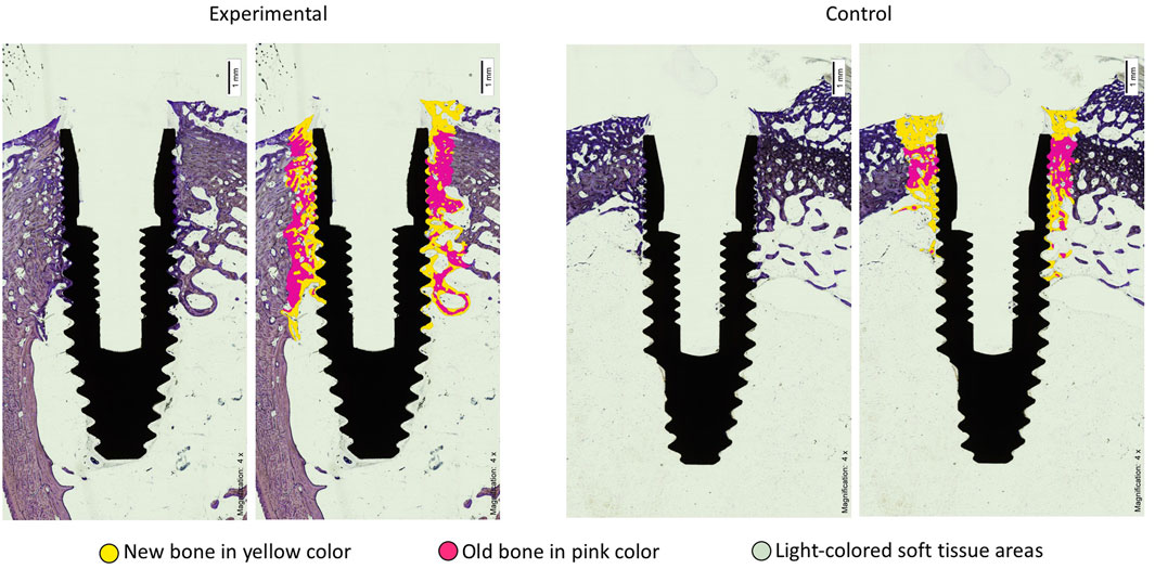
FIGURE 1. Histomorphometric section images of the BIC of implants in the Acid-BMP-7 (experimental) and SLA (control) groups after four weeks. The yellow color is new bone, more abundant and with greater contact with the implant surface in the experimental group compared to the control group.
3.2 Statistical results
The means, medians and SD values for BIC (%) histomorphometric parameter of the study groups are displayed in Table 1 and Table 2 and Figure 2.
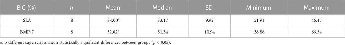
TABLE 1. Descriptive statistics of the BIC (%) histomorphometric parameters.
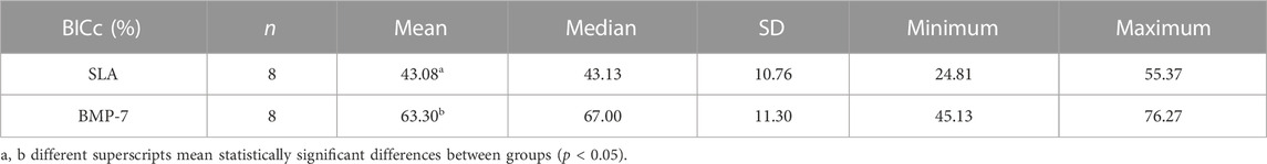
TABLE 2. Descriptive statistics of the BICc (%) histomorphometric parameters.
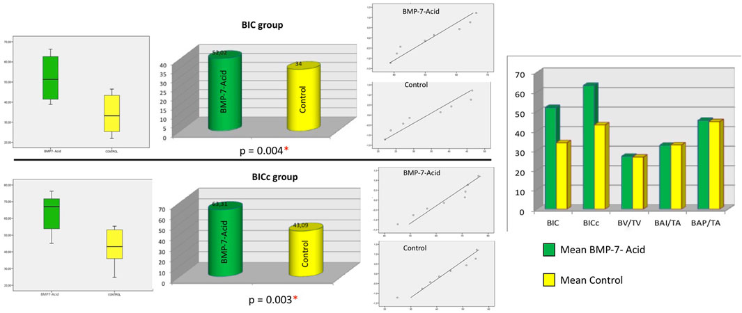
FIGURE 2. For both BIC and BICc, the box plot shows that the mean value of the BMP7-acid group (test group) was higher than that of the control group. In the Q-Q plots, the points are within the confidence interval and are randomly distributed on both sides of the axis, indicating that the distribution is normal for each group and variable. The bar chart shows the means of the compared groups (statistically significant for the variables BIC and BICc). * Statistical significance.
Student’s t-test revealed statistically significant differences in histomorphometric parameters BIC and BICc between the experimental and control groups (p = 0.004 and p = 0.003, respectively). No statistical significance was found between the groups for the other parameters studied; the BAI/TA group obtained values of 32.71 ± 10.87 for the experimental group versus 32.91 ± 7.76 for the control group (p = 0.967); the BAP/TA group, 45.47 ± 11.07 for the experimental group versus 44.79 ± 8.67 for the control group (p = 0.894) and the BV/TV group, 27.01 ± 6.00 for the experimental group versus 26.63 ± 7.90 for the control group (p = 0.951).
4 Discussion
Early osseointegration has been shown to be influenced by both the roughness and surface coatings of Ti (Le Guéhennec et al., 2007; Sakka et al., 2012).
Organic coatings on inorganic surfaces, such as Ti, have been widely used to improve their biocompatibility and induce a specific biological response and different in vivo investigations demonstrated increased adhesion and proliferation of osteoblastic cells with BMP-2 based coatings (Schliephake et al., 2005; Schliephake et al., 2009).
BMPs act as potent regulators during bone and cartilage formation and repair and both recombinant human BMP-2 and BMP-7 have been approved for clinical use in bone regeneration, fracture healing and spinal fusion (Carreira et al., 2014). Although BMP-2 has been the most studied, certain studies have demonstrated in vitro the potential of BMP-7 as a stimulator of bone regeneration (Zhang et al., 2012), it has also been documented that BMP-7 stimulates the maturation of osteoblastic progenitors and induces the differentiation of undifferentiated non-osteogenic cells to osteoblasts (Shen et al., 2010). Busuttil Naudi et al. reported bone regeneration in critical rabbit mandibular defects using beta-tricalcium phosphate (β-TCP) scaffolds and recombinant human bone morphogenetic protein 7 (rhBMP-7) (Busuttil Naudi et al., 2012).
Currently, biomimetic coatings have been developed to induce the formation of a biologically active surface layer of apatite on the implant surface, by developing various techniques capable of creating functional groups (-OH, -COOH, -CH = CH2, etc.). Liu et al. (2002) suggested that pre-deposition of hydroxyapatite (HA) on functionalized self-assembled monolayer surfaces, could be an efficient and fast way to prepare biomimetic apatite coatings on surgical implants. On the other hand, it has been described that the use of acids for peptide anchoring further stimulates the activity of the implant surface, increasing its initial osseointegration; Aresti et al. (2021) demonstrated that the treatment of the surface with CEPA provides functional anchoring groups such as carboxyl groups, and increases the roughness of the dental implant surface, due to the corrosive action of the acid, which causes structural modifications on the surface as a result of the erosive action of the acid, thus showing a more complex surface structure, which induces a greater surface hydrophilicity, which would guarantee a greater affinity for the adhesion and proliferation of osteoblasts to the implant surface. These results would coincide with those found in our study, in which the parameters BIC and BICc were statistically significant between the groups experimental and control.
Other studies reported on the immobilization of BMP-2 after using 11-(hydroxydecyl) phosphonic acid and 12-(carboxydecyl) phosphonic acid to evaluate the reactivity of different surfaces using trifluoroethylamine hydrochloride, since these reactive molecules, can immobilize on the implant surface under the same conditions as a protein, demonstrating a higher covalent immobilization of fluorine molecules in the -COOH group with respect to the -OH group (Adden et al., 2006).
The inducing effect of BMPs on the differentiation of mesenchymal stem cells into osteoblasts has also been demonstrated, which would lead to an improvement in local bone growth, in addition to stimulating the formation of calcium deposits (Hoffmann et al., 2001; Karageorgiou et al., 2004); therefore, these molecules have been extensively studied in dental implantology, in particular BMP-2 (Sykaras et al., 2001; Stadlinger et al., 2008; Wölfle et al., 2014), mainly for their ability to improve osseointegration of dental implants, since BMP-2, BMP-4 and BMP-7 appear to induce comparable levels of bone augmentation (Kirkwood et al., 2003; Leknes et al., 2008; Susin et al., 2010) and in recent years several studies have focused on the role of BMPs in osteoblastic differentiation and their inducing function in collagenous and non-collagenous protein synthesis (Constantinescu-Bercu et al., 2022), however, few studies have focused on analyzing BMP-derived peptides for their osteogenic capacity; Kirkwook et al. (2003), analyzed in vitro the minimal bioactive sequences of BMP-7, to evaluate its potential for biomaterial immobilization and its use in controlling osteoblast functions, demonstrating the potential of this molecule to induce osteoblast-specific genes associated with non-collagenous matrix formation and mineralization, along with gene expression, which would suggest some role of BMP-7 in mineralization.
However, there are no uniform results regarding the osteogenic power of BMP-7-functionalized Ti; an in vitro study by Togashi et al. (2009) found no effect of the addition of rhBMP-7 to the culture medium on the viability, proliferation, or differentiation of osteoblast-like cells, whereas others have found superior osteogenic power of samples with BMP-7 (Cirano et al., 2014). Zuardi et al. (2022) in a recent study, demonstrated that the effects of BMP-7 on osteogenic differentiation in vitro, would be conditioned by the topography of the surface on which the cell cultures are grown, the concentration of the growth factor in the culture medium and the stage of differentiation of the osteoblastic cells. Chen et al. (2013) developed a BMP-7-activated Ti gene delivery system for in vitro osteoblast culture and found enhanced differentiation capacity. This demonstrates that strategies for the development of metallic implants functionalized with BMP-7 (and probably with other bioactive molecules) should take into account the release profile during the repair process, depending on the concentration used for functionalization, and the physicochemical characteristics of the implant surface, all with the aim of guaranteeing a specific cellular behavior and/or generating an appropriate tissue phenotype. Nemcakova et al. (2022) in a combined in vitro and in vivo study found, after 4 and 12 weeks, a more pronounced osteogenic cell maturation and increased mineralization of the extracellular matrix around Ti6Al4V implants functionalized with BMP-7; in contrast, in our study we found no statistical significance in the BAI/TA and BAP/TA parameters. These results coincided with those obtained by Stenport et al. (2003) who, in a study in rabbit tibiae and femurs, found more areas of osteoid-like tissue in implants treated with BMP-7. Susin et al. (2010), Leknes et al. (2008) attribute the results to the concentration of rhBMP-7 used and agree that the observed bone remodeling would be proportional to higher doses of rhBMP-7. Leknes et al. (2008) are in agreement with our study and show robust bone formation after 4 weeks and remodeling of newly formed bone with characteristics similar to those of old bone. As in our study, Hunziker et al. (2021) in an experimental osteoporotic model, found at the third postoperative week, a significantly higher BIC value in the BMP-7 group compared to the Ti metal surface only control group.
Finally, it should be noted that the potential of rhBMP-7 to promote bone formation has been evaluated in a wide variety of large preclinical animal models (Phillips et al., 2006; Hunziker et al., 2021; Schierano et al., 2021) and in our study we used an experimental model with comparable bone anatomy and healing to humans and which has been used extensively in research related to maxillofacial surgery and dental implants. The anatomic and physiologic similarities of the pig to humans in terms of size, physiology, and bone biology have contributed to the successful use of this animal to understand and treat various bone situations and our experimental design was based on previous studies with this animal model, which considered the anatomical characteristics of the tibia to be similar to those of the human mandibular bone in terms of cortical/cancellous ratio and bone quality (Okubo et al., 2002; Mardas et al., 2014).
Our results rejected H0 and demonstrated that Ti dental implants exposed to CEPA and coated with BMP-7 exhibit a favorable biological response prone to osseointegration.
Further preclinical studies are needed to investigate the beneficial effects of BMP-7 on bone repair in contact with biomaterials (Senna et al., 2021).
Data availability statement
The original contributions presented in the study are included in the article/Supplementary Material, further inquiries can be directed to the corresponding author.
Ethics statement
The animal study was reviewed and approved by Ethics Committee in Animal Experimentation (ECAE) of the Puerta de Hierro University Hospital (Madrid, Spain) (ECAE Code: 017/2013).
Author contributions
Conceptualization, NL-V and JA; methodology, JA and NL-V; software, CR; validation, JMA and AL-V; investigation, NL-V and JA; data curation, CR; writing—original draft preparation, AL-V; writing—review and editing, JA; supervision, JA. All authors have read and agreed to the published version of the manuscript.
Conflict of interest
The authors declare that the research was conducted in the absence of any commercial or financial relationships that could be construed as a potential conflict of interest.
Publisher’s note
All claims expressed in this article are solely those of the authors and do not necessarily represent those of their affiliated organizations, or those of the publisher, the editors and the reviewers. Any product that may be evaluated in this article, or claim that may be made by its manufacturer, is not guaranteed or endorsed by the publisher.
References
Adden, N., Gamble, L. J., Castner, D. G., Hoffmann, A., Gross, G., and Menzel, H. (2006). Phosphonic acid monolayers for binding of bioactive molecules to titanium surfaces. Langmuir 22, 8197–8204. doi:10.1021/la060754c
Alghamdi, H. S., and Jansen, J. A. (2020). The development and future of dental implants. Dent. Mater J. 39, 167–172. doi:10.4012/dmj.2019-140
Aragoneses, J., Suárez, A., López-Valverde, N., Martínez-Martínez, F., and Aragoneses, J. M. (2021). Assessment of the tissue response to modification of the surface of dental implants with carboxyethylphosphonic acid and basic fibroblastic growth factor immobilization (Fgf-2): An experimental study on minipigs. Biol. (Basel) 10, 358. doi:10.3390/biology10050358
Aresti, A., Aragoneses, J., López-Valverde, N., Suárez, A., and Aragoneses, J. M. (2021). Effectiveness of biofunctionalization of titanium surfaces with phosphonic acid. Biomedicines 9, 1663. doi:10.3390/biomedicines9111663
Balik, V., Uberall, I., Sulla, I., Ehrmann, J., Kato, Y., Sulla, I. J., et al. (2019). Variability in wall thickness and related structures of major dural sinuses in posterior cranial fossa: A microscopic anatomical study and clinical implications. Oper. Neurosurg. Hagerst. 17, 88–96. doi:10.1093/ons/opy287
Busuttil Naudi, K., Ayoub, A., McMahon, J., Di Silvio, L., Lappin, D., Hunter, K. D., et al. (2012). Mandibular reconstruction in the rabbit using beta-tricalcium phosphate (β-TCP) scaffolding and recombinant bone morphogenetic protein 7 (rhBMP-7) - histological, radiographic and mechanical evaluations. J. Craniomaxillofac Surg. 40, e461-e469. doi:10.1016/j.jcms.2012.03.005
Carreira, A. C., Lojudice, F. H., Halcsik, E., Navarro, R. D., Sogayar, M. C., and Granjeiro, J. M. (2014). Bone morphogenetic proteins: Facts, challenges, and future perspectives. J. Dent. Res. 93, 335–345. doi:10.1177/0022034513518561
Chen, S., Yang, J., Wang, H., Chao, Y., Zhang, C., Shen, J., et al. (2013). Adenovirus encoding BMP-7 immobilized on titanium surface exhibits local delivery ability and regulates osteoblast differentiation in vitro. Arch. Oral Biol. 58, 1225–1231. doi:10.1016/j.archoralbio.2013.03.019
Cirano, F. R., Togashi, A. Y., Marques, M. M., Pustiglioni, F. E., and Lima, L. A. (2014). Role of rhBMP-2 and rhBMP-7 in the metabolism and differentiation of osteoblast-like cells cultured on chemically modified titanium surfaces. J. Oral Implant. 40, 655. doi:10.1563/aaid-joi-d-12-00071
Constantinescu-Bercu, A., Wang, Y. A., Woollard, K. J., Mangin, P., Vanhoorelbeke, K., Crawley, J. T. B., et al. (2022). The GPIbα intracellular tail - role in transducing VWF- and collagen/GPVI-mediated signaling. Haematologica 107, 933–946. doi:10.3324/haematol.2020.278242
Esposito, M., Hirsch, J. M., Lekholm, U., and Thomsen, P. (1998). Biological factors contributing to failures of osseointegrated oral implants. (I). Success criteria and epidemiology. Eur. J. Oral Sci. 106, 527–551. doi:10.1046/j.0909-8836.t01-2-.x
Ferraris, S., Spriano, S., Pan, G., Venturello, A., Bianchi, C. L., Chiesa, R., et al. (2011). Surface modification of Ti-6Al-4V alloy for biomineralization and specific biological response: Part I, inorganic modification. J. Mater Sci. Mater Med. 22, 533–545. doi:10.1007/s10856-011-4246-2
Fini, M., Savarino, L., Nicoli Aldini, N., Martini, L., Giavaresi, G., Rizzi, G., et al. (2003). Biomechanical and histomorphometric investigations on two morphologically differing titanium surfaces with and without fluorohydroxyapatite coating: An experimental study in sheep tibiae. Biomaterials 24, 3183–3192. doi:10.1016/s0142-9612(03)00164-9
Gittens, R. A., Scheideler, L., Rupp, F., Hyzy, S. L., Geis-Gerstorfer, J., Schwartz, Z., et al. (2014). A review on the wettability of dental implant surfaces II: Biological and clinical aspects. Acta Biomater. 10, 2907–2918. doi:10.1016/j.actbio.2014.03.032
Hall, J., Sorensen, R. G., Wozney, J. M., and Wikesjö, U. M. (2007). Bone formation at rhBMP-2-coated titanium implants in the rat ectopic model. J. Clin. Periodontol. 34, 444–451. doi:10.1111/j.1600-051X.2007.01064.x
Hoffmann, A., Weich, H. A., Gross, G., and Hillmann, G. (2001). Perspectives in the biological function, the technical and therapeutic application of bone morphogenetic proteins. Appl. Microbiol. Biotechnol. 57, 294–308. doi:10.1007/s002530100746
Hunziker, E. B., Liu, Y., Muff, M., Haegi, T., Shintani, N., and Lippuner, K. (2021). The slow release of BMP-7 at a low dose accelerates dental implant healing in an osteopenic environment. Eur. Cell Mater 41, 170–183. doi:10.22203/eCM.v041a12
Jemat, A., Ghazali, M. J., Razali, M., and Otsuka, Y. (2015). Surface modifications and their effects on titanium dental implants. Biomed. Res. Int. 2015, 1–11. doi:10.1155/2015/791725
Karageorgiou, V., Meinel, L., Hofmann, S., Malhotra, A., Volloch, V., and Kaplan, D. (2004). Bone morphogenetic protein-2 decorated silk fibroin films induce osteogenic differentiation of human bone marrow stromal cells. J. Biomed. Mater Res. A 71, 528–537. doi:10.1002/jbm.a.30186
Kaur, M., and Singh, K. (2019). Review on titanium and titanium-based alloys as biomaterials for orthopaedic applications. Mater Sci. Eng. C Mater Biol. Appl. 102, 844–862. doi:10.1016/j.msec.2019.04.064
Khang, W., Feldman, S., Hawley, C. E., and Gunsolley, J. (2001). A multi-center study comparing dual acid-etched and machined-surfaced implants in various bone qualities. J. Periodontol. 72, 1384–1390. doi:10.1902/jop.2001.72.10.1384
Kim, S., Park, C., Moon, B. S., Kim, H. E., and Jang, T. S. (2017). Enhancement of osseointegration by direct coating of rhBMP-2 on target-ion induced plasma sputtering treated SLA surface for dental application. J. Biomater. Appl. 31, 807–818.doi:10.1177/0885328216679761
Kirkwood, K., Rheude, B., Kim, Y. J., White, K., and Dee, K. C. (2003). In vitro mineralization studies with substrate-immobilized bone morphogenetic protein peptides. J. Oral Implantol. 29, 57–65. doi:10.1563/1548-1336(2003)029<0057:IVMSWS>2.3.CO;2
Le Guéhennec, L., Soueidan, A., Layrolle, P., and Amouriq, Y. (2007). Surface treatments of titanium dental implants for rapid osseointegration. Dent. Mater 23, 844–854. doi:10.1016/j.dental.2006.06.025
Leknes, K. N., Yang, J., Qahash, M., Polimeni, G., Susin, C., and Wikesjö, U. M. (2008). Alveolar ridge augmentation using implants coated with recombinant human bone morphogenetic protein-7 (rhBMP-7/rhOP-1): Radiographic observations. J. Clin. Periodontol. 35, 914–919. doi:10.1111/j.1600-051X.2008.01308.x
Leon-Ramos, J. R., Diosdado-Cano, J. M., López-Santos, C., Barranco, A., Torres-Lagares, D., and Serrera-Figallo, M. Á. (2019). Influence of titanium oxide pillar array nanometric structures and ultraviolet irradiation on the properties of the surface of dental implants-A pilot study. Nanomater. (Basel). 9, 1458. doi:10.3390/nano9101458
Liu, Q., Ding, J., Mante, F. K., Wunder, S. L., and Baran, G. R. (2002). The role of surface functional groups in calcium phosphate nucleation on titanium foil: A self-assembled monolayer technique. Biomaterials 23, 3103–3111. doi:10.1016/s0142-9612(02)00050-9
Liu, Y., Hunziker, E. B., Layrolle, P., De Bruijn, J. D., and De Groot, K. (2004). Bone morphogenetic protein 2 incorporated into biomimetic coatings retains its biological activity. Tissue Eng. 10, 101–108. doi:10.1089/107632704322791745
Liu, Y., Huse, R. O., de Groot, K., Buser, D., and Hunziker, E. B. (2007). Delivery mode and efficacy of BMP-2 in association with implants. J. Dent. Res. 86, 84–89. doi:10.1177/154405910708600114
López-Valverde, N., Aragoneses, J., López-Valverde, A., Quispe-López, N., Rodríguez, C., and Aragoneses, J. M. (2022). Effectiveness of biomolecule-based bioactive surfaces, on osseointegration of titanium dental implants: A systematic review and meta-analysis of in vivo studies. Front. Bioeng. Biotechnol. 10, 986112. doi:10.3389/fbioe.2022.986112
López-Valverde, N., Flores-Fraile, J., Ramírez, J. M., Sousa, B. M., Herrero-Hernández, S., and López-Valverde, A. (2020). Bioactive surfaces vs. Conventional surfaces in titanium dental implants: A comparative systematic review. J. Clin. Med. 9, 2047. doi:10.3390/jcm9072047
Mardas, N., Dereka, X., Donos, N., and Dard, M. (2014). Experimental model for bone regeneration in oral and cranio-maxillo-facial surgery. J. Invest. Surg. 27, 32–49. doi:10.3109/08941939.2013.817628
Matinlinna, J. P., Tsoi, J. K., de Vries, J., and Busscher, H. J. (2013). Characterization of novel silane coatings on titanium implant surfaces. Clin. Oral Implants Res. 24, 688–697. doi:10.1111/j.1600-0501.2012.02504.x
Nemcakova, I., Litvinec, A., Mandys, V., Potocky, S., Plencner, M., Doubkova, M., et al. (2022). Coating Ti6Al4V implants with nanocrystalline diamond functionalized with BMP-7 promotes extracellular matrix mineralization in vitro and faster osseointegration in vivo. Sci. Rep. 28, 5264. doi:10.1038/s41598-022-09183-z
Okubo, Y., Bessho, K., Fujimura, K., Kusumoto, K., Ogawa, Y., and Iizuka, T. (2002). Expression of bone morphogenetic protein in the course of osteoinduction by recombinant human bone morphogenetic protein-2. Clin. Oral Implants Res. 13, 80–85. doi:10.1034/j.1600-0501.2002.130110.x
Phillips, F. M., Turner, A. S., Seim, H. B., MacLeay, J., Toth, C. A., Pierce, A. R., et al. (2006). In vivo BMP-7 (OP-1) enhancement of osteoporotic vertebral bodies in an ovine model. Spine J. 6, 500–506. doi:10.1016/j.spinee.2006.01.014
Rupp, F., Liang, L., Geis-Gerstorfer, J., Scheideler, L., and Hüttig, F. (2018). Surface characteristics of dental implants: A review. Dent. Mater 34, 40–57. doi:10.1016/j.dental.2017.09.007
Sakka, S., Baroudi, K., and Nassani, M. Z. (2012). Factors associated with early and late failure of dental implants. J. Investig. Clin. Dent. 3, 258–261. doi:10.1111/j.2041-1626.2012.00162.x
Schierano, G., Canuto, R. A., Mauthe von Degerfeld, M., Navone, R., Peirone, B., Preti, G., et al. (2021). Role of rhBMP-7, fibronectin, and type I collagen in dental implant osseointegration process: An initial pilot study on minipig animals. Mater. (Basel) 14, 2185. doi:10.3390/ma14092185
Schliephake, H., Aref, A., Scharnweber, D., Bierbaum, S., Roessler, S., and Sewing, A. (2005). Effect of immobilized bone morphogenic protein 2 coating of titanium implants on peri-implant bone formation. Clin. Oral Implants Res. 16, 563–569. doi:10.1111/j.1600-0501.2005.01143.x
Schliephake, H., Aref, A., Scharnweber, D., Bierbaum, S., and Sewing, A. (2009). Effect of modifications of dual acid-etched implant surfaces on peri-implant bone formation. Part I: Organic coatings. Clin. Oral Implants Res. 20, 31–37. doi:10.1111/j.1600-0501.2008.01603.x
Senna, P. M., de Almeida Barros Mourão, C. F., Mello-Machado, R. C., Javid, K., Montemezzi, P., Del Bel Cury, A. A., et al. (2021). Silane-coating strategy for titanium functionalization does not impair osteogenesis in vivo. Mater. (Basel). 14, 1814. doi:10.3390/ma14071814
Shen, B., Wei, A., Whittaker, S., Williams, L. A., Tao, H., Ma, D. D., et al. (2010). The role of BMP-7 in chondrogenic and osteogenic differentiation of human bone marrow multipotent mesenchymal stromal cells in vitro. J. Cell Biochem. 109, 406–416. doi:10.1002/jcb.22412
Stadlinger, B., Pilling, E., Huhle, M., Mai, R., Bierbaum, S., Scharnweber, D., et al. (2008). Evaluation of osseointegration of dental implants coated with collagen, chondroitin sulphate and BMP-4: An animal study. Int. J. Oral Maxillofac. Surg. 37, 54–59. doi:10.1016/j.ijom.2007.05.024
Stenport, V. F., Johansson, C., Heo, S. J., Aspenberg, P., and Albrektsson, T. (2003). Titanium implants and BMP-7 in bone: An experimental model in the rabbit. J. Mater Sci. Mater Med. 14, 247–254. doi:10.1023/a:1022884724059
Susin, C., Qahash, M., Polimeni, G., Lu, P. H., Prasad, H. S., Rohrer, M. D., et al. (2010). Alveolar ridge augmentation using implants coated with recombinant human bone morphogenetic protein-7 (rhBMP-7/rhOP-1): Histological observations. J. Clin. Periodontol. 37, 574–581. doi:10.1111/j.1600-051X.2010.01554.x
Sykaras, N., Triplett, R. G., Nunn, M. E., Iacopino, A. M., and Opperman, L. A. (2001). Effect of recombinant human bone morphogenetic protein-2 on bone regeneration and osseointegration of dental implants. Clin. Oral Implants Res. 12, 339–349. doi:10.1034/j.1600-0501.2001.012004339.x
Szmukler-Moncler, S., Perrin, D., Ahossi, V., Magnin, G., and Bernard, J. P. (2004). Biological properties of acid etched titanium implants: Effect of sandblasting on bone anchorage. J. Biomed. Mater Res. B Appl. Biomater. 68, 149–159. doi:10.1002/jbm.b.20003
Togashi, A. Y., Cirano, F. R., Marques, M. M., Pustiglioni, F. E., Lang, N. P., and Lima, L. A. (2009). Effect of recombinant human bone morphogenetic protein-7 (rhBMP-7) on the viability, proliferation and differentiation of osteoblast-like cells cultured on a chemically modified titanium surface. Clin. Oral Implants Res. 20, 452–457. doi:10.1111/j.1600-0501.2008.01669.x
Wennerberg, A., and Albrektsson, T. (2009). Effects of titanium surface topography on bone integration: A systematic review. Clin. Oral Implants Res. 20, 172–184. doi:10.1111/j.1600-0501.2009.01775.x
Wölfle, J. V., Fiedler, J., Dürselen, L., Reichert, J., Scharnweber, D., Förster, A., et al. (2014). Improved anchorage of Ti6Al4V orthopaedic bone implants through oligonucleotide mediated immobilization of BMP-2 in osteoporotic rats. PLoS One 9, e86151. doi:10.1371/journal.pone.0086151
Zhang, F., Ren, L. F., Lin, H. S., Yin, M. N., Tong, Y. Q., and Shi, G. S. (2012). The optimal dose of recombinant human osteogenic protein-1 enhances differentiation of mouse osteoblast-like cells: An in vitro study. Arch. Oral Biol. 57, 460–468. doi:10.1016/j.archoralbio.2011.10.008
Keywords: dental implants, biological parameters, BMP-7, osseointegration, minipig model
Citation: López-Valverde N, Aragoneses J, López-Valverde A, Rodríguez C and Aragoneses JM (2023) Role of BMP-7 on biological parameters osseointegration of dental implants: Preliminary results of a preclinical study. Front. Bioeng. Biotechnol. 11:1153631. doi: 10.3389/fbioe.2023.1153631
Received: 29 January 2023; Accepted: 20 February 2023;
Published: 28 February 2023.
Edited by:
Juncen Zhou, Stony Brook University, United StatesReviewed by:
Kakyung Kim, Stony Brook University, United StatesYuyun Yang, Harbin Engineering University, China
Copyright © 2023 López-Valverde, Aragoneses, López-Valverde, Rodríguez and Aragoneses. This is an open-access article distributed under the terms of the Creative Commons Attribution License (CC BY). The use, distribution or reproduction in other forums is permitted, provided the original author(s) and the copyright owner(s) are credited and that the original publication in this journal is cited, in accordance with accepted academic practice. No use, distribution or reproduction is permitted which does not comply with these terms.
*Correspondence: Antonio López-Valverde, YWxvcGV6dmFsdmVyZGVAdXNhbC5lcw==
†These authors have contributed equally to this work