 Elia Ciani
 Kai-Inge Lie2
 Marit Stormoen
 Even Hjalmar Jørgensen
- 1Department of Production Animal Clinical Sciences, Norwegian University of Life Sciences, Ås, Norway
- 2Pharmaq Analytiq AS, Bergen, Norway
- 3Seacalx AS, Gratangen, Norway
- 4Department of Arctic and Marine Biology, The Arctic University of Norway, Tromsø, Norway
Particulate calcium oxide (CaO) has been successfully used for the control of sea urchin and starfish populations. Recent studies have proven its efficacy in killing planktonic copepods, making it a promising and cheap option for salmon louse control in the Atlantic salmon (Salmo salar) farming industry. Additionally, preliminary laboratory tests demonstrated that exposure to 0.2 g/L–0.6 g/L of fine CaO particles induced significant mortality of salmon louse (Lepeophtheirus salmonis) in the planktonic stages. The present study investigated the effects of water treatment with fine CaO particles [0.2 g/L or 127.4 g/m2; 0.1 mm–0.3 mm], conducted twice a week for three consecutive weeks at 5°C and 12°C on Atlantic salmon post-smolt in a flow-through system. The study compared mortality rates and histopathology of skin, eyes, gills, and intestines between treated and untreated control salmon. The results indicated that CaO exposure did not induce fish mortality or histopathological damages in the skin, eyes, or intestines. Although there were no significant effects of CaO exposure on gill inflammation or hyperplasia, the exposure did increase the occurrence of gill vascular injuries and necrosis in small portions of the tissue (< 10% of the respiratory gill tissue in most of the samples) by 60% and 35%, respectively. The effect was not modulated by temperature or time. The results indicate that the CaO treatment protocol used in this study is not lethal to Atlantic salmon and is safe for most analyzed tissues. Nonetheless, the induction of vascular damage and necrosis in the gill, albeit in small tissue portions, emphasizes the need for further research. It is crucial to develop a protocol that avoids such lesions, ensuring the secure application of CaO as an antiparasitic treatment in salmon aquaculture.
Highlights
● To our knowledge, this is the first study investigating the histopathological effects of calcium oxide exposure on Atlantic salmon.
● Calcium oxide exposure increases the occurrence of vascular injuries and necrosis in small portions of the gills.
● Calcium oxide exposure does not induce gill inflammation or hyperplasia.
● Calcium oxide exposure does not cause skin, eye, or intestinal lesions.
1 Introduction
The use of quicklime, or calcium oxide (CaO), as a controlling agent for echinoderm populations, has been documented back to the beginning of the 20th century (Wood, 1908). CaO particles have also been successfully used in Nova Scotia and California for the management of sea urchins and starfish in commercial kelp and oyster beds (Bernstein and Welsford, 1982) and in Norwegian fjords for the control of sea urchin populations (Strand et al., 2020).
A recent laboratory study (Brooks et al., 2020) ranked several marine species according to their vulnerability to fine CaO particles (<0.8 mm mm). The most sensitive was the copepod Tisbe battagliai [Crustacea; lethal concentration for 50% of the fish (LC50) 3.14 g/g/m2], followed by the green sea urchin (Strongylocentrotus droebachiensis, echinoderms; LC50 20.1 g/g/m2), the common starfish (Asterias ruben, echinoderms; LC50 22.2 g/g/m2), the slender ragworm (Nereis pelagica, polychaete; LC50 26.6 g/m2), and the netted dog whelk (Hinia reticulata; Mollusca; LC50 41.9 g/m2). The fish species tested, the lumpsucker (Cyclopterus lumpus), was affected only by a high concentration of particles (LC50 226 g/m2). Three species were unaffected by CaO particle exposure: the shore crab (Carcinus maenas, Crustacea), the blue mussel (Mytilus edulis, Mollusca), and seaweed germlings (Fucus vesiculosus). Given the high efficacy of CaO particles against copepods, there is a growing interest in the use of quicklime as a natural antiparasitic treatment against sea lice in the Atlantic salmon (Salmo salar) farming industry. Recent laboratory trials (Seacalx AS, unpublished) showed that the application of CaO treatment (0.2 g/L–0.6 g/L; particle size 0.1 mm–0.6 mm; 2 min–10 min) had detrimental effects during the free-living planktonic stages of the salmon louse (Lepeophtheirus salmonis). The treatment led to a significant 60%–70% reduction in the number of nauplii progressing to the copepodid stage due to decreased survival and molting rates. Furthermore, the treatment resulted in up to a 90% mortality rate among free-living copepodids and a 15% decrease in the egg-hatching rate. No significant effects were reported on sessile and motile stages once attached to fish.
CaO reacts with water, producing calcium hydroxide, Ca(OH)2, in an exothermic and alkalinizing reaction (increasing temperature and pH), which causes epidermal burns and lesions when in contact with the surface of the target organisms. This results in both acute and delayed mortality due to osmotic imbalances and bacterial infections in echinoderms (Bernstein and Welsford, 1982). The speed and duration of the exothermic reaction are heavily influenced by particle size (Strand et al., 2020). On the one hand, smaller particles react faster and sink slower in water columns due to increased surface-to-volume ratio and buoyancy. On the other hand, bigger particles react slower and sink much faster. The optimization of particle size composition is therefore crucial to maintain particles long enough in water columns so that the exothermic reaction is not dissipated before reaching the target organisms (Bernstein and Welsford, 1982; Strand et al., 2020).
To date, very limited knowledge is available on the impact of CaO on the health and welfare of fish. The aim of this study was to conduct a histopathological assessment of the effects of fine CaO particle exposure at different water temperatures (5°C and 12°C) on Atlantic salmon in flow-through systems.
2 Materials and methods
2.1 Ethics statement
The experiment was performed in accordance with the EU regulations concerning the protection of experimental animals (Directive 2010/63/EU). Appropriate measures were taken to minimize pain and discomfort. The experiment was approved by the Norwegian Food and Safety Authority (FOTS id. number 19906).
2.2 Experimental setup
The experiment setup is summarized in Table 1. The exposure experiment was carried out at the Aquaculture Research Station in Tromsø (ARST), Norway. Atlantic salmon (Salmo salar) post-smolts (sexually immature; mixed sex) of the AquaGen strain (AquaGen, Trondheim, Norway) were kept in flow-through tanks. A total of 120 fish (average fish weight 1.048 kg) were evenly distributed among six circular tanks (500 L; 1-m diameter; 0.8-m tank height; ≈ 0.6-m water height; water flow ≈ 600 L/hour; stocking density 41,10 kg fish/m3) and were kept at constant light and salinity of 32‰. Three tanks were kept at a water temperature of 5°C and three at 12°C. In each temperature group, one tank was used as an untreated control and two for the treatment, as technical replicates. The fish were fed ad libitum with dry pellets (Skretting, Stavanger, Norway) and acclimatized for a period of 8 days prior to the start of the experiment. The oxygen saturation was kept above 90% during the whole experiment.
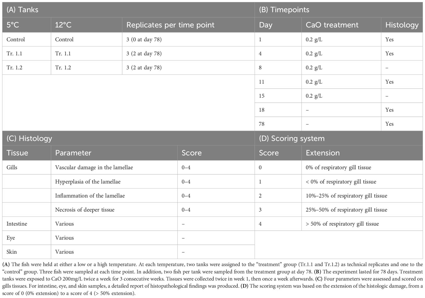
Table 1 Summary of experimental design.
The treatment tanks were exposed to 0.2 g/L (127.4 g/m2) calcium oxide (CaO; Miljøkalk AS Norway; particle size 0.1 mm–0.3 mm) for 10 min twice a week for three consecutive weeks (at days 1, 4, 8, 11, and 15). The dose of CaO (100g) was applied through a small sieve (to avoid clumping) evenly over the entire water surface over a period of 10 min. The water flow in each tank was not interrupted during treatment to mimic field conditions, in which water masses are constantly replaced. As the reaction of CaO with water to form Ca(OH)2 is alkaline, the change in pH was monitored during CaO exposure and for the following 3 h in the treatment tanks (Supplementary Figure 1). After dosing, the product was diluted over time due to the efflux of the water.
2.3 Sampling procedure
The histologic sampling setup is summarized in Table 1. Tissues were collected from three fish per tank twice in the first week, then once a week (at days 1, 4, 11, and 18). The sampling was performed immediately before CaO exposure at all-time points. In addition, tissues from two fish per tank in the treatment group were collected 3 months after the first treatment, at day 78. The fish were euthanized by an overdose of benzocaine (150 ppm). The gill, intestine, eye, and skin samples were collected as follows:
● Skin: the tissue was excised below the dorsal fin, at least 1 cm × 1 cm in size with subcutaneous muscles.
● Gill: the tissue was excised from all four gill arches by cutting in two places, one at the lower attachment and the other at the mid curvature of the arch.
● Intestine: the tissue was excised from the anterior part of the small intestine, about 2 cm behind the end of the pylorus and at approximately 2 cm in length. The intestine was rinsed in seawater before preservation in formalin.
● Eye: the whole left eye was extracted from each fish. If the procedure damaged the tissue, the eye in question was discarded and replaced with the right eye.
2.4 Histopathology
The histopathology assessment was carried out according to Østevik et al. (2021, with minor adjustments. Sampled tissues were fixed in buffered formalin (4% formalin, 0.08 M sodium phosphate, pH 7.0) and processed in a Thermo Scientific Excelsior tissue processor (Thermo Fisher Scientific, Runcorn, UK). The tissues were embedded in paraffin using a Tissue-Tek®, TEC 5 (Sakura Finetek Europe B.V., Alphen aan den Rijn, the Netherlands) embedding center. The tissue sections (1.5 μm–2 μm) were cut using a Leica RM2255 microtome, mounted on glass slides, and stained with hematoxylin–eosin (HE). The slides were scanned in an Aperio ScanScope® AT Turbo slide scanner and read using Aperio ImageScope software (Leica Biosystems, Buffalo Grove, IL, USA). All counting and measuring were done using the annotation tools in the ImageScope software. The pathologist was “blinded” in regard to the group (control vs. treatment), time point, and temperature. Four different histopathological lesion types were examined and scored in the gill tissues (Table 1; Figure 1):
(i) lamellar inflammatory cell infiltration.
(ii) lamellar epithelial hyperplasia.
(iii) subacute/chronic lamellar vascular lesions (comprising thrombi and thrombosing aneurysms–telangiectasia/organizing thrombi and aneurysms).
(iv) necrosis involving one or more lamella and/or the filament.
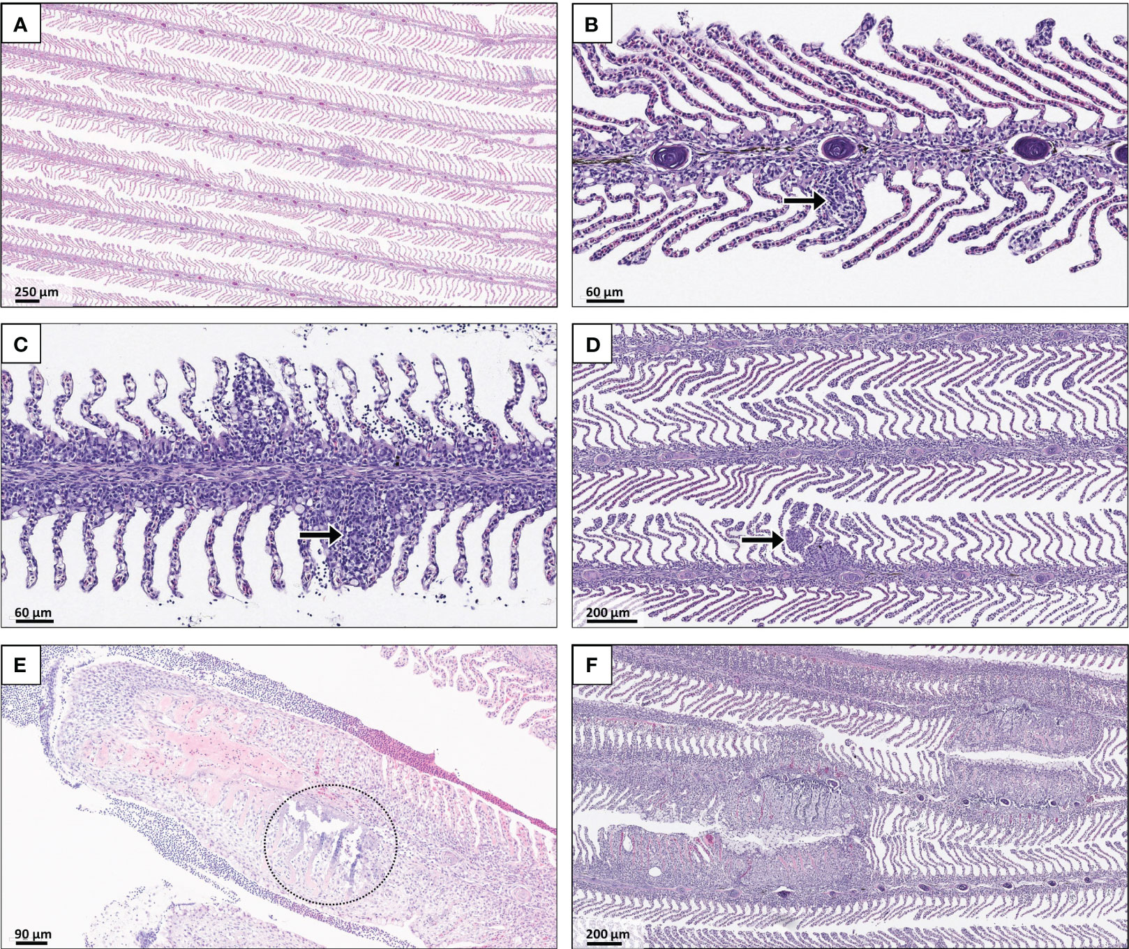
Figure 1 Normal gill tissue and histopathological lesions. All tissues were stained with hematoxylin and eosin. (A) Healthy gill tissue. The four types of lesions scored in the study were (B) inflammation, (C) hyperplasia, (D) vascular damages, and (E) necrosis (indicated with arrows and a circle). The gill tissue in (F) shows multifocal changes in which lamellar vascular lesions (thrombosis), epithelial and mucus cell hyperplasia, lamellar inflammation, and necrosis extend into the filament.
A five-level scoring protocol was used based on the extent of the gill tissue affected: score 0, no damage detected; score 1, < 10%; score 2, 10%–25%; score 3, 25%–50%; and score 4, > 50% of the respiratory gill tissue (Table 1).
Additionally, intestines, eyes, and skin were evaluated for histopathological lesions. The findings in these tissues were noted, but no scoring was performed.
2.5 Data analysis and statistics
Data were analyzed using Stata 17 (StataCorp. 2021; Stata Statistical Software: Release 17.0; StataCorp LLC, College Station, TX, USA). The descriptive data are presented as summary tables.
Mixed models, allowing the inclusion of fixed and random effects, were used, as all observations were clustered by tank. Different regressions were used to assess the histopathological effects of treatment between the gills and other tissues.
- Gill histology. A multilevel ordinal logistic regression (MOLR) model was built for each of the four investigated parameters, as the outcome of interest (histologic score) was an ordinal variable with a clear ordering of the category levels (from score 0 “no effects” to score 4 “severe effects”). When a reliable MOLR could not be built, a multilevel logistic regression (MLR) was produced instead, using the histologic score as the dichotomous outcome variable (comparing 0 “no effects—score 0” vs.1 “any affect—score > 0”). When needed, the risk was calculated using the “csi” command in Stata.
- Eye, intestine, and skin histology. Two different models were built for each tissue. The first was a multinomial regression (MR) model in which the outcome “no evidence of histopathological changes” was used as a reference value versus multiple histopathological changes detected. The second was a multilevel logistic regression (MLR) model with the dichotomous outcome “no evidence of histopathological changes” versus “any histopathological changes”.
The explanatory variables “treatment” (dichotomous variable, treatment vs. control), “time” (in days, continuous variable), and “temperature” (dichotomous variable, 5°C vs. 12°C) were included as fixed effects. The variable “cage” was included as a random effect. The interactions between “treatment” and “temperature” were included in the full model to account for the differential effects of the treatment at different temperatures. The fixed-effects variables were first screened for multicollinearity by correlation matrix and exclusion of predictors with correlation > 0.6 (Supplementary Table 1). Then, they were screened in a univariable model by applying a liberal cut-off value (p <0.2) and controlling for the tank. Finally, multivariable models were built to assess the association of filtered predictors while still controlling for the tank. For the final models, the cut-off value for statistical significance was set to a p-value < 0.05.
The models were built by backward selection, including initially all predictors that passed the screening stage and by removing one by one those that were not statistically significant (p > 0.05). The interaction between temperature and treatment was also assessed during the model-building procedure.
The assumption of proportional odds in the ordered logistic model was validated using the Brant test and the “omodel” command in Stata.
The effects of the statistically significant variables on the outcome were investigated by using the Stata “margins” command after each model. These data were visualized using the “marginsplot” command.
3 Results
3.1 Mortality
No fish died or showed sign of abnormal behavior during the experiment in any of the tanks.
3.2 Gill histopathology
Four histopathological parameters—inflammation, hyperplasia, vascular injuries, and necrosis—were examined and scored (range from 0 to 4) for 80 fish divided between the control (24 fish) and treatment groups (56 fish) at 5°C and 12°C water temperature.
The mean scores were low, mostly between 0 and 1, in both the control and treatment fish groups at the different water temperatures (Figure 2). Notably, none of the biological replicates indicated scores exceeding 2 for any of the parameters.
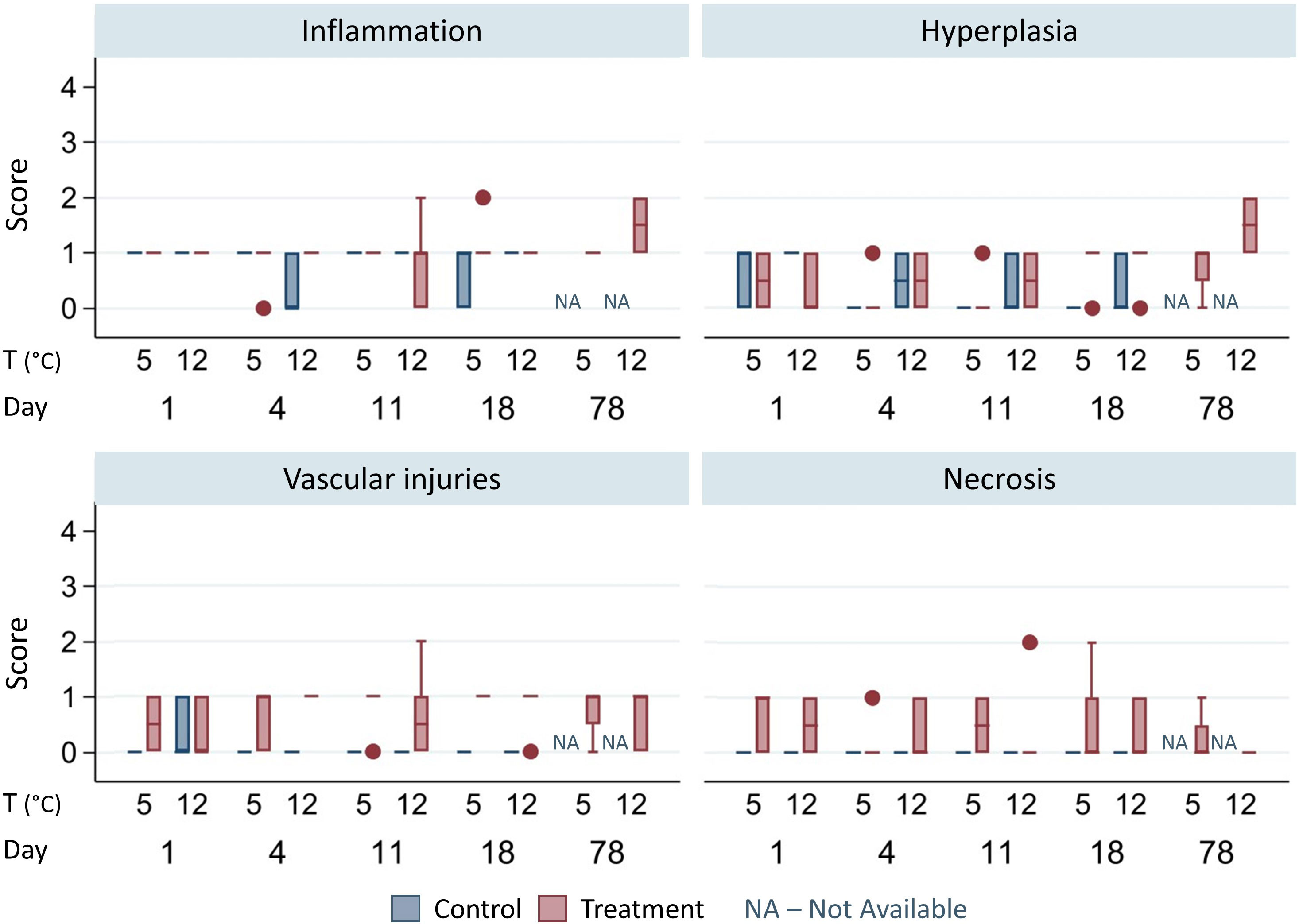
Figure 2 Histopathological parameter scores for the gills. The lower and upper box boundaries represent the 25th and 75th percentiles; the line inside the box is the median; the lower and upper error lines represent the lower and upper adjacent values; and the dots represent outliers. The data are clustered between groups [control (blue columns)—treatment (red columns)], temperature [(T) 5°C—12°C], and over time (day 1 to 78). NA, not available; –, no control samples were available at day 78. The number of replicates per group is available in Table 2. Although the mean inflammation and hyperplasia scores did not differ between treatment and control groups, a noteworthy increase in vascular injuries and necrosis was observed in treated fish compared with the control fish.
3.2.1 Inflammation
The statistical model (Table 2A) indicates that exposure to CaO did not induce gill inflammation, which occurred equally in both the control and treated fish groups and increased over time.
Table 2 Multilevel ordinal logistic regression models (MOLR).
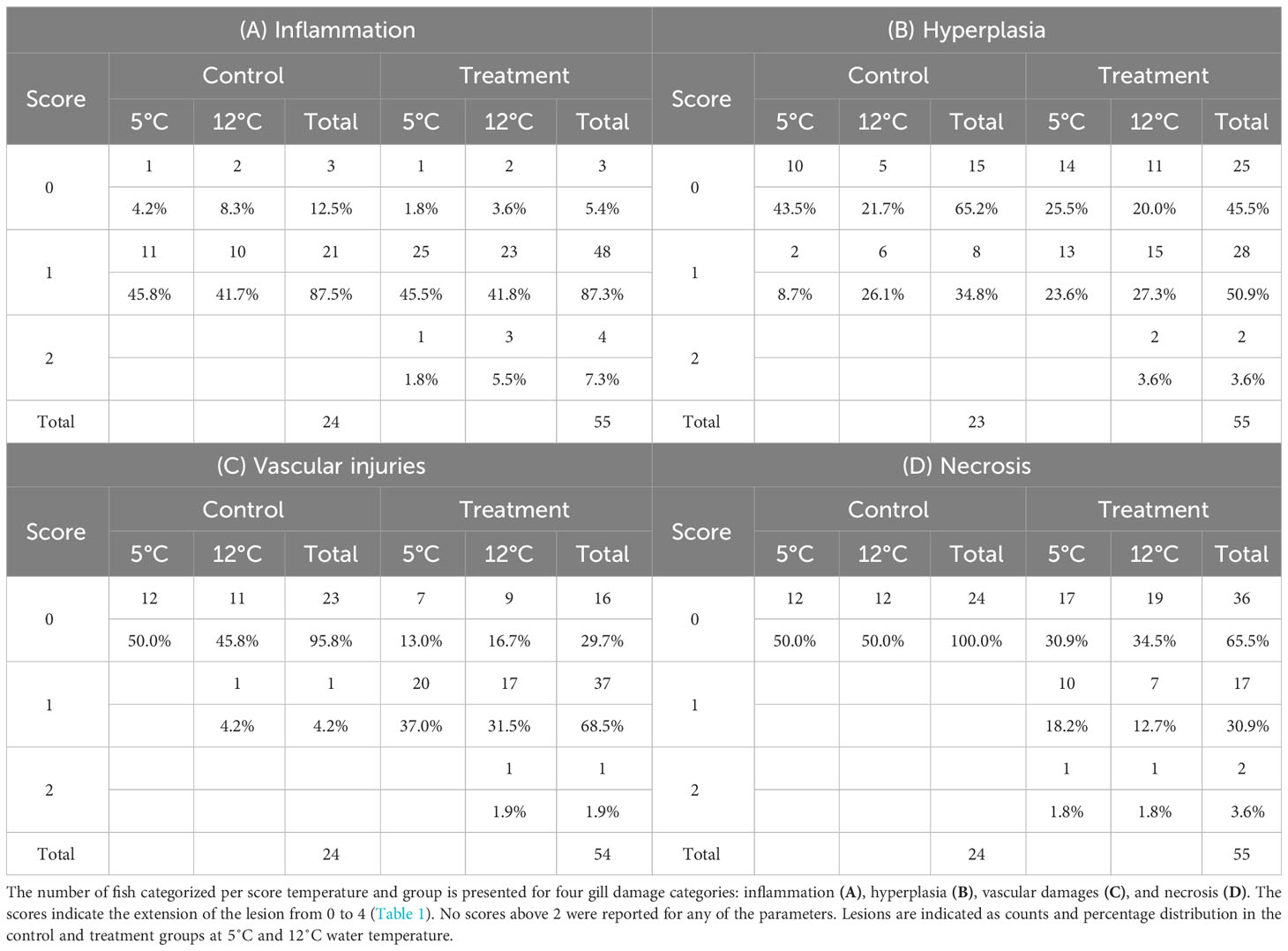
Table 3 Overview of histopathology lesion types and severity in gills.
Most of the samples analyzed showed some degree of inflammation (Table 3A). In the control group, 87.5% (21/24) of the samples were scored 1, while in the treatment group, 87.3% (48/55) scored 1, and 7.3% (4/55) scored 2 for inflammation. Healthy (score 0) and damaged (score ≥ 1) tissues were equally distributed between the temperature groups.
The MOLR model using “inflammation score” as the outcome, “time” as the fixed effect, and “cage” as the random effect was composed of 79 observations. The Wald chi-squared value was equal to 5.77 (p = 0.016), thus indicating that at least one of the predictors’ regression coefficients was not equal to zero in the model (Table 2A). The assumption of proportional odds was confirmed by the Brandt and Omodel tests. The random-effects section indicated no variation between tanks (coefficient 1.23−33). The fixed-effects section showed a positive ordered log-odds (logit) regression coefficient of 0.04 (p = 0.016) for time (in days). The standard interpretation of such a coefficient is that for a one-unit increase in the predictor, the response variable level is expected to change by the regression coefficient in the ordered log-odds scale while the other variables in the model are held constant. The marginal effect analysis (Figure 3A; Supplementary Table 2) showed that, at the beginning of the experiment, the probability of detecting an inflammation score of 0 was 11%, a score of 1 was 87%, and a score of 2 was 2%. From day 35 onward, the probability of detecting a score of 2 increased constantly, while the probability of detecting scores 0 and 1 decreased.

Figure 3 Marginal effects with 95%CI. (A) The probability of detecting inflammation scores 0, 1, and 2 over time. (B) The marginal effects over time of a higher water temperature (12°C) on the probability of detection of hyperplasia scores 0, 1, and 2. (C) The marginal effects of CaO treatment on the probability of detecting vascular injuries scores 0, 1, and 2.
3.2.2 Hyperplasia
The statistical model (Table 2B) reveals that CaO exposure did not lead to gill hyperplasia, a condition observed equally in both control and treated fish. The occurrence of hyperplasia increased with higher temperatures, and this effect was further amplified over time.
A total of 34.8% (8/23) of the control samples were scored 1, while 50.9% (28/55) and 3.6% (2/55) of treatment samples were scored 1 and 2, respectively (Table 3B). The 12°C group had more replicates that scored ≥ 1 than the 5°C group (24 vs. 15) and fewer samples that scored 0 (16 vs. 24).
The MOLR model using “hyperplasia score” as the outcome, “temperature” and “time” as the fixed effects, and “cage” as the random effect was composed of 78 observations. The highly significant (p = 0.0018) Wald chi-squared value of 12.6 indicated that at least one of the predictors’ regression coefficients was not equal to zero (Table 2B). The assumption of proportional odds was confirmed by the Brandt and Omodel tests. The random-effects section of the model indicated no variation between tanks (coefficient 5.11−33). The fixed-effects section showed a positive coefficient of 1.06 (p = 0.028) for temperature and 0.04 (p = 0.003) for time. The marginal effects analysis showed that keeping the water temperature at 12°C, rather than at 5°C, increased the occurrence of hyperplasia (score 1) by 20% between day 0 and day 40 (Figure 3B; Supplementary Table 2).
3.2.3 Lamellar vascular injuries
The statistical model (Table 2C) shows that CaO exposure led to vascular damage in the gills (+60% occurrence of score 1), irrespective of the water temperature.
Only one sample in the control group was scored 1 for lamellar vascular injuries. All other control samples were scored 0. On the other hand, 68% (37/54) of the treatment samples were scored 1, and 1.9% (1/54) were scored 2 with respect to lamellar vascular injuries (Table 3C). The healthy (score 0) and damaged (score ≥ 1) tissues were equally distributed between the temperature groups.
The MOLR model using “vascular injury” score as the outcome, “treatment” as the fixed effect, and “cage” as the random effect was composed of 78 observations. The highly significant (p < 0.001) Wald chi-squared value of 14.14 indicated that at least one of the predictors’ regression coefficients was not equal to zero in the model (Table 2C). The assumption of proportional odds was confirmed by the Brandt and Omodel tests. The random-effects section of the model indicated no variation between tanks (coefficient 4.28−33). The model showed a positive coefficient of 4.00 (p < 0.001) for the treatment. The marginal effects analysis (Figure 3C; Supplementary Table 2) indicated that the treatment increased the occurrence of damage (score 1) by almost 60% and decreased by the same margin the incidence of no damage (score 0). The treatment had no effect on the probability of detecting more extensive damage (score 2).
3.2.4 Necrosis
The statistical model (Table 4) shows that CaO exposure induced gills necrosis (+35% occurrence of scores 1 and 2), irrespective of the water temperature.
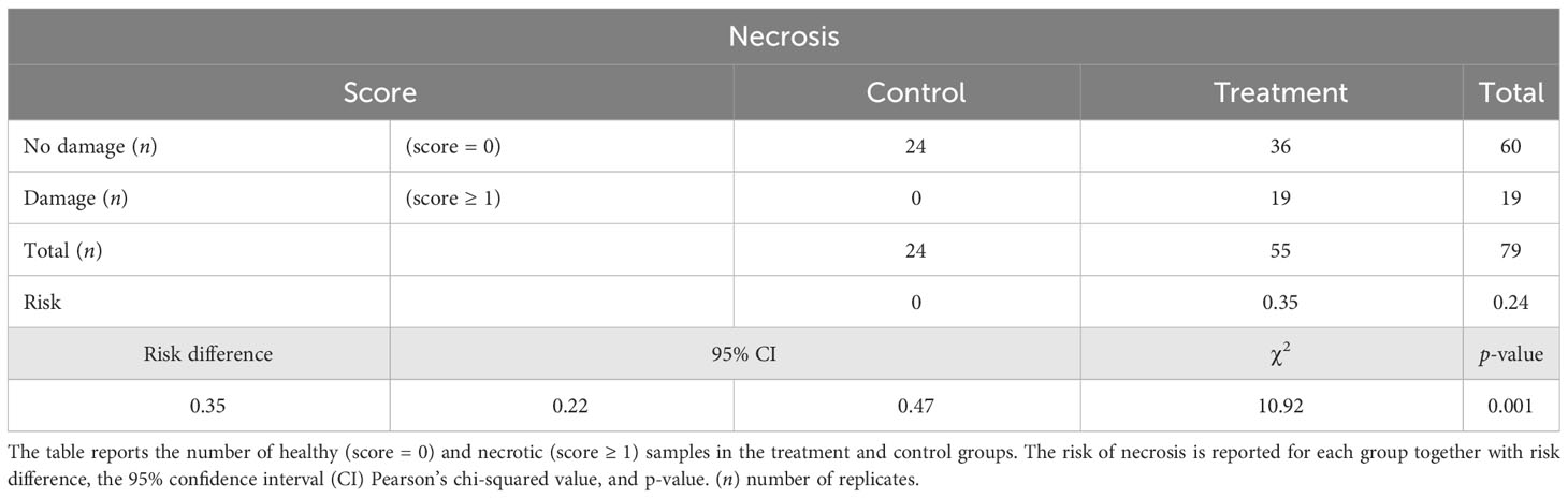
Table 4 Risk of necrosis.
None of the control samples (24 out of 24) showed sign of gill necrosis, while 30.9% (17/55) and 3.6% (2/55) of treatment samples received a necrosis score of 1 and 2, respectively (Table 3D). The healthy (score 0) and damaged (score ≥ 1) tissues were equally distributed between the temperature groups.
The variable “treatment” could not be included in the MOLR or MLR models, as all necrotic tissues (score ≥ 1) belonged to the treatment group (thus predicting failure perfectly). The risk of necrosis was significantly higher in the treatment group (risk difference = 0.35; p = 0.001, Table 4).
The MOLR model using “necrosis score” as the outcome, “temperature” and “time” as the fixed effects, and “cage” as the random effect was composed of 79 observations. The model indicated no statistically significant effects of temperature or time on the necrosis histologic score. Also, a MLR model using necrosis score as a binary variable (score 0 vs. score ≥ 1) indicated no significant effects of temperature or time on necrosis.
3.3 Other tissues
3.3.1 Skin
A total of 78 skin samples were processed in the present study (Tables 5A, B). No signs of histopathological changes were detected in most of the control (22/24) and treatment (48/54) samples. One control sample showed a thin epidermis and mild inflammation, while one showed focal superficial inflammation. The treatment samples showed focal mild inflammation of the dermis (1/54) and epidermis (4/54) and multifocal mild inflammation of the epidermis (1/54).
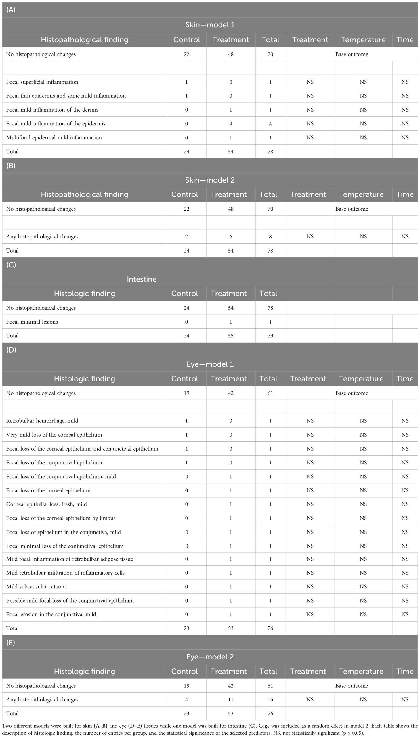
Table 5 Skin, intestine, and eye histopathology.
The multinomial logistic regression (MNLR) model showed that none of the predictors (i.e., treatment, temperature, and time) had a statistically significand coefficient for any of the histopathological damages reported in the skin. To further investigate any correlation between treatment and skin histopathology, a MLR model was then built using skin histopathology as a dichotomous outcome (damage present vs. absent) and adjusting for clustering at tank level. This model reported no statistically significant coefficient for any of the independent variables.
3.3.2 Intestine
Only one sample (1/54) in the treatment group showed focal minimal lesions (Table 5C). No control samples showed signs of histopathology. Given the presence of only one record for histopathology, no regression models were built.
3.3.3 Eye
Most of the samples in both the control (19/23) and treatment (42/53) groups showed no histopathological anomalies (Tables 5D,E). The lesions reported varied between fish and included focal loss of corneal and conjunctival epithelium, subcapsular cataract, and infiltration of inflammatory cells, among other injuries. Both the MNLR and MLR models showed no statistically significant coefficient for any of the potential predictors investigated.
4 Discussion
This study describes the effects of particulate CaO exposure on the health and welfare of Atlantic salmon. It is important to clearly differentiate particulate from dissolved exposure. Dissolved CaO is generally not harmful to marine organisms and could contribute to resistance against divalent metal toxicity (Das and Das, 2005). On the other hand, particulate CaO causes severe wounds in sensitive species through the exothermal reaction when in contact with seawater (Brooks et al., 2020). As no toxic residues form when CaO reacts with water, the U.S. Environmental Protection Agency removed it from its list of hazardous substances (Shumway et al., 1988), and it is registered in the list of chemicals used and discharged offshore that are considered to pose little or no risk to the environment (OSPAR, 2021). Despite its potential applications as a natural antiparasitic treatment in aquaculture, very limited information is available on the effects of CaO particles in fish to date.
In the present study, Atlantic salmon post-smolts were exposed to fine CaO particles [0.1 mm–0.3 mm; 0.2 g/L (127.4 g/m2)] twice a week for three consecutive weeks. The concentration and particle size applied was selected in accordance with previous optimization studies conducted by Seacalx AS for the treatment of the salmon louse (Lepeophtheirus salmonis). These earlier studies exposed lice to fine CaO particle concentrations ranging from 0.2 g/L to 0.6 g/L for 10 min. Although there was a clear “dose-dependent” effect, minimal concentrations of 0.2 g/L were sufficient to induce significant mortality in nauplii and copepodids. The present study deliberately applied the minimal effective concentration to salmon to assess the fish’s tolerance to the treatment. Moving forward, it is essential for future studies to explore the effects of higher CaO concentrations to establish the boundaries of a safe concentration for salmon. The optimization of particle size and concentration is the most important factor contributing to the efficacy of CaO treatment, as finer particles (<0.8 mm) seem to be most harmful to marine organisms (Brooks et al., 2020; Strand et al., 2020). Interestingly, no mortality was reported during the whole observation period of 3 months. This is in contrast to the mortality observed in other fish species. The lumpsucker exhibited significant mortality within 10 days of a single exposure to fine CaO particles, with an estimated LC50 (lethal concentration for 50% of the fish) of 226 g/m2 (≈ 0.6 g/L) (Brooks et al., 2020). An acute exposure to CaO nanoparticles (≈ 36 nm) induces significant mortality in the common carp (Cyprinus carpio) with a 96-hour LC50 of 0.23 g/L (Bhavya et al., 2016). In zebrafish (Danio rerio), CaO nanoparticles (< 160 nm) have a 96-h LC50 of 0.26 g/L and a 96-h LC100 of 0.4 g/L (Kovrižnych et al., 2013). The nanoparticles might possess different properties and higher toxicity because of their greater surface-to-volume ratio than larger particles (Colvin, 2003). Hence, the results in the present study indicate that Atlantic salmon have a higher tolerance to particulate CaO than the other fish species tested in the aforementioned investigations. Anatomical differences might contribute to the varying survival rates. When combined with water, CaO produces a reaction that can cause epidermal burns and lesions leading to acute mortalities by osmotic imbalances or delayed mortality caused by infections (Bernstein and Welsford, 1982). Bacterial infection is one of the major constraints in lumpsucker farming, especially when fish are exposed to a stressor (Patel and Brinchmann, 2017; Norwegian Veterinary Institute report, 2019). Most fish studies have primarily focused on measuring mortality as the sole outcome, making it challenging to speculate on the physiological implications of CaO exposure or the underlying causes of mortality. In contrast, this study further assessed the histopathological consequences of CaO exposure on various tissues, including the skin, eyes, gills, and the intestine.
Considering that CaO damage is a consequence of its reaction with water, it might be expected that tissues in direct contact with the external environment would experience the greatest physical stress. While the pH in treatment tanks increased from 8 up to ≈ 10 in the 3 hours following the CaO administration, most of the fish in both the control and treatment groups showed no histologic signs of skin damage. The few instances of skin injuries observed (11% in the treated and 8.3% in the control samples) mainly consisted of mild focal inflammation in the dermis and epidermis. Our study did not find any correlation between CaO exposure and skin tissue damage, suggesting that salmon skin is resilient to the stress caused by particulate CaO. On visual examination, it was noticed that CaO tended to accumulate at the base of the dorsal fin and was washed away from other surfaces during swimming. Since the skin samples were obtained from the side of the fish, just above the lateral line, they might not fully represent the most vulnerable area of salmon skin. Future sampling procedures should consider including the base of the dorsal fin for a more comprehensive evaluation. In line with the findings on skin, no indications of CaO-induced lesions were detected in eye samples. Mild erosion of conjunctival and corneal epithelium was reported in both treatment and control groups, without any statistically significant difference between the groups.
The gills are delicate organs that are extremely sensitive to all types of internal and external stressors, thus the study of gill morphology provides insights on fish health status and on the possible environmental health hazards (Strzyzewska et al., 2016). Four categories of lesions—inflammation, hyperplasia, vascular injuries, and necrosis—were analyzed and scored according to the extension of the damage across the gill. For instance, scores of 1 and 2 refer to any type of lesions, within a specific category, extending to less than 10% and between 10% and 25% of the respiratory gill tissue, respectively.
The statistical model showed that CaO exposure and temperature had no effect on gill inflammation, whereas time was a significant predictor. This indicates that the longer the fish were kept in the tanks, the more likely they were to develop gill inflammation, regardless of tank, rearing temperature, or CaO exposure. Inflammation is a generic response to a wide array of stressors including chemical and natural toxic substances (Scott and Rogers, 1980; Kantham and Richards, 1995; Authman and Abbas, 2007), viruses, bacteria, unicellular parasites, and changes in water pH (Strzyzewska et al., 2016). It is therefore difficult to identify the cause of such a diffuse mild inflammation among the study population. Indeed, prolonged gill inflammation might lead to, or be associated with, other tissue imbalances such as hyperplasia (Strzyzewska et al., 2016).
The exposure to CaO had no effect on gill hyperplasia, while keeping the fish at 12°C increased the occurrence of score 1 hyperplasia by 20% between day 0 and day 40 compared with 5°C. The exposure to hazardous substances or drastic changes in pH often induce proliferation of mucous, and epithelial and chloride cells in the gills, especially after chronic exposure (Kantham and Richards, 1995; Singhadach et al., 2009; Strzyzewska et al., 2016). While this mechanism initially protects against an excessive penetration of toxins to the blood vessels (Mallatt, 1985; Fernandes, 2003), prolonged hyperplasia might decrease the respiratory–excretory function of the gills. The data in the present study show that particulate CaO do not act as a toxic substance, while prolonged exposure to higher temperature accentuates the physiological response to stressors in gills.
Regarding vascular injuries, the statistical model confirmed a significant effect of CaO, increasing the occurrence of a score of 1 for vascular damage by 60% in exposed fish regardless of water temperature. This is in line with previous studies on sea urchins, in which the impact of CaO treatment was temperature independent, with comparable effects in spring and autumn, when the water temperature was at 2°C and 10°C, respectively (Strand et al., 2020). Vascular lesions are an unspecific response to a vast array of stressful events. For instance, a higher prevalence of thrombi, hemorrhages, and aneurysms have been reported after in situ net washing (Østevik et al., 2021), non-medicinal delousing (Østevik et al., 2022), and after exposure to urticant hydroids (Baxter et al., 2011; Mitchell et al., 2011; Marcos-López et al., 2016; Bloecher et al., 2018; Powell et al., 2018). Edema of respiratory lamellae may arise after exposure to therapeutics, heavy metals, pesticides, and drastic changes in water pH (Walsh and Ribelin, 1975; Roberts, 2002), and the accumulation of particles in the gills (Strzyzewska et al., 2016). While the exact mechanism is not known, it is possible that the mechanical effect of entrapped CaO particles together with its alkaline nature could contribute to the insurgence of vascular damages in lamellae. The physiological consequences include disorder in osmoregulation and respiration depending on the extension, severity, and duration of the damage (Movahedinia et al., 2012; Strzyzewska et al., 2016). In the present study, the extension of the damage was generally low, occurring in less than 10% of the gill tissue, but it also persisted in the gills 2 months after suspension of the treatment. While the impact of such damages was not severe enough to cause mortality, further studies are required to evaluate the physiological consequences on the respiration and osmoregulation of exposed fish.
The most prominent finding in the examined gills was focal to multifocal necrotic lesions detected in treated fish. Although no statistically significant effects of temperature or time were indicated from the model, the exposure to CaO increased the risk of gill necrosis by 35%. Gill necrosis is a severe pathology, resulting from prolonged exposure to irritants and/or pathogens (Rodger, 2007). The focal or multifocal presence of single or groups of lamellae with hyperplastic epithelium was detected in salmon gills during in situ net cleaning (Østevik et al., 2021). In the most advanced stages, it may result in complete atrophy of respiratory tissues (Noga, 1996). In the present study the extension was limited to less than 10% of the respiratory gill tissue in most of the samples analyzed. While no mortality was reported, the physiological and welfare consequences are yet to be evaluated.
No fish in the control group showed any sign of tissue damage in the intestine, while only one sample in the treatment group presented with focal minimal lesions. This was expected, as the intestine is not in direct contact with water, and suggests that CaO might have limited or absent adverse reactions on internal organs.
Some considerations on the experimental design are worth mentioning. One potential limitation of the present study could be the limited number of replicate tanks allocated to each experimental group: four tanks for the “treatment” group and two tanks for the “control” group, with an equal distribution between the 5°C and 12°C conditions. To address this issue, all statistical models included tank as a random effect, to adjust for the clustering of biological replicates and to quantify the variation between replicates due to unknown, or “random”, causes. The models consistently showed no significant variation between replicate tanks, indicating a consistent effect of CaO on the fish. In light of these results, it is reasonable to conclude that the relatively low number of replicate tanks should not undermine the reliability of the conclusions drawn from the study. In addition, technical adjustments were made to ensure a uniform exposure to CaO during treatment, complementing the statistical tools used in the study. The experiment was designed to expose the fish to reactive CaO without the access to refuges, which might offer significant protection to the treatment. Previous research has demonstrated that sea urchins exposed to CaO particles in protected areas, with access to refuges, experienced significantly fewer adverse effects than those in unprotected areas (Strand et al., 2020). The relatively small size of the tanks (i.e., in comparison to an open-sea pen) meant there was only a 60-cm water column, leaving little room for fish to avoid contact with the CaO. This setup ensured that all fish were consistently exposed to a comparable amount of CaO. Furthermore, the treatments were replicated multiple times over the course of three consecutive weeks, helping to compensate for any potential variations in exposure during a single-treatment session. Considering the study’s duration, the repetition of the treatments, the number of biological replicates, and the statistical assessment confirming the absence of inter-cage variation, it is reasonable to conclude that the reliability of the results is well founded.
5 Conclusion
The present study investigated the effects of water treatments using fine CaO particles [0.2 g/L (127.4 g/m2); 0.1–0.3 mm] on Atlantic salmon post-smolts in a flow-through system. The results demonstrated that conducting the treatment twice a week for three consecutive weeks did not induce mortality or abnormal behavior in the fish. Moreover, no CaO-induced damage to the skin, eyes, or intestines was detected. Although CaO exposure did not induce inflammation or hyperplasia in the gills, it did increase the occurrence of vascular injuries by 60% and necrosis by 35%. Indeed, the damaged areas were limited, affecting less than 10% of the respiratory gill tissue in most of the analyzed samples. Follow-up studies are necessary to further evaluate the impact of water CaO treatment on Atlantic salmon health and welfare in a commercial setup at sea.
Data availability statement
The raw data supporting the conclusions of this article will be made available by the authors, without undue reservation.
Ethics statement
The animal study was approved by the Norwegian Food and Safety Authority (FOTS id. 19,906). The study was conducted in accordance with the local legislation and institutional requirements.
Author contributions
EC: Data curation, Formal analysis, Visualization, Writing – original draft, Writing – review & editing. KL: Data curation, Formal analysis, Methodology, Writing – review & editing. MS: Data curation, Formal analysis, Writing – review & editing. SA: Conceptualization, Funding acquisition, Project administration, Resources, Writing – review & editing. EJ: Conceptualization, Investigation, Methodology, Project administration, Resources, Supervision, Writing – review & editing.
Funding
The author(s) declare financial support was received for the research, authorship, and/or publication of this article. The study was financed by Seacalx AS.
Acknowledgments
The authors thank the personnel at ARST for the professional care of the fish and help during the sampling, and Siri Svenning for her help with the sampling. The authors are thankful to Professor Ian Dohoo (at the University of Prince Edward Island, Canada) for his feedback on the statistical analysis.
Conflict of interest
SA is the CEO of Seacalx AS. The postdoc position of EC is 50% financed from Seacalx AS. KL is employed as veterinary pathologist at Pharmaq Analytics AS.
The remaining authors declare that the research was conducted in the absence of any commercial or financial relationships that could be construed as a potential conflict of interest.
Publisher’s note
All claims expressed in this article are solely those of the authors and do not necessarily represent those of their affiliated organizations, or those of the publisher, the editors and the reviewers. Any product that may be evaluated in this article, or claim that may be made by its manufacturer, is not guaranteed or endorsed by the publisher.
Supplementary material
The Supplementary Material for this article can be found online at: https://www.frontiersin.org/articles/10.3389/faquc.2023.1307835/full#supplementary-material
Supplementary Figure 1 | pH in a treatment tank during CaO exposure. The distribution of CaO lasted for 10 min. The treatment end is indicated with the vertical dotted line.
References
Authman M. M. N., Abbas H. H. H. (2007). Accumulation and distribution of copper and zinc in both water and some vital tissues of two fish species (Tilapia zillii and Mugil cephalus) of Lake Qarun, Fayoum Province, Egypt. Pakistan J. Biol. Sci. 10, 2106–2122. doi: 10.3923/pjbs.2007.2106.2122
Baxter E. J., Rodger H. D., McAllen R., Doyle T. K. (2011). Gill disorders in marine-farmed salmon: Investigating the role of hydrozoan jellyfish. Aquac Environ. Interact. 1, 245–257. doi: 10.3354/AEI00024
Bernstein B. B., Welsford R. W. (1982). An assessment of feasibility of using high-calcium quicklime as an experimental tool for research into kelp bed/sea urchin ecosystems in Nova Scotia. Can. Tech Rep. Fish Aquat Sci. 968, ix+51.
Bhavya C., Yogendra K., Mahadevan K. M., Madhusudhana N. (2016). Synthesis of calcium oxide nanoparticles and its mortality study on fresh water fish cyprinus carpio. IOSR J. Environ. Sci. 10 (12), 55–60. doi: 10.9790/2402-1012015560
Bloecher N., Powell M., Hytterød S., Gjessing M., Wiik-Nielsen J., Mohammad S. N., et al. (2018). Effects of cnidarian biofouling on salmon gill health and development of amoebic gill disease. PloS One 13 (7), e0199842. doi: 10.1371/JOURNAL.PONE.0199842
Brooks S. J., Georgantzopoulou A., Johansen J. T., Mengede M. (2020). Determining the risk of calcium oxide (CaO) particle exposure to marine organisms. Mar. Environ. Res. 156, 104917. doi: 10.1016/j.marenvres.2020.104917
Colvin V. L. (2003). The potential environmental impact of engineered nanomaterials. Nat. Biotechnol. 21 (10), 1166–1170. doi: 10.1038/nbt875
Das B. K., Das N. (2005). Impacts of quicklime (CaO) on the toxicity of copper (CuSO4, 5H2O) to fish and fish food organisms. Chemosphere 61, 186–191. doi: 10.1016/j.chemosphere.2005.02.064
Kantham K. P. L., Richards R. H. (1995). Effect of buffers on the gill structure of common carp, Cyprinus carpio L., and rainbow trout, Oncorhynchus mykiss (Walbaum). J. Fish Dis. 18, 411–423. doi: 10.1111/J.1365-2761.1995.TB00333.X
Kovrižnych J. A., Sotńikóva R., Zeljenková D., Rollerová E., Szabová E., Wimmerovía S. (2013). Acute toxicity of 31 different nanoparticles to zebrafish (Danio rerio) tested in adulthood and in early life stages - Comparative study. Interdiscip Toxicol. 6, 67–73. doi: 10.2478/intox-2013-0012
Mallatt J. (1985). Fish gill structural changes induced by toxicants and other irritants: A statistical review. Can. J. Fisheries Aquat. Sci. 42, 630–648. doi: 10.1139/F85-083
Marcos-López M., Mitchell S. O., Rodger H. D. (2016). Pathology and mortality associated with the mauve stinger jellyfish Pelagia noctiluca in farmed Atlantic salmon Salmo salar L. J. Fish Dis. 39, 111–115. doi: 10.1111/JFD.12267
Mitchell S. O., Baxter E. J., Rodger H. D. (2011). Gill pathology in farmed salmon associated with the jellyfish Aurelia aurita. Vet Rec. 169 (23), 609. doi: 10.1136/vr.100045
Movahedinia A., Abtahi B., Bahmani M. (2012). Gill histopathological lesions of the sturgeons. Asian J. Anim. Vet. Adv. 7, 710–717. doi: 10.3923/AJAVA.2012.710.717
Noga E. J. (1996). Fish disease: diagnosis and treatment (St Louis, Missouri: Mosby-Year Book Inc.).
Norwegian Veterinary Institute report (2019) The health situation in norwegian aquaculture 2019 [WWW document]. Available at: https://www.vetinst.no/rapporter-og-publikasjoner/rapporter/2020/fish-health-report-2019/_/attachment/download/7507c5b2-df54-4028-aee8-da5ae3010d0d:728b5711c9fb60a0ba16803064780727f5bc3b6b/Fish%20health%20report%202019.pdf (Accessed 9.13.22).
Østevik L., Stormoen M., Hellberg H., Kraugerud M., Manji F., Lie K. I., et al. (2022). A cohort study of gill infections, gill pathology and gill-related mortality in sea-farmed Atlantic salmon (Salmo salar L.): A descriptive analysis. J. Fish Dis. 45, 1301–1321. doi: 10.1111/jfd.13662
Østevik L., Stormoen M., Nødtvedt A., Alarcón M., Lie K. I., Skagøy A., et al. (2021). Assessment of acute effects of in situ net cleaning on gill health of farmed Atlantic salmon (Salmo salar L). Aquaculture 545, 737203. doi: 10.1016/J.AQUACULTURE.2021.737203
Patel D. M., Brinchmann M. F. (2017). Skin mucus proteins of lumpsucker (Cyclopterus lumpus). Biochem. Biophys. Rep. 9, 217–225. doi: 10.1016/J.BBREP.2016.12.016
Powell M. D., Åtland, Dale T. (2018). Acute lion’s mane jellyfish, Cyanea capillata (Cnideria: Scyphozoa), exposure to Atlantic salmon (Salmo salar L.). J. Fish Dis. 41, 751–759. doi: 10.1111/JFD.12771
Roberts R. J. (2012). Fish pathology. Fourth edition. (Chichester, West Sussex UK: John Wiley & Sons). doi: 10.1046/j.1365-2761.2002.00335.x
Rodger H. D. (2007). Erythrocytic inclusion body syndrome virus in wild Atlantic salmon, Salmo salar L. J. Fish Dis. 30, 411–418. doi: 10.1111/J.1365-2761.2007.00831.X
Scott A. L., Rogers W. A. (1980). Histological effects of prolonged sublethal hypoxia on channel catfish Ictalurus punctatus (Rafinesque). J. Fish Dis. 3, 305–316. doi: 10.1111/J.1365-2761.1980.TB00401.X
Shumway S. E., Card D., Getchell R., Newell C. (1988). Effects of calcium oxide (quicklime) on non-target organisms in mussel beds. Bull. Environ. Contam Toxicol. 40, 503–509. doi: 10.1007/BF01688373
Singhadach P., Jiraungkoorskul W., Tansatit T., Kosai P., Ariyasrijit C. (2009). Calcium pre-exposure reducing histopathological alteration in Nile tilapia (Oreochromis niloticus) after lead exposure. J. Fish Aquat Sci. 4, 228–237. doi: 10.3923/JFAS.2009.228.237
Strand H. K., Christie H., Fagerli C. W., Mengede M., Moy F. (2020). Optimizing the use of quicklime (CaO) for sea urchin management — A lab and field study. Ecol. Engineering: X 6, 100018. doi: 10.1016/j.ecoena.2020.100018
Strzyzewska E., Szarek J., Babinska I. (2016). Morphologic evaluation of the gills as a tool in the diagnostics of pathological conditions in fish and pollution in the aquatic environment: a review. Vet. Med. (Praha) 61, 123–132. doi: 10.17221/8763-VETMED
Walsh A. H., Ribelin W. E. (1975). The pathology of pesticide poisoning. University of Wisconsin Press, Madison (USA) p515–557.
Keywords: Atlantic salmon (Salmo salar L.), calcium oxide (CaO), quicklime (CaO), salmon lice control, delousing salmon farming
Citation: Ciani E, Lie K-I, Stormoen M, Antonsen SI and Jørgensen EH (2024) Histopathological assessment of Atlantic salmon exposed to calcium oxide particles: a controlled clinical study. Front. Aquac. 2:1307835. doi: 10.3389/faquc.2023.1307835
Received: 05 October 2023; Accepted: 06 December 2023;
Published: 12 January 2024.
Edited by:
Prabhugouda Siriyappagouder, Nord University, NorwayReviewed by:
Alberto Falco, Miguel Hernández University of Elche, SpainM. Camino Ordás, Rey Juan Carlos University, Spain
Usha Kumari, Banaras Hindu University, India
Copyright © 2024 Ciani, Lie, Stormoen, Antonsen and Jørgensen. This is an open-access article distributed under the terms of the Creative Commons Attribution License (CC BY). The use, distribution or reproduction in other forums is permitted, provided the original author(s) and the copyright owner(s) are credited and that the original publication in this journal is cited, in accordance with accepted academic practice. No use, distribution or reproduction is permitted which does not comply with these terms.
*Correspondence: Elia Ciani, ZWxpYS5jaWFuaUBubWJ1Lm5v