- 1Laboratory for Biomedical Neurosciences, Neurocenter of Southern Switzerland, Ente Ospedaliero Cantonale, Torricella-Taverne, Switzerland
- 2Department of Neurosurgery, Neurocenter of Southern Switzerland, Ente Ospedaliero Cantonale, Lugano, Switzerland
- 3Faculty of Biomedical Neurosciences, Università Della Svizzera Italiana, Lugano, Switzerland
- 4Medical Faculty, University of Bern, Bern, Switzerland
- 5Faculty of Medicine, Graduate School for Cellular and Biomedical Sciences, University of Bern, Bern, Switzerland
- 6Carl Zeiss Meditec AG, Oberkochen, Germany
A Corrigendum on
Quantitative Modulation of PpIX Fluorescence and Improved Glioma Visualization
by Reinert, M., Piffaretti, D., Wilzbach, M., Hauger, C., Guckler, R., Marchi, F., et al. (2019). Front. Surg. 6:41. doi: 10.3389/fsurg.2019.00041
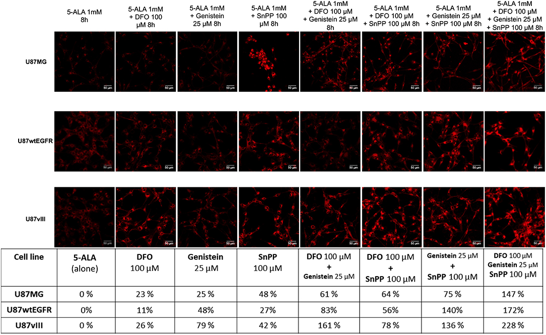
In the published article there is an error in Figure 4. In the images the third and fifth column of the first row are the same. The image in the third column of the first row (Genistein 25 μM) has been corrected. The image in the fifth column of the first row (DFO 100 μM + Genistein 25 μM) remains as it is.

Figure 4. PpIX fluorescence accumulation after single and combined treatments. Confocal images showing the increment in PpIX fluorescence (represented in red, excitation 405 nm and emission 635 nm) in GBM cells after single and combined treatment with two or three drugs compared to 5-ALA alone (represented as 0%). Scale bars represent 50 μm. Table summarizes the increment of PpIX fluorescence in percentage. DFO (deferoxamine), SnPP (tin protoporphyrin IX).
The authors apologize for these errors and state that this does not change the scientific conclusions of the article in any way. The original article has been updated.
Keywords: GBM—glioblastoma multiforme, 5-ALA=5-aminolevulinic acid, protoporphyin IX, quantification, breakdown, visualization, microscope
Citation: Reinert M, Piffaretti D, Wilzbach M, Hauger C, Guckler R, Marchi F and D'Angelo ML (2020) Corrigendum: Quantitative Modulation of PpIX Fluorescence and Improved Glioma Visualization. Front. Surg. 7:14. doi: 10.3389/fsurg.2020.00014
Received: 12 February 2020; Accepted: 10 March 2020;
Published: 02 April 2020.
Edited and reviewed by: Mark Preul, Barrow Neurological Institute (BNI), United States
Copyright © 2020 Reinert, Piffaretti, Wilzbach, Hauger, Guckler, Marchi and D'Angelo. This is an open-access article distributed under the terms of the Creative Commons Attribution License (CC BY). The use, distribution or reproduction in other forums is permitted, provided the original author(s) and the copyright owner(s) are credited and that the original publication in this journal is cited, in accordance with accepted academic practice. No use, distribution or reproduction is permitted which does not comply with these terms.
*Correspondence: Michael Reinert, bWljaGFlbC5yZWluZXJ0QGVvYy5jaA==
 Michael Reinert
 Deborah Piffaretti
 Marco Wilzbach
 Christian Hauger
 Roland Guckler6
 Francesco Marchi