94% of researchers rate our articles as excellent or good
Learn more about the work of our research integrity team to safeguard the quality of each article we publish.
Find out more
CORRECTION article
Front. Pharmacol., 16 June 2020
Sec. Inflammation Pharmacology
Volume 11 - 2020 | https://doi.org/10.3389/fphar.2020.00871
This article is a correction to:
Resolvin D1 Modulates the Intracellular VEGF-Related miRNAs of Retinal Photoreceptors Challenged With High Glucose
 Rosa Maisto1
 Maria Consiglia Trotta1
 Francesco Petrillo2
 Sara Izzo3
 Giovanna Cuomo4
 Roberto Alfano5
 Anca Hermenean6
 Jorge Miquel Barcia7
 Marilena Galdiero2
 Chiara Bianca Maria Platania8
 Claudio Bucolo8
 Michele D’Amico1*
A Corrigendum on
Resolvin D1 Modulates the Intracellular VEGF-Related miRNAs of Retinal Photoreceptors Challenged With High Glucose
By Maisto R, Trotta MC, Petrillo F, Izzo S, Cuomo G, Alfano R, Hermenean A, Barcia JM, Galdiero M, Platania CBM, Bucolo C and D’Amico M (2020) Resolvin D1 Modulates the Intracellular VEGF-Related miRNAs of Retinal Photoreceptors Challenged With High Glucose. Front. Pharmacol. 11:235. doi: 10.3389/fphar.2020.00235
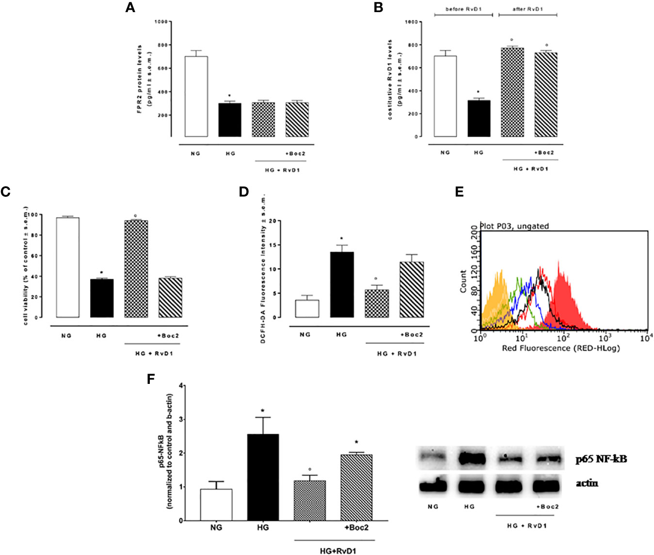
In the original article, there was a mistake in Figure 2F as published. The wrong image was included due to the incorrect labeling of a file. The correct Figure 2 appears below.

Figure 2 FPR2 and RvD1 levels, cell viability, ROS content, NF-kb protein expression. (A) ELISA detecting the levels of FPR2 receptor and (B) constitutive Resolvin D1 before and after RvD1 addition to photoreceptors exposed to high glucose; (C) XTT assay for determination of total cell number; (D, E) average intensity from DCFH-DA for total intracellular ROS levels compared to a negative control (yellow) and a positive control (fill red, 100 μM H2O2). Green = normal glucose; black = high glucose; blue = HG + RvD1 and red = HG + RvD1 + Boc2; (F) Western Blotting determination and representative images of NFκB protein levels into photoreceptors stimulated with normal glucose (5 mM D-glucose); high glucose (30 mM D-glucose); HG + RvD1 (RvD1, 50 nM); HG + RvD1 + Boc2 (20 μM). Values are expressed as mean ± s.e.m. of n = 9 values, obtained from the triplicates of three independent experiments. They were analyzed by one-way ANOVA followed by Bonferroni’s test for each panel, except for panel (B) were ANOVA for repeated measures was applied. NG, normal glucose; HG, high glucose; RvD1, Resolvin D1; Boc-2, selective FPR2 inhibitor. *P > 0.01 vs. NG; °P > 0.01 vs. HG.
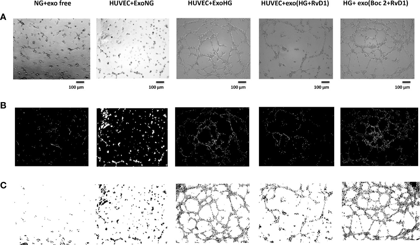
In addition, there was a mistake in Figure 7. Incorrect panels were included in Figures 7B, C. The corrected Figure 7 appears below.

Figure 7 Representative images of the tubular structures from non-transfected HUVEC cells. (A) Matrigel natural views, (B) dark field Matrigel views and (C) Matrigel graphical images of HUVEC cells grown in normal glucose (NG, 5 mM) seeded with: exosome-free medium (NG-exofree); standard medium containing exosomes released after stimulation of photoreceptors with Normal Glucose (NG, 5 mM) (NG + exoNG); standard medium containing exosomes released after stimulation of photoreceptors with High Glucose (HG, 35 mM) (NG + exoHG); standard medium containing exosomes released after stimulation of photoreceptors with HG + RvD1 (50 nM) (NG + exoHG-RvD1); standard medium containing exosomes released after stimulation of photoreceptors with HG + RvD1 + Boc2 (20 μM) (NG + exoHG-RvD1 + Boc2). Scale bar 100 μm. Magnification 100X.
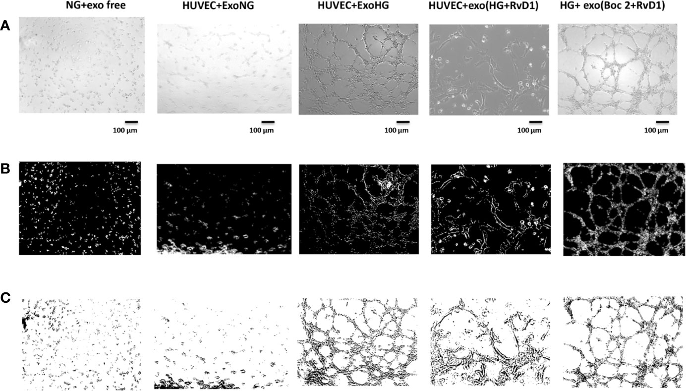
Finally, there was a mistake in Figure 9 where the first two and last columns were mislabelled. The corrected Figure 9 appears below.

Figure 9 Representative images of the tubular structures and node formation from transfected HUVEC cells. (A) Matrigel natural views, (B) dark field Matrigel views, and (C) Matrigel graphical images of HUVEC cells grown in normal glucose (NG, 5 mM) after the silencing of miR-20a-5p, miR-20a-3p, miR-20b, and miR-106a-5p in these cells. Cell seeded with exosome-free medium (NG-exofree); standard medium containing exosomes released after stimulation of primary cells with Normal Glucose (NG, 5 mM) (NG + exoNG); standard medium containing exosomes released after stimulation of photoreceptors with High Glucose (HG, 35 mM) (NG + exoHG); standard medium containing exosomes released after stimulation of photoreceptors with HG + RvD1 (50 nM) (NG + exoHG-RvD1); standard medium containing exosomes released after stimulation of photoreceptors with HG + RvD1 + Boc2 (20 μM) (NG + exoHG-RvD1 + Boc2). Scale bar 100 μm. Magnification 100X.
The authors apologize for these errors and state that these do not change the scientific meaning and conclusions of the article in any way. The original article has been updated.
Keywords: retinal photoreceptors, exosomes, miRNAs, resolvin D1, VEGF
Citation: Maisto R, Trotta MC, Petrillo F, Izzo S, Cuomo G, Alfano R, Hermenean A, Barcia JM, Galdiero M, Platania CBM, Bucolo C and D’Amico M (2020) Corrigendum: Resolvin D1 Modulates the Intracellular VEGF-Related miRNAs of Retinal Photoreceptors Challenged With High Glucose. Front. Pharmacol. 11:871. doi: 10.3389/fphar.2020.00871
Received: 12 May 2020; Accepted: 27 May 2020;
Published: 16 June 2020.
Edited and reviewed by: Fulvio D’Acquisto, University of Roehampton London, United Kingdom
Copyright © 2020 Maisto, Trotta, Petrillo, Izzo, Cuomo, Alfano, Hermenean, Barcia, Galdiero, Platania, Bucolo and D’Amico. This is an open-access article distributed under the terms of the Creative Commons Attribution License (CC BY). The use, distribution or reproduction in other forums is permitted, provided the original author(s) and the copyright owner(s) are credited and that the original publication in this journal is cited, in accordance with accepted academic practice. No use, distribution or reproduction is permitted which does not comply with these terms.
*Correspondence: Michele D’Amico, bWljaGVsZS5kYW1pY29AdW5pY2FtcGFuaWEuaXQ=
Disclaimer: All claims expressed in this article are solely those of the authors and do not necessarily represent those of their affiliated organizations, or those of the publisher, the editors and the reviewers. Any product that may be evaluated in this article or claim that may be made by its manufacturer is not guaranteed or endorsed by the publisher.
Research integrity at Frontiers

Learn more about the work of our research integrity team to safeguard the quality of each article we publish.