- 1Department of Orthodontics, Peking University School and Hospital of Stomatology & National Engineering Laboratory for Digital and Material Technology of Stomatology & Beijing Key Laboratory of Digital Stomatology, Beijing, China
- 2Department of Geriatric Dentistry, Peking University School and Hospital of Stomatology, Beijing, China
Bone defects caused by injury, disease, or congenital deformity remain a major health concern, and efficiently regenerating bone is a prominent clinical demand worldwide. However, bone regeneration is an intricate process that requires concerted participation of both cells and bioactive factors. Mimicking physiological bone healing procedures, the sustained release of bioactive molecules plays a vital role in creating an optimal osteogenic microenvironment and achieving promising bone repair outcomes. The utilization of biomaterial scaffolds can positively affect the osteogenesis process by integrating cells with bioactive factors in a proper way. A high water content, tunable physio-mechanical properties, and diverse synthetic strategies make hydrogels ideal cell carriers and controlled drug release reservoirs. Herein, we reviewed the current advancements in hydrogel-based drug sustained release systems that have delivered osteogenesis-inducing peptides, nucleic acids, and other bioactive molecules in bone tissue engineering (BTE).
Introduction
Bone defects may be caused by various events, including trauma, inflammation, neoplasm resection, congenital deformity, and degeneration (Crane et al., 1995; Spicer et al., 2012). Despite numerous solutions being applied to tackle this issue, clinical demands remain unmet.
To date, autologous bone grafts are still the gold standard and most considered therapeutic strategy for critical-sized bone defects among all restoration methods due to their remarkable osteoconductive and osteoinductive properties. However, de novo problems might arise, such as a limited amount of donor tissue, an excessive harvest procedure, and the possibility of postoperative infection of the donor site (Langer and Vacanti, 1993; Betz, 2002; Ahlfeld et al., 2019). Allografts or xenografts usually serve as secondary alternatives, as slower incorporation, immune rejection, and pathogen transmission might occur (Crane et al., 1995; Haugen et al., 2019). Utilizing biocompatible scaffold materials, such as mesenchymal stem cells (MSCs) and/or bioactive factors (Meijer et al., 2007), bone tissue engineering can offer more possibilities. Achieving sufficient and qualified bone formation via artificial composites is the grand aim of bone tissue engineering.
Compared with bone harvest operations, MSCs are relatively easy to obtain. These cells exhibit self-renewal, multipotentiality (Prockop, 1997), and immunomodulatory properties (Keating, 2008), which are imperative for bone regeneration. In addition, bioactive factors, for example, cytokines and growth factors (GFs), play a crucial role in new bone formation. Bone morphogenetic proteins (BMPs) are a group of GFs that have been substantially investigated. Recombinant human BMP-2 and BMP-7 is commercially available for limited clinical usage (Nauth et al., 2011). However, naked GFs are vulnerable in vivo, and to achieve optimal osteogenic effects, a supraphysiological dose of GFs is required. Paradoxically, diffusion or uncontrolled release of GFs may lead to ectopic bone formation and other complications, including carcinogenicity (Carragee et al., 2011; de Melo Pereira and Habibovic, 2018). Hence, attaining sustained release of bioactive factors is an essential objective for scaffold design to promote the therapeutic efficacy of bone tissue engineering. The scaffold materials not only create a congenial microenvironment to promote MSC biological behaviors but also help to maintain bioactive molecules in situ. To date, the controlled release of bioactive factors in bone tissue engineering has been realized by a wide range of biomaterials of different natures and configurations, which provide diverse release profiles in different treatment scenarios (Lee and Shin, 2007).
Hydrogels are a category of highly hydrated 3-dimensional (3D) crosslinked homopolymer, copolymer, or macromer networks that can be cast into different shapes and sizes (Slaughter et al., 2009; Seliktar, 2012). The application of hydrogels in tissue engineering, bone tissue engineering in particular, has been garnering increasing attention. Laden with osteogenic-inducing drugs and sustained release profiles, hydrogels have been suggested to be promising bone tissue engineering biomaterials. In this review, we discuss the progress and limitations of current bone tissue engineering, the advantages of hydrogel-based bone regeneration biomaterials and recent advancements in hydrogel-based drug sustained release systems for bone tissue engineering.
The Present Challenges of Bone Tissue Engineering
To date, substantial progress has been made in bone regenerative medicine. A variety of biomimetic polymers and inorganic materials with bone-like microarchitecture have been designed with advanced manufacturing methods (Wei et al., 2011; Kim et al., 2017b; Yin et al., 2019), including 3D printing, aiming to achieve superb osteogenic properties as well as accuracy and spatial fitness of critical-sized defects. Light-cured, thermal-setting, pH- or enzyme-sensitive, and other smart biomaterials enable bone tissue engineering to serve in many on-demand circumstances. Varieties of seed cells from different origins including umbilical cord MSCs (UCMSCs), induced pluripotent stem cell-derived MSCs (iPSC-MSCs), and embryonic stem cell-derived MSCs (ESC-MSCs) are successfully applied (Xie et al., 2016; Chen et al., 2018). Multifarious drugs or bioactive factors are delivered in situ with different strategies and tailored release profiles, offering osteogenic-friendly environments for relevant cells. Noteworthy, it was reported that MSC-derived exosomes combining scaffolds achieved preferable osteogenesis outcomes (Li et al., 2018), indicating the promising prospect of exosomes-based cell-free bone regeneration.
MSCs from different sources, such as bone marrow and dental tissue, are available for bone tissue engineering. The stem cell niche, 3D microenvironments containing specific biophysical and biochemical signals, maintains the stemness of stem cells in vivo (Scadden, 2006; Jones and Wagers, 2008). However, maintaining the viability and stemness of MSCs as well as controlling stem cell fate is a fairly critical issue in regenerative medicine. Substrate-derived stimuli are able to prolong the stemness of stem cells and guide stem cell fate into specific lineages (Fisher et al., 2010; Marklein and Burdick, 2010; Lee et al., 2015). Moreover, as the proliferation and differentiation of MSCs may drive into specific lineages depending on different microenvironmental cues, biochemical stimuli, including cytokines and GFs, are used in a spatiotemporal sequence during the complex and continuous reparative procedure (Samorezov and Alsberg, 2015; Farokhi et al., 2016). Successful bone regeneration requires the proper combination of stimuli that can trigger MSC differentiation and matrix deposition. As the scaffold material itself is capable of combining substrate-derived and biochemical stimuli, biomimetic and bioinspired synthetic materials with sustained drug release systems should be designed to facilitate bone tissue regeneration. Due to the constraints of current knowledge in this field, the research is far from sufficient.
Natural bone fracture healing requires the coordinated participation of osteogenesis and angiogenesis (Collin-Osdoby, 1994; Marsell and Einhorn, 2011). Bioactive factors and signal pathway crosstalk, which mediates the interplay between epithelial cells and osteoprogenitors, has been well summarized (Ramasamy et al., 2016). Likewise, vascularization in bone substitutes is vital for successful bone tissue engineering. Insufficient blood supply may result in undernutrition, hypoxia, and inadequate cell recruitment, leading to the failure of bone tissue engineering. Varieties of assessments and solutions have been summarized (Rouwkema et al., 2008; Das and Botchwey, 2011), yet there is no convincing evidence that the strategies are ample to sustain large tissue constructs, encouraging the proposal of more promising methods.
The Preponderance of Hydrogels in Bone Tissue Engineering
Ideal bone tissue engineering scaffolds should meet the following criteria: (1) biocompatible, nontoxic and nonimmunogenic; (2) porous-structured; (3) proper mechanical properties, load-bearing ability, and sufficient dimensional stability; and (4) fully degradable, with a degradation rate that matches neotissue formation (Lee and Shin, 2007; Slaughter et al., 2009; Haugen et al., 2019). Numerous inorganic scaffolds, such as metals and bioceramics, have been applied in bone regeneration, yet their lack of cell affinity, unbalanced mechanical properties, and rather poor degradation cannot be ignored (Pearlin et al., 2018).
According to types of raw materials, hydrogels can be briefly categorized into natural and synthetic. It is usually considered that natural hydrogels are more biocompatible and bioactive, while synthetic ones possess more tunable mechanical and degradation properties. 3D-structured, highly water-containing, and biocompatible hydrogels act as excellent extracellular matrix (ECM) analogs. The porous structure of the hydrogel enables substance exchange and cell entrance at the initial stage as well as vascular ingrowth in the follow-up stage. It has been substantially shown that cells are easily suspended within hydrogels, and the viability of the encapsulated cells is highly preserved (Gao et al., 2020; Paez et al., 2020).
MSCs are highly sensitive to physical parameters (Higuchi et al., 2013), including viscoelasticity Engler et al. (2006) and topography (Fiedler et al., 2013), in the surrounding milieus. The stiffness (elastic modulus) of the matrix is believed to contribute greatly to determining stem cell fate. As Engler et al. (2006) demonstrated, 2D-cultured MSCs exhibited osteogenic characteristics when the microenvironmental stiffness was relatively rigid, at 20–40 kPa. However, osteogenesis occurred at 11–30 kPa when MSCs were cultivated 3-dimensionally (Huebsch et al., 2010). Due to flexible synthetic strategies and the range of constituents, hydrogels possess tunable physio-mechanical properties, which could match the desirable ranges of material elasticity, porosity, and degradation rate required for bone tissue engineering (Slaughter et al., 2009). Meanwhile, photodegradable (Lunzer et al., 2018), thermal-sensitive, or pH-sensitive (GhavamiNejad et al., 2016) linkages as well as other advantageous materials could be subtly introduced into hydrogels, which may fabricate a versatile and intelligent composite system to fulfill the growing clinical demands.
On the other hand, bioactive molecules play an important role in bone regenerative medicine. During bone formation, numerous cytokines and GFs are orchestrated in a spatiotemporal manner (Farokhi et al., 2016), which would provide a suitable microenvironment for MSC proliferation and differentiation, as well as recruit progenitors from surrounding tissue and peripheral blood for further restoration. Apart from competent cell carriers, hydrogels can also be employed as promising local drug reservoirs. Multiple schemes have been applied to reach desirable and smart drug delivery kinetics (Lee and Shin, 2007; Slaughter et al., 2009). Non-covalent immobilization strategies are the most commonly used in hydrogel-based drug depots, the drug release rate was mainly determined by parameters such as crosslink density, carrier affinity for drugs, and the matrix degradation profile (Dimatteo et al., 2018). Bioactive factors also could be linked covalently to polymers by which a longer drug retention time would be achieved, and covalent linkages could be broken as reactions of specific external cues, leading an on-demand drug controlled release. Moreover, other sustained release systems like microspheres could be introduced to hydrogel matrix, enabling multiple drug molecules sustained release in sequential or spatiotemporal manners (Chen et al., 2010).
Hydrogel-Based Drug Sustained Release Systems for Bone Tissue Engineering
Extensive drug and sustained release strategies have been designed for bone tissue engineering. Herein, we introduce studies on hydrogel-based controlled release systems according to the category of bioactive molecule loaded within.
Peptides
The majority of cytokines, GFs, and hormones that stimulate bone formation are peptides. These biomolecules are produced through the autocrine, paracrine, and endocrine systems, acting concertedly to regulate the complex cascade of bone-related gene expression (Lee and Shin, 2007; Farokhi et al., 2016). Hence, a well-orchestrated sustained release system of these peptides has been pursued in order to present a more biomimetic approach.
BMP
With the promoted understanding of the underlying mechanism of osteogenesis (Chen et al., 2012), BMP, as a prominent member of the TGF-β superfamily, has always been a favored candidate for bone tissue engineering applications.
Since some hydrogels are believed to possess inferior osteoconductive properties, Olthof et al. (2018) modified an oligo[poly(ethylene glycol) (PEG) fumarate] (OPF) hydrogel with bisphosphate. BMP-2 was encapsulated in poly(lactic-co-glycolic acid) (PLGA) microspheres. The additional BMP-2 and drug-laden PLGA microspheres were entrapped in the hydrogel matrix. The researchers believed that the anionic groups would produce a strong interaction between the matrix and inorganic phase of the bone as well as enhance BMP-2-induced bone formation. The hydrogel matrix could be functionalized by peptides, which might be beneficial to reduce the dose of encapsulated BMP. In addition, nanofibrous mesh-hydrogel hybrid composites have been applied to reach a proper spatiotemporal release profile (Kolambkar et al., 2011). Shekaran et al. (2014) modified a matrix metalloproteinase (MMP)-degradable peptide crosslinked PEG with an α2β1 integrin-specific peptide (GFOGER). The interaction between integrin and collagen I has been proposed to be vital in osteogenic differentiation and mineralization. It was suggested that the modified matrix is able to support cell adhesion and proliferation and upregulate osteogenic gene expression. Laden with the low dose of BMP-2, robust bone healing was achieved. Along with BMP-2, BMP-7 is considered to be a promising GF in bone formation. An injectable chitosan/β-glycerophosphate (CS/β-GP) hydrogel laden with BMP-7 and antibiotic exhibited preferable reparative effects towards infection-induced bone loss (Zang et al., 2019). Growth differentiation factor-5 (GDF-5), also known as BMP-14, regulates the development of numerous tissue and cell types, including limbs and teeth. Bae et al. (2014) mixed different concentrations of GDF-5 with a light-cured hydrogel matrix. The results showed that GDF-5 improved the osteogenic ability in a dose-dependent manner, as the strongest augmentation was achieved by the hydrogel loaded with the highest concentration.
Apart from adsorption or physical entrapment, electrostatic, hydrophobic, or other interactions have been introduced into the systems to prolong the release of BMPs. Heparin was reported to be a strong binder to BMPs, yet the side effects were not negligible. Heparin mimics, which are usually negatively charged, are supposed to be capable of controlling BMP release. Chondroitin sulfate (Anjum et al., 2016), 2-N,6-O-sulfated chitosan (26SCS)-based nanoparticles (Cao et al., 2014), alginate sulfate (Park et al., 2018) were synthesized by researchers, and satisfactory results were achieved both in vitro and in vivo. When higher concentrations of heparin mimics were introduced, the release rate of BMP became slower. Seo et al. (2015, 2017) harnessed the ionic and hydrophobic interactions provided by polyphosphazene nanoparticles. They found that release rate of BMPs were controlled by the types and amounts of pendants. Thus, the optimal release profile and osteogenesis outcomes rely on a reasonable proportion of BMP-tethering molecules.
Genetic engineering is another option to obtain long-lasting BMP release. As Lin et al. (2019) described in a manuscript, the BMP gene was transduced into human bone marrow-derived stem cells (BMMSCs), obtaining a continuous (up to 56 days) and economical BMP supply. Using visible light-based projection stereolithography (VL-PSL) technology to encapsulate the transduced cells, the researchers were able to fabricate more structurally and geometrically compatible constructs for individualized bone defects, which would be conducive to achieving tissue fusion and bone tissue engineering long-term success.
Vascular Epithelial Growth Factor (VEGF)
Vascularization plays a crucial role in both bone development and bone regeneration (Collin-Osdoby, 1994; Olsen et al., 2000). Blood vessels do not solely work as substance exchange pathways; they are also regarded as highly active paracrine organs targeting various progenitors during bone formation and reconstruction (Collin-Osdoby, 1994). VEGF, a key angiogenic growth factor (Carmeliet and Jain, 2011), has been widely used in bone tissue engineering.
The cooperation between VEGF receptors and integrin adhesion receptors has been elucidated in angiogenic regulation. Garcia et al. (2016) engineered a protease-degradable, GFOGER-modified PEG hydrogel as a VEGF depot. They found that covalently linked VEGF remained highly bioactive during a prolonged release period. Whereas it was shown that a GFOGER hydrogel augmented neovascularization regardless of exogenous VEGF, micro computed tomography (micro-CT) showed delivering exogenous VEGF failed to enhance critical-sized bone repair. Heterogenous material composites are manufactured by which we can juggle both timed drug release and osteoconduction. Composed of a 3D multichannel calcium phosphate cement (CPC) and alginate/gellan gum (AlgGG) hydrogel, the CPC/AlgGG biphasic scaffold tethers VEGF via the interaction with heparin (Ahlfeld et al., 2019). Despite some remarkable properties observed in vitro, significant enhancement by VEGF on new bone formation has not been detected. Amirian et al. (2015) coated VEGF and BMP-2 separately onto gelatin-pectin-biphasic calcium phosphate composites. The results revealed that composites coated with VEGF mainly aided in woven bone formation, and the percent of new bone formation was not greater than those coated with BMP-2.
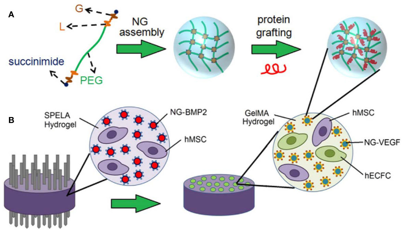
Since exclusive delivery of VEGF performed barely satisfactorily in GF-induced osteogenesis, dual or multidrug delivery is warranted. When accompanied by BMP-2, VEGF exhibited a significant improvement in bone formation compared with hydrogels encapsulating BMP-2 alone. VEGF combined with BMP-2 has been used routinely as a GF formula in bone tissue engineering. Similar loading strategies were applied by Barati et al. (2016) and Kader et al. (2019) for spatiotemporal release of BMP-2 and VEGF. MSCs and BMP-tethered nanoparticles were embedded in the outer space, while endothelial colony-forming cells (ECFCs) and VEGF-tethered nanoparticles were dispersed inside the microchannel-patterned hydrogel, as illustrated in Figure 1. Degradation and GF release could be tuned by altering stoichiometric ratio chain-extended molecules and proteolysis. According to the data, the release of VEGF and BMP-2 could last over 10 days and 21 days, respectively. It was observed that the patterned hydrogel dual delivery system performed significantly better than that of single delivery systems, which was attributed to paracrine crosstalk. During bone repair, VEGF expression peaks appear in the early period, while BMP peaks later. Thus, consisting of a PLGA microsphere-incorporating poly(propylene fumarate) (PPF) rod surrounded by a rapidly degrading gelatin hydrogel, the composite was designed as a GF delivery vehicle (Kempen et al., 2009). VEGF was encapsulated in the hydrogel, whereas BMP-2 was immobilized by microspheres inside the rod in order to achieve an ideal GF sequential release pattern. VEGF exhibited a large initial burst release within the first 3 days, and BMP-2 showed sustained release over 8 weeks. Likewise, although VEGF did not induce neo-bone formation, it significantly enhanced BMP-induced osteogenesis. Organic-inorganic modular scaffolds are able to optimally orchestrate dual GF release and serve as an “anatomy-structure-function” trinity system in regenerating weight-bearing bones (Bao et al., 2017). Mesoporous bioactive glass (MBG) with hollowed channels and hierarchical porous structures was introduced in a controlled release system as a scaffold (Tang et al., 2020). VEGF was carried by hydrogel inside the channel, and BMP-2 was adsorbed by the MBG scaffolds. 26SCS acted as an analog of ECM, which exhibits super-affinity to GFs. In vitro experiments showed that 26SCS promoted the bioactivity of BMP-2 and VEGF. It could be assumed that the VEGF hydrogel column in the hollowed channels might induce chemotaxis of vascular endothelial cells, thus regulating cell migration and vascular infiltration. Moreover, increased type H vessels and neotissue ingrowth were observed.

Figure 1 Schematic illustration of (A) nanogel (NG) assembly and peptide grafting. (B) Achievement of BMP-2 and VEGF spatiotemporal release profiles via a patterned hydrogel-based sustained release system. Reprinted from a previous article by Barati et al. (2016) with permission.
Fibroblast Growth Factor (FGF)
FGF signaling is a dominant regulator during bone development and fracture repair (Bourque et al., 1993; Kronenberg, 2003). However, contradictory results have implied that FGF signaling may exert dual-directional effects on osteogenic procedures, probably in a dose-dependent manner (Kato et al., 1998; Quarto and Longaker, 2006). Thus, sustained release should be achieved when FGF is delivered in bone tissue engineering.
Two Japanese groups encapsulated FGF in gelatin hydrogels for controlled release (Kodama et al., 2009; Furuya et al., 2014). A longer FGF release period may improve cell proliferation, the expression levels of osteogenic markers and BMP-2 as well as bone mineral density (BMD) at defect sites. However, these enhancements vanished, and side effects occurred when a high dose of FGF was delivered (Kodama et al., 2009). In order to achieve bone-like biomechanical properties and slower release of FGF, a stiffer hydrogel matrix, poly(2-hydroxyethyl methacrylate) copolymerized with 2-vinyl pyrrolidone, was engineered (Mabilleau et al., 2008). The data suggested that in the first 4 days, the FGF release rate was approximately 1% per day, which was relevant to hydrogel swelling. Unfortunately, no significant difference between the FGF and control groups was noted in bone mass, but the poorly mineralized woven bone area was significantly larger in the FGF group.
It is a preferable strategy for other GFs to accompany FGF in order to obtain a promising outcome. Chen et al. (2016) chose gelatin microspheres as BMP-2 and basic FGF (bFGF) carriers, which were further embedded in a commercialized injectable thermal-sensitive hydrogel. The hydrogel was injected into a porous cell-loading scaffold before use. Micro-CT revealed that the dual-loaded composites achieved the best reparative results. As expected, composites loading bFGF alone regenerated less bone and neobone at the margin of the defect areas, while the dual-loaded composites showed much more central area bone formation. FGF9 has been indicated to be a stabilizing factor for neovessels, thus, Yuan et al. (2016) introduced FGF9 as an assistant for VEGF, exerting synergetic effects on angiogenesis in bone tissue engineering. A specific peptide segment was fused to VEGF and FGF9 to obtain a covalent connection with the fibrin hydrogel. BMP-2 was transfected into BMSCs, endowing a greater osteogenic ability and resistance of the osteogenic differentiation inhibition induced by fusion with FGF9. Less bone was formed in the FGF9 groups compared to the groups treated with only VEGF, whereas VEGF/FGF9-loaded composites performed the best among the groups.
Other Peptides
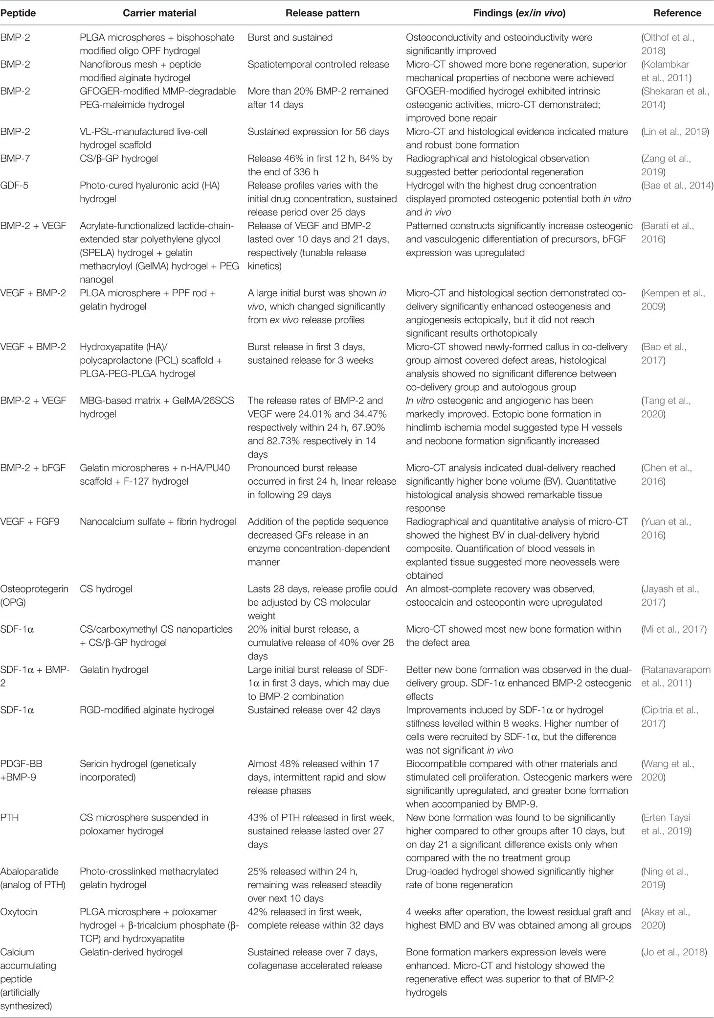
Other peptides that regulate the bone regeneration cascade, including osteoprotegerin (OPG) (Jayash et al., 2017), stromal cell-derived factor-1α (SDF-1α) (Ratanavaraporn et al., 2011; Cipitria et al., 2017; Mi et al., 2017), platelet-derived growth factor (PDGF) (Wang et al., 2020), and parathyroid hormone (PTH) (Erten Taysi et al., 2019), etc., might also be worthy of an attempt. The selected studies of hydrogel-based peptide sustained release systems for bone regeneration and their findings are concluded in Table 1.
Nucleic Acids
Since GFs and cytokines are required for weeks during new bone formation, gene therapy might be a feasible alternative. Delivering DNA or RNA locally to increase or knockdown target gene expression, gene therapy is capable of manipulating the microenvironment and determining cell fate in bone regenerative medicine.
Fang et al. (1996) utilized collagen sponges as BMP-4 and PTH plasmid DNA carriers to regenerate nonunion rat femur defects early in 1996. Bonadio et al. (1999) confirmed that non-viral DNA delivery possesses numerous advantages compared with the protein strategy. Hydrophilic nucleic acids and hydrogels could provide stable and sequestered environments for gene delivery. Komatsu et al. (2016) demonstrated that gelatin hydrogels could transduce BMP-2 plasmid DNA efficiently, facilitating local bone regeneration. CS or polyethyleneimine (PEI) is usually introduced as the carrier due to the electrostatic interaction between the negatively charged nucleic acids and the polycations. It was reported that branched PEI-HA-DNA complexes were entrapped in bilayered OPF hydrogels to restore osteochondral defects (Needham et al., 2014). Moreover, BMP-2 plasmid DNA conjugated with CS nanoparticles exhibited significant augmentation in hydrogel-mediated rat calvaria bone regeneration (Li et al., 2017). Due to the low stability of liposomes and electrostatic disturbance of other charged compounds, calcium phosphate (CaP) can also be used for DNA incorporation and transfection in bone tissue engineering (Krebs et al., 2010).
MicroRNAs (miRNAs) and small interfering RNAs (siRNAs) are groups of short single-stranded RNA fragments that downregulate target gene expression post-translationally. Various miRNAs associated with bone formation have been reported (Fang et al., 2015), shedding new light on future bone tissue engineering. Nguyen et al. (2014) synthesized an 8-arm PEG in situ-forming hydrogel loaded with siRNA/PEI nanocomplexes. siRNA remained bioactive during the prolonged release period. The in vitro results showed that siNoggin and siNoggin/miRNA-20a sustained release promoted hMSC osteogenic differentiation in 3D hydrogel cultivation. As mentioned previously, a stiffer substrate may lead to MSC osteogenic differentiation. Carthew et al. (2020) incorporated PEG/gelatin norbornene hydrogels with mechanosensitive miRNAs. MSCs encapsulated in hydrogels were transfected in situ, which predominantly enhanced osteogenic gene expression and mineralization. Researchers presumed that the higher transfection efficacy might be ascribed to longer cell exposure times to the transfection agent.
Ions or Small Molecules
To date, a number of metal ions and artificially synthesized compounds have been found to be beneficial in bone regeneration. Achieving a sustained release pattern and longer duration of drug function may lead to promising therapeutic outcomes.
Metal Ions
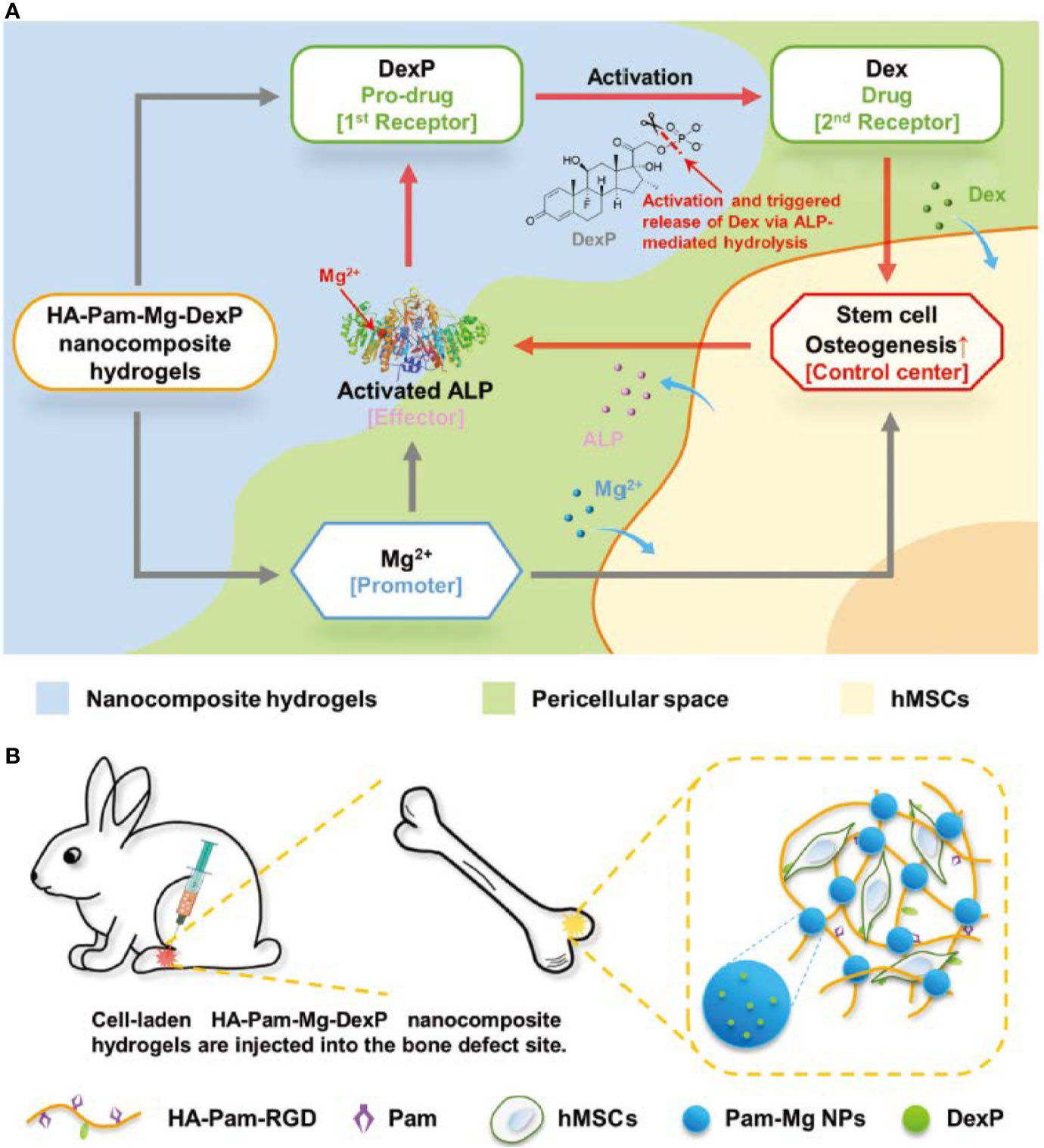
Since magnesium ions (Mg2+) play an important role in bone metabolism and mineralization, a variety of strategies for the sustained delivery of Mg2+ have been applied to hydrogel-based scaffolds. Lin et al. (2018b) coated MgO nanoparticles with PLGA and an alginate hydrogel, constructing a monodisperse core-shell delivery system. The release profile of Mg2+ revealed a significant suppression of the initial burst, and its release rate was stable and programmed. Enhancement of progenitor cell viability and proliferation, upregulation of osteogenic gene expression levels, and increased bone regeneration volume in vivo were attributed to the stable and precise Mg2+ supply. Bisphosphonates (BPs) possess two adjacent phosphonic groups, which are propitiously bind to divalent metal ions. Zhang and colleagues (Zhang et al., 2017) developed acrylated-BP-Mg nanoparticles to deliver Mg2+ as well as strengthen the acellular hydrogel composite, which serves as a matrix for in situ bone formation, via multivalent crosslinked domains. They also utilized Mg2+ to fulfill on-demand intelligent drug release in bone tissue engineering (Zhang et al., 2018). Intriguingly, Mg2+ played multiple roles in this research. First, BP-Mg nanoparticles enabled hydrogel formation and stabilized the prodrug. Second, Mg2+ promoted osteogenic differentiation, resulting in increased alkaline phosphatase (ALP) expression. However, and more importantly, Mg2+ is also a critical cofactor of ALP. ALP enzymatic hydrolysis was promoted; thus, more bioactive drug molecules were generated, which introduced positive feedback (Figure 2). According to the results, this strategy significantly enhanced bone regeneration.

Figure 2 Schematic illustration of (A) positive feedback mediated by a cofactor-assisted smart hydrogel drug release system and (B) in situ application to promote bone regeneration. Reprinted from a previous article by Zhang et al. (2018) with permission.
Other metal ions, such as strontium ions (Sr2+) and cobalt ions (Co2+), may act synergistically in bone reconstruction. A Sr2+-crosslinked RGD-alginate hydrogel combined with Sr-doped hydroxyapatite microspheres was engineered, showing a sustained release of Sr2+ from two sources (Lourenco et al., 2019). The researchers elaborated that this Sr-hybrid system facilitated MSC osteogenic differentiation, inhibited the functions of osteoclasts and modulated the inflammatory response. As a pro-vasculogenic factor, Co2+ was incorporated into the alginate hydrogel shell, while BMP-2 was laden into the collagen core (Perez et al., 2015). Co2+ released relatively rapidly, as expected. VEGF secretion and qPCR revealed that Co2+ not only stimulated angiogenesis but also elevated osteogenic gene expression. These results indicated an appealing prospect for applying metal ions bone tissue engineering in the future.
Small Molecules
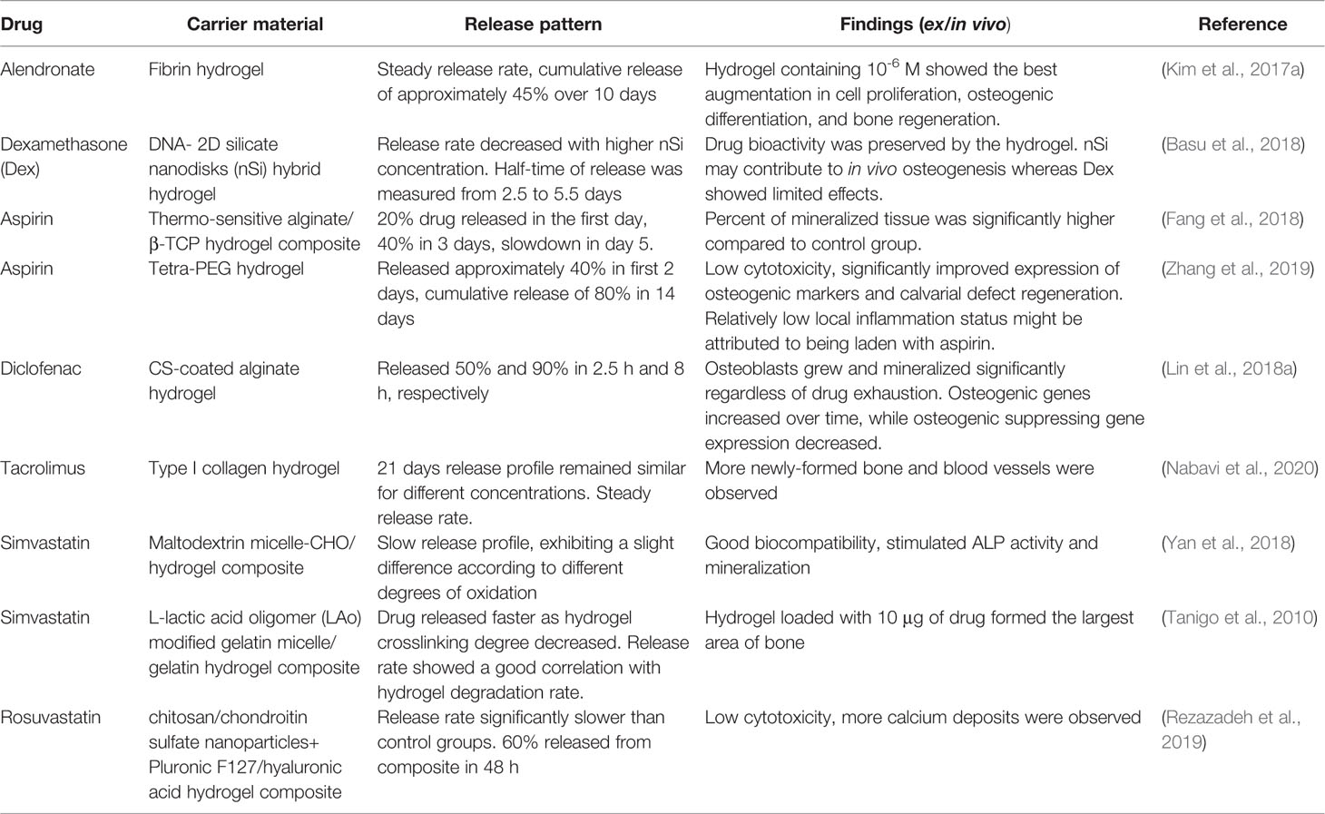
A range of pharmaceutical molecules were designed or discovered to be effective in bone regeneration. Highlighted as chelating agents, BPs are utilized as antiresorptive drugs frequently in clinics. BPs mainly target osteoclasts, impeding the differentiation and maturation of osteoclast progenitors. Increasing evidence has shown that BPs directly or indirectly take part in other bone-forming mechanisms and are capable of targeting various cells (Corrado et al., 2017). Since bone healing and regeneration is known to consist of three consecutive phases of inflammation, repair, and remodeling, a proper scale of immune response is indispensable (Claes et al., 2012). However, excess or aberrant immune activation may jeopardize bone repair procedures (Claes et al., 2012; Gibon et al., 2017). Therefore, immunomodulatory drugs, such as nonsteroidal anti-inflammatory drugs (NSAIDs), have been applied in bone tissue engineering. Evidence has shown that aspirin elevates MSC osteogenic potency by inhibiting the tumor necrosis factor-α (TNF-α) and interferon-γ (IFN-γ) pathways (Liu et al., 2011). Statins are inhibitors of a key enzyme of cholesterol synthesis and are widely used to lower serum lipids. Researchers have reported that osteogenesis was enhanced concomitant with promoted BMP-2 expression in bone cells when treating cells and rodents with statins (Mundy et al., 1999). Localized and sustained delivery of these drugs via hydrogels has pointed to a new direction in bone tissue engineering. Many relevant studies are listed and outlined in Table 2.

Table 2 Summary of selected studies on hydrogel-based small bioactive compound sustained release systems.
Conclusion and Future Perspectives
In this review, we summarized a series of investigations focused on hydrogel-based drug sustained release systems in bone tissue engineering. The hydrogels possess a porous microarchitecture, tunable biophysical parameters, and an adjustable degradation rate, which makes them qualified bone tissue engineering scaffolds. Due to their high water content, chemical inertness and relatively sequestered and stable internal environment, they are also excellent in preserving the viabilities of the laden cells and bioactive factors. With the combination of biophysical and biochemical cues, researchers are able to facilely establish an osteo-friendly microenvironment, which would be beneficial for osteoprogenitors to obtain better bone regeneration. Thus, hydrogel-based biomaterials are strong candidates for current or future bone tissue engineering.
Evidence has shown that hydrogel-based drug sustained release systems are highly biocompatible and versatile drug deliverers, obtaining satisfactory osteogenesis results both in vitro and in vivo. The drug release profile varies according to the loading strategy, degradation ability of the matrix and drug concentration. Among these studies, physical entrapment and diffusion are the most applied drug loading and release strategies, respectively. In particular, the dispersion of drugs, ions and small molecules largely depends on hydrogel pore size and crosslinking density. Although it is quite simple and easy to operate, there are difficulties in initial burst release management. Swelling or degradation of hydrogel matrices contributes to polymer mesh size enlargement, resulting in drug release acceleration, especially for macromolecular drugs. Stronger interactions between matrices and drugs, such as electrostatic interactions and covalent bonds, and other drug reservoirs could be introduced into hydrogels, providing more efficacious drug protection and immobilization. However, negative results have been reported from sustained release systems that did not facilitate bone formation mainly because the carrier exhibited an extremely strong affinity towards the growth factor, resulting in a low level of drug concentration in the surrounding tissue (Hettiaratchi et al., 2017). Thus, optimal drug concentrations should also be determined to achieve a more reasonable and effective release profile.
As mentioned above, cells from different origins are involved in the bone formation process. A vast number of GFs and cytokines collaboratively trigger the repair cascade. Extensive studies have already been conducted on multiple bioactive factors controlled release. Spatiotemporal sequence release of bioactive factors might be a better mimic of complex regeneration procedures as well as exert extraordinary synergistic effects on bone regeneration. Various of multiple GFs delivery strategies was coherently summarized (Chen et al., 2010). Nevertheless, controlling dose ratio of drugs to maximize the synergistic effects and manipulating multiple bioactive factors release kinetics to mimic physiological release profile in different phases of bone regeneration are obstacles in nowadays bone tissue regeneration which needs further investigation.
Author Contributions
YZ and TY contributed equally to this review. TY, YW, and BH designed and revised this article. YZ, TY, LP, and QS collected the literatures, arranged the outline of collected documents, and wrote the articles. All authors reviewed and commented on the entire manuscript.
Funding
This work was supported by the National Natural Science Foundation of China (51972005, 51672009, 51903003), National Natural Science Foundation of China Youth Fund (81922019), and National Youth Top-notch Talent Support Program (QNBJ2019-3).
Conflict of Interest
The authors declare that the research was conducted in the absence of any commercial or financial relationships that could be construed as a potential conflict of interest.
References
Ahlfeld, T., Schuster, F. P., Forster, Y., Quade, M., Akkineni, A. R., Rentsch, C., et al. (2019). 3D Plotted Biphasic Bone Scaffolds for Growth Factor Delivery: Biological Characterization In Vitro and In Vivo. Adv. Healthc. Mater. 8 (7), e1801512. doi: 10.1002/adhm.201801512
Akay, A. S., Arisan, V., Cevher, E., Sessevmez, M., Cam, B. (2020). Oxytocin-loaded sustained-release hydrogel graft provides accelerated bone formation: An experimental rat study. J. Orthop. Res. 1–12. doi: 10.1002/jor.24607
Amirian, J., Linh, N. T., Min, Y. K., Lee, B. T. (2015). Bone formation of a porous Gelatin-Pectin-biphasic calcium phosphate composite in presence of BMP-2 and VEGF. Int. J. Biol. Macromol. 76, 10–24. doi: 10.1016/j.ijbiomac.2015.02.021
Anjum, F., Lienemann, P. S., Metzger, S., Biernaskie, J., Kallos, M. S., Ehrbar, M. (2016). Enzyme responsive GAG-based natural-synthetic hybrid hydrogel for tunable growth factor delivery and stem cell differentiation. Biomaterials 87, 104–117. doi: 10.1016/j.biomaterials.2016.01.050
Bae, M. S., Ohe, J. Y., Lee, J. B., Heo, D. N., Byun, W., Bae, H., et al. (2014). Photo-cured hyaluronic acid-based hydrogels containing growth and differentiation factor 5 (GDF-5) for bone tissue regeneration. Bone 59, 189–198. doi: 10.1016/j.bone.2013.11.019
Bao, X., Zhu, L., Huang, X., Tang, D., He, D., Shi, J., et al. (2017). 3D biomimetic artificial bone scaffolds with dual-cytokines spatiotemporal delivery for large weight-bearing bone defect repair. Sci. Rep. 7 (1), 7814. doi: 10.1038/s41598-017-08412-0
Barati, D., Shariati, S. R. P., Moeinzadeh, S., Melero-Martin, J. M., Khademhosseini, A., Jabbari, E. (2016). Spatiotemporal release of BMP-2 and VEGF enhances osteogenic and vasculogenic differentiation of human mesenchymal stem cells and endothelial colony-forming cells co-encapsulated in a patterned hydrogel. J. Control Release 223, 126–136. doi: 10.1016/j.jconrel.2015.12.031
Basu, S., Pacelli, S., Feng, Y., Lu, Q., Wang, J., Paul, A. (2018). Harnessing the Noncovalent Interactions of DNA Backbone with 2D Silicate Nanodisks To Fabricate Injectable Therapeutic Hydrogels. ACS Nano. 12 (10), 9866–9880. doi: 10.1021/acsnano.8b02434
Betz, R. R. (2002). Limitations of autograft and allograft: new synthetic solutions. Orthopedics 25 (5 Suppl), s561–s570.
Bonadio, J., Smiley, E., Patil, P., Goldstein, S. (1999). Localized, direct plasmid gene delivery in vivo: prolonged therapy results in reproducible tissue regeneration. Nat. Med. 5 (7), 753–759. doi: 10.1038/10473
Bourque, W. T., Gross, M., Hall, B. K. (1993). Expression of four growth factors during fracture repair. Int. J. Dev. Biol. 37 (4), 573–579.
Cao, L., Werkmeister, J. A., Wang, J., Glattauer, V., McLean, K. M., Liu, C. (2014). Bone regeneration using photocrosslinked hydrogel incorporating rhBMP-2 loaded 2-N, 6-O-sulfated chitosan nanoparticles. Biomaterials 35 (9), 2730–2742. doi: 10.1016/j.biomaterials.2013.12.028
Carmeliet, P., Jain, R. K. (2011). Molecular mechanisms and clinical applications of angiogenesis. Nature 473 (7347), 298–307. doi: 10.1038/nature10144
Carragee, E. J., Hurwitz, E. L., Weiner, B. K. (2011). A critical review of recombinant human bone morphogenetic protein-2 trials in spinal surgery: emerging safety concerns and lessons learned. Spine J. 11 (6), 471–491. doi: 10.1016/j.spinee.2011.04.023
Carthew, J., Donderwinkel, I., Shrestha, S., Truong, V. X., Forsythe, J. S., Frith, J. E. (2020). In situ miRNA delivery from a hydrogel promotes osteogenesis of encapsulated mesenchymal stromal cells. Acta Biomater. 101, 249–261. doi: 10.1016/j.actbio.2019.11.016
Chen, F. M., Zhang, M., Wu, Z. F. (2010). Toward delivery of multiple growth factors in tissue engineering. Biomaterials 31 (24), 6279–6308. doi: 10.1016/j.biomaterials.2010.04.053
Chen, G., Deng, C., Li, Y. P. (2012). TGF-beta and BMP signaling in osteoblast differentiation and bone formation. Int. J. Biol. Sci. 8 (2), 272–288. doi: 10.7150/ijbs.2929
Chen, T., Gomez, A. W., Zuo, Y., Li, X., Zhang, Z., Li, Y., et al. (2016). Osteogenic potential and synergistic effects of growth factors delivered from a bionic composite system. J. BioMed. Mater. Res. A 104 (3), 659–668. doi: 10.1002/jbm.a.35605
Chen, W., Liu, X., Chen, Q., Bao, C., Zhao, L., Zhu, Z., et al. (2018). Angiogenic and osteogenic regeneration in rats via calcium phosphate scaffold and endothelial cell co-culture with human bone marrow mesenchymal stem cells (MSCs), human umbilical cord MSCs, human induced pluripotent stem cell-derived MSCs and human embryonic stem cell-derived MSCs. J. Tissue Eng. Regenerative Med. 12 (1), 191–203. doi: 10.1002/term.2395
Cipitria, A., Boettcher, K., Schoenhals, S., Garske, D. S., Schmidt-Bleek, K., Ellinghaus, A., et al. (2017). In-situ tissue regeneration through SDF-1alpha driven cell recruitment and stiffness-mediated bone regeneration in a critical-sized segmental femoral defect. Acta Biomater. 60, 50–63. doi: 10.1016/j.actbio.2017.07.032
Claes, L., Recknagel, S., Ignatius, A. (2012). Fracture healing under healthy and inflammatory conditions. Nat. Rev. Rheumatol. 8 (3), 133–143. doi: 10.1038/nrrheum.2012.1
Collin-Osdoby, P. (1994). Role of vascular endothelial cells in bone biology. J. Cell Biochem. 55 (3), 304–309. doi: 10.1002/jcb.240550306
Corrado, A., Sanpaolo, E. R., Di Bello, S., Cantatore, F. P. (2017). Osteoblast as a target of anti-osteoporotic treatment. Postgrad. Med. 129 (8), 858–865. doi: 10.1080/00325481.2017.1362312
Crane, G. M., Ishaug, S. L., Mikos, A. G. (1995). Bone tissue engineering. Nat. Med. 1 (12), 1322–1324. doi: 10.1038/nm1295-1322
Das, A., Botchwey, E. (2011). Evaluation of angiogenesis and osteogenesis. Tissue Eng. Part B. Rev. 17 (6), 403–414. doi: 10.1089/ten.TEB.2011.0190
de Melo Pereira, D., Habibovic, P. (2018). Biomineralization-Inspired Material Design for Bone Regeneration. Adv. Healthc. Mater. 7 (22), e1800700. doi: 10.1002/adhm.201800700
Dimatteo, R., Darling, N. J., Segura, T. (2018). In situ forming injectable hydrogels for drug delivery and wound repair. Adv. Drug Delivery Rev. 127, 167–184. doi: 10.1016/j.addr.2018.03.007
Engler, A. J., Sen, S., Sweeney, H. L., Discher, D. E. (2006). Matrix elasticity directs stem cell lineage specification. Cell 126 (4), 677–689. doi: 10.1016/j.cell.2006.06.044
Erten Taysi, A., Cevher, E., Sessevmez, M., Olgac, V., Mert Taysi, N., Atalay, B. (2019). The efficacy of sustained-release chitosan microspheres containing recombinant human parathyroid hormone on MRONJ. Braz. Res. 33, e086. doi: 10.1590/1807-3107bor-2019.vol33.0086
Fang, J., Zhu, Y. Y., Smiley, E., Bonadio, J., Rouleau, J. P., Goldstein, S. A., et al. (1996). Stimulation of new bone formation by direct transfer of osteogenic plasmid genes. Proc. Natl. Acad. Sci. U. S. A. 93 (12), 5753–5758. doi: 10.1073/pnas.93.12.5753
Fang, S., Deng, Y., Gu, P., Fan, X. (2015). MicroRNAs regulate bone development and regeneration. Int. J. Mol. Sci. 16 (4), 8227–8253. doi: 10.3390/ijms16048227
Fang, X., Lei, L., Jiang, T., Chen, Y., Kang, Y. (2018). Injectable thermosensitive alginate/beta-tricalcium phosphate/aspirin hydrogels for bone augmentation. J. BioMed. Mater. Res. B. Appl. Biomater. 106 (5), 1739–1751. doi: 10.1002/jbm.b.33982
Farokhi, M., Mottaghitalab, F., Shokrgozar, M. A., Ou, K. L., Mao, C., Hosseinkhani, H. (2016). Importance of dual delivery systems for bone tissue engineering. J. Control Release 225, 152–169. doi: 10.1016/j.jconrel.2016.01.033
Fiedler, J., Ozdemir, B., Bartholoma, J., Plettl, A., Brenner, R. E., Ziemann, P. (2013). The effect of substrate surface nanotopography on the behavior of multipotnent mesenchymal stromal cells and osteoblasts. Biomaterials 34 (35), 8851–8859. doi: 10.1016/j.biomaterials.2013.08.010
Fisher, O. Z., Khademhosseini, A., Langer, R., Peppas, N. A. (2010). Bioinspired materials for controlling stem cell fate. Acc. Chem. Res. 43 (3), 419–428. doi: 10.1021/ar900226q
Furuya, H., Tabata, Y., Kaneko, K. (2014). Bone regeneration for murine femur fracture by gelatin hydrogels incorporating basic fibroblast growth factor with different release profiles. Tissue Eng. Part A 20 (9-10), 1531–1541. doi: 10.1089/ten.TEA.2012.0763
Gao, F., Li, J., Wang, L., Zhang, D., Zhang, J., Guan, F., et al (2020). Dual-enzymatically crosslinked hyaluronic acid hydrogel as a long-time 3D stem cell culture system. BioMed. Mater. doi: 10.1088/1748-605X/ab712e
Garcia, J. R., Clark, A. Y., Garcia, A. J. (2016). Integrin-specific hydrogels functionalized with VEGF for vascularization and bone regeneration of critical-size bone defects. J. BioMed. Mater. Res. A 104 (7), 1845. doi: 10.1002/jbm.a.35777
GhavamiNejad, A., SamariKhalaj, M., Aguilar, L. E., Park, C. H., Kim, C. S. (2016). pH/NIR Light-Controlled Multidrug Release via a Mussel-Inspired Nanocomposite Hydrogel for Chemo-Photothermal Cancer Therapy. Sci. Rep. 6, 33594. doi: 10.1038/srep33594
Gibon, E., Lu, L. Y., Nathan, K., Goodman, S. B. (2017). Inflammation, ageing, and bone regeneration. J. Orthop. Translat. 10, 28–35. doi: 10.1016/j.jot.2017.04.002
Haugen, H. J., Lyngstadaas, S. P., Rossi, F., Perale, G. (2019). Bone grafts: which is the ideal biomaterial? J. Clin. Periodontol. 46 Suppl 21, 92–102. doi: 10.1111/jcpe.13058
Hettiaratchi, M. H., Rouse, T., Chou, C., Krishnan, L., Stevens, H. Y., Li, M. A., et al. (2017). Enhanced in vivo retention of low dose BMP-2 via heparin microparticle delivery does not accelerate bone healing in a critically sized femoral defect. Acta Biomater. 59, 21–32. doi: 10.1016/j.actbio.2017.06.028
Higuchi, A., Ling, Q. D., Chang, Y., Hsu, S. T., Umezawa, A. (2013). Physical cues of biomaterials guide stem cell differentiation fate. Chem. Rev. 113 (5), 3297–3328. doi: 10.1021/cr300426x
Huebsch, N., Arany, P. R., Mao, A. S., Shvartsman, D., Ali, O. A., Bencherif, S. A., et al. (2010). Harnessing traction-mediated manipulation of the cell/matrix interface to control stem-cell fate. Nat. Mater. 9 (6), 518–526. doi: 10.1038/nmat2732
Jayash, S. N., Hashim, N. M., Misran, M., Baharuddin, N. A. (2017). Formulation and in vitro and in vivo evaluation of a new osteoprotegerin-chitosan gel for bone tissue regeneration. J. BioMed. Mater. Res. A 105 (2), 398–407. doi: 10.1002/jbm.a.35919
Jo, B. S., Lee, Y., Suh, J. S., Park, Y. S., Lee, H. J., Lee, J. Y., et al. (2018). A novel calcium-accumulating peptide/gelatin in situ forming hydrogel for enhanced bone regeneration. J. BioMed. Mater. Res. A 106 (2), 531–542. doi: 10.1002/jbm.a.36257
Jones, D. L., Wagers, A. J. (2008). No place like home: anatomy and function of the stem cell niche. Nat. Rev. Mol. Cell Biol. 9 (1), 11–21. doi: 10.1038/nrm2319
Kader, S., Monavarian, M., Barati, D., Moeinzadeh, S., Makris, T. M., Jabbari, E. (2019). Plasmin-Cleavable Nanoparticles for On-Demand Release of Morphogens in Vascularized Osteogenesis. Biomacromolecules 20 (8), 2973–2988. doi: 10.1021/acs.biomac.9b00532
Kato, T., Kawaguchi, H., Hanada, K., Aoyama, I., Hiyama, Y., Nakamura, T., et al. (1998). Single local injection of recombinant fibroblast growth factor-2 stimulates healing of segmental bone defects in rabbits. J. Orthop. Res. 16 (6), 654–659. doi: 10.1002/jor.1100160605
Keating, A. (2008). How do mesenchymal stromal cells suppress T cells? Cell Stem Cell 2 (2), 106–108. doi: 10.1016/j.stem.2008.01.007
Kempen, D. H., Lu, L., Heijink, A., Hefferan, T. E., Creemers, L. B., Maran, A., et al. (2009). Effect of local sequential VEGF and BMP-2 delivery on ectopic and orthotopic bone regeneration. Biomaterials 30 (14), 2816–2825. doi: 10.1016/j.biomaterials.2009.01.031
Kim, B. S., Shkembi, F., Lee, J. (2017a). In Vitro and In Vivo Evaluation of Commercially Available Fibrin Gel as a Carrier of Alendronate for Bone Tissue Engineering. BioMed. Res. Int. 2017, 6434169. doi: 10.1155/2017/6434169
Kim, H. D., Amirthalingam, S., Kim, S. L., Lee, S. S., Rangasamy, J., Hwang, N. S. (2017b). Biomimetic Materials and Fabrication Approaches for Bone Tissue Engineering. Adv. Healthc. Mater. 6 (23), 1700612. doi: 10.1002/adhm.201700612
Kodama, N., Nagata, M., Tabata, Y., Ozeki, M., Ninomiya, T., Takagi, R. (2009). A local bone anabolic effect of rhFGF2-impregnated gelatin hydrogel by promoting cell proliferation and coordinating osteoblastic differentiation. Bone 44 (4), 699–707. doi: 10.1016/j.bone.2008.12.017
Kolambkar, Y. M., Boerckel, J. D., Dupont, K. M., Bajin, M., Huebsch, N., Mooney, D. J., et al. (2011). Spatiotemporal delivery of bone morphogenetic protein enhances functional repair of segmental bone defects. Bone 49 (3), 485–492. doi: 10.1016/j.bone.2011.05.010
Komatsu, K., Shibata, T., Shimada, A., Ideno, H., Nakashima, K., Tabata, Y., et al. (2016). Cationized gelatin hydrogels mixed with plasmid DNA induce stronger and more sustained gene expression than atelocollagen at calvarial bone defects in vivo. J. Biomater. Sci. Polym. Ed. 27 (5), 419–430. doi: 10.1080/09205063.2016.1139486
Krebs, M. D., Salter, E., Chen, E., Sutter, K. A., Alsberg, E. (2010). Calcium phosphate-DNA nanoparticle gene delivery from alginate hydrogels induces in vivo osteogenesis. J. BioMed. Mater. Res. A 92 (3), 1131–1138. doi: 10.1002/jbm.a.32441
Kronenberg, H. M. (2003). Developmental regulation of the growth plate. Nature 423 (6937), 332–336. doi: 10.1038/nature01657
Langer, R., Vacanti, J. P. (1993). Tissue engineering. Science 260 (5110), 920–926. doi: 10.1126/science.8493529
Lee, S. H., Shin, H. (2007). Matrices and scaffolds for delivery of bioactive molecules in bone and cartilage tissue engineering. Adv. Drug Delivery Rev. 59 (4-5), 339–359. doi: 10.1016/j.addr.2007.03.016
Lee, J., Abdeen, A. A., Kim, A. S., Kilian, K. A. (2015). Influence of Biophysical Parameters on Maintaining the Mesenchymal Stem Cell Phenotype. ACS Biomater. Sci. Eng. 1 (4), 218–226. doi: 10.1021/ab500003s
Li, H., Ji, Q., Chen, X., Sun, Y., Xu, Q., Deng, P., et al. (2017). Accelerated bony defect healing based on chitosan thermosensitive hydrogel scaffolds embedded with chitosan nanoparticles for the delivery of BMP2 plasmid DNA. J. BioMed. Mater. Res. A 105 (1), 265–273. doi: 10.1002/jbm.a.35900
Li, W., Liu, Y., Zhang, P., Tang, Y., Zhou, M., Jiang, W., et al. (2018). Tissue-Engineered Bone Immobilized with Human Adipose Stem Cells-Derived Exosomes Promotes Bone Regeneration. ACS Appl. Mater. Interf. 10 (6), 5240–5254. doi: 10.1021/acsami.7b17620
Lin, H. Y., Chang, T. W., Peng, T. K. (2018a). Three-dimensional plotted alginate fibers embedded with diclofenac and bone cells coated with chitosan for bone regeneration during inflammation. J. BioMed. Mater. Res. A 106 (6), 1511–1521. doi: 10.1002/jbm.a.36357
Lin, Z., Wu, J., Qiao, W., Zhao, Y., Wong, K. H. M., Chu, P. K., et al. (2018b). Precisely controlled delivery of magnesium ions thru sponge-like monodisperse PLGA/nano-MgO-alginate core-shell microsphere device to enable in-situ bone regeneration. Biomaterials 174, 1–16. doi: 10.1016/j.biomaterials.2018.05.011
Lin, H., Tang, Y., Lozito, T. P., Oyster, N., Wang, B., Tuan, R. S. (2019). Efficient in vivo bone formation by BMP-2 engineered human mesenchymal stem cells encapsulated in a projection stereolithographically fabricated hydrogel scaffold. Stem Cell Res. Ther. 10 (1), 254. doi: 10.1186/s13287-019-1350-6
Liu, Y., Wang, L., Kikuiri, T., Akiyama, K., Chen, C., Xu, X., et al. (2011). Mesenchymal stem cell-based tissue regeneration is governed by recipient T lymphocytes via IFN-gamma and TNF-alpha. Nat. Med. 17 (12), 1594–1601. doi: 10.1038/nm.2542
Lourenco, A. H., Torres, A. L., Vasconcelos, D. P., Ribeiro-Machado, C., Barbosa, J. N., Barbosa, M. A., et al. (2019). Osteogenic, anti-osteoclastogenic and immunomodulatory properties of a strontium-releasing hybrid scaffold for bone repair. Mater. Sci. Eng. C. Mater. Biol. Appl. 99, 1289–1303. doi: 10.1016/j.msec.2019.02.053
Lunzer, M., Shi, L., Andriotis, O. G., Gruber, P., Markovic, M., Thurner, P. J., et al. (2018). A Modular Approach to Sensitized Two-Photon Patterning of Photodegradable Hydrogels. Angew. Chem. Int. Ed. Engl. 57 (46), 15122–15127. doi: 10.1002/anie.201808908
Mabilleau, G., Aguado, E., Stancu, I. C., Cincu, C., Basle, M. F., Chappard, D. (2008). Effects of FGF-2 release from a hydrogel polymer on bone mass and microarchitecture. Biomaterials 29 (11), 1593–1600. doi: 10.1016/j.biomaterials.2007.12.018
Marklein, R. A., Burdick, J. A. (2010). Controlling stem cell fate with material design. Adv. Mater. 22 (2), 175–189. doi: 10.1002/adma.200901055
Marsell, R., Einhorn, T. A. (2011). The biology of fracture healing. Injury 42 (6), 551–555. doi: 10.1016/j.injury.2011.03.031
Meijer, G. J., de Bruijn, J. D., Koole, R., van Blitterswijk, C. A. (2007). Cell-based bone tissue engineering. PloS Med. 4 (2), e9. doi: 10.1371/journal.pmed.0040009
Mi, L., Liu, H., Gao, Y., Miao, H., Ruan, J. (2017). Injectable nanoparticles/hydrogels composite as sustained release system with stromal cell-derived factor-1alpha for calvarial bone regeneration. Int. J. Biol. Macromol. 101, 341–347. doi: 10.1016/j.ijbiomac.2017.03.098
Mundy, G., Garrett, R., Harris, S., Chan, J., Chen, D., Rossini, G., et al. (1999). Stimulation of bone formation in vitro and in rodents by statins. Science 286 (5446), 1946–1949. doi: 10.1126/science.286.5446.1946
Nabavi, M. H., Salehi, M., Ehterami, A., Bastami, F., Semyari, H., Tehranchi, M., et al. (2020). A collagen-based hydrogel containing tacrolimus for bone tissue engineering. Drug Delivery Transl. Res. 10 (1), 108–121. doi: 10.1007/s13346-019-00666-7
Nauth, A., Ristevski, B., Li, R., Schemitsch, E. H. (2011). Growth factors and bone regeneration: how much bone can we expect? Injury 42 (6), 574–579. doi: 10.1016/j.injury.2011.03.034
Needham, C. J., Shah, S. R., Dahlin, R. L., Kinard, L. A., Lam, J., Watson, B. M., et al. (2014). Osteochondral tissue regeneration through polymeric delivery of DNA encoding for the SOX trio and RUNX2. Acta Biomater. 10 (10), 4103–4112. doi: 10.1016/j.actbio.2014.05.011
Nguyen, M. K., Jeon, O., Krebs, M. D., Schapira, D., Alsberg, E. (2014). Sustained localized presentation of RNA interfering molecules from in situ forming hydrogels to guide stem cell osteogenic differentiation. Biomaterials 35 (24), 6278–6286. doi: 10.1016/j.biomaterials.2014.04.048
Ning, Z., Tan, B., Chen, B., Lau, D. S. A., Wong, T. M., Sun, T., et al. (2019). Precisely Controlled Delivery of Abaloparatide through Injectable Hydrogel to Promote Bone Regeneration. Macromol. Biosci. 19 (6), e1900020. doi: 10.1002/mabi.201900020
Olsen, B. R., Reginato, A. M., Wang, W. (2000). Bone development. Annu. Rev. Cell Dev. Biol. 16, 191–220. doi: 10.1146/annurev.cellbio.16.1.191
Olthof, M. G. L., Tryfonidou, M. A., Liu, X., Pouran, B., Meij, B. P., Dhert, W. J. A., et al. (2018). Phosphate Functional Groups Improve Oligo[(Polyethylene Glycol) Fumarate] Osteoconduction and BMP-2 Osteoinductive Efficacy. Tissue Eng. Part A 24 (9-10), 819–829. doi: 10.1089/ten.TEA.2017.0229
Paez, J. I., Farrukh, A., Valbuena-Mendoza, R., Wlodarczyk-Biegun, M. K., Del Campo, A. (2020). Thiol-Methylsulfone-Based Hydrogels for 3D Cell Encapsulation. ACS Appl. Mater. Interf. 12 (7), 8062–8072. doi: 10.1021/acsami.0c00709
Park, J., Lee, S. J., Lee, H., Park, S. A., Lee, J. Y. (2018). Three dimensional cell printing with sulfated alginate for improved bone morphogenetic protein-2 delivery and osteogenesis in bone tissue engineering. Carbohydr. Polym. 196, 217–224. doi: 10.1016/j.carbpol.2018.05.048
Pearlin, N. S., Manivasagam, G., Sen, D. (2018). Progress of Regenerative Therapy in Orthopedics. Curr. Osteoporos. Rep. 16 (2), 169–181. doi: 10.1007/s11914-018-0428-x
Perez, R. A., Kim, J. H., Buitrago, J. O., Wall, I. B., Kim, H. W. (2015). Novel therapeutic core-shell hydrogel scaffolds with sequential delivery of cobalt and bone morphogenetic protein-2 for synergistic bone regeneration. Acta Biomater. 23, 295–308. doi: 10.1016/j.actbio.2015.06.002
Prockop, D. J. (1997). Marrow stromal cells as stem cells for nonhematopoietic tissues. Science 276 (5309), 71–74. doi: 10.1126/science.276.5309.71
Quarto, N., Longaker, M. T. (2006). FGF-2 inhibits osteogenesis in mouse adipose tissue-derived stromal cells and sustains their proliferative and osteogenic potential state. Tissue Eng. 12 (6), 1405–1418. doi: 10.1089/ten.2006.12.1405
Ramasamy, S. K., Kusumbe, A. P., Itkin, T., Gur-Cohen, S., Lapidot, T., Adams, R. H. (2016). Regulation of Hematopoiesis and Osteogenesis by Blood Vessel-Derived Signals. Annu. Rev. Cell Dev. Biol. 32, 649–675. doi: 10.1146/annurev-cellbio-111315-124936
Ratanavaraporn, J., Furuya, H., Kohara, H., Tabata, Y. (2011). Synergistic effects of the dual release of stromal cell-derived factor-1 and bone morphogenetic protein-2 from hydrogels on bone regeneration. Biomaterials 32 (11), 2797–2811. doi: 10.1016/j.biomaterials.2010.12.052
Rezazadeh, M., Parandeh, M., Akbari, V., Ebrahimi, Z., Taheri, A. (2019). Incorporation of rosuvastatin-loaded chitosan/chondroitin sulfate nanoparticles into a thermosensitive hydrogel for bone tissue engineering: preparation, characterization, and cellular behavior. Pharm. Dev. Technol. 24 (3), 357–367. doi: 10.1080/10837450.2018.1484765
Rouwkema, J., Rivron, N. C., van Blitterswijk, C. A. (2008). Vascularization in tissue engineering. Trends Biotechnol. 26 (8), 434–441. doi: 10.1016/j.tibtech.2008.04.009
Samorezov, J. E., Alsberg, E. (2015). Spatial regulation of controlled bioactive factor delivery for bone tissue engineering. Adv. Drug Delivery Rev. 84, 45–67. doi: 10.1016/j.addr.2014.11.018
Scadden, D. T. (2006). The stem-cell niche as an entity of action. Nature 441 (7097), 1075–1079. doi: 10.1038/nature04957
Seliktar, D. (2012). Designing cell-compatible hydrogels for biomedical applications. Science 336 (6085), 1124–1128. doi: 10.1126/science.1214804
Seo, B. B., Choi, H., Koh, J. T., Song, S. C. (2015). Sustained BMP-2 delivery and injectable bone regeneration using thermosensitive polymeric nanoparticle hydrogel bearing dual interactions with BMP-2. J. Control Release 209, 67–76. doi: 10.1016/j.jconrel.2015.04.023
Seo, B. B., Koh, J. T., Song, S. C. (2017). Tuning physical properties and BMP-2 release rates of injectable hydrogel systems for an optimal bone regeneration effect. Biomaterials 122, 91–104. doi: 10.1016/j.biomaterials.2017.01.016
Shekaran, A., Garcia, J. R., Clark, A. Y., Kavanaugh, T. E., Lin, A. S., Guldberg, R. E., et al. (2014). Bone regeneration using an alpha 2 beta 1 integrin-specific hydrogel as a BMP-2 delivery vehicle. Biomaterials 35 (21), 5453–5461. doi: 10.1016/j.biomaterials.2014.03.055
Slaughter, B. V., Khurshid, S. S., Fisher, O. Z., Khademhosseini, A., Peppas, N. A. (2009). Hydrogels in regenerative medicine. Adv. Mater. 21 (32-33), 3307–3329. doi: 10.1002/adma.200802106
Spicer, P. P., Kretlow, J. D., Young, S., Jansen, J. A., Kasper, F. K., Mikos, A. G. (2012). Evaluation of bone regeneration using the rat critical size calvarial defect. Nat. Protoc. 7 (10), 1918–1929. doi: 10.1038/nprot.2012.113
Tang, W., Yu, Y., Wang, J., Liu, H., Pan, H., Wang, G., et al. (2020). Enhancement and orchestration of osteogenesis and angiogenesis by a dual-modular design of growth factors delivery scaffolds and 26SCS decoration. Biomaterials 232, 119645. doi: 10.1016/j.biomaterials.2019.119645
Tanigo, T., Takaoka, R., Tabata, Y. (2010). Sustained release of water-insoluble simvastatin from biodegradable hydrogel augments bone regeneration. J. Control Release 143 (2), 201–206. doi: 10.1016/j.jconrel.2009.12.027
Wang, F., Hou, K., Chen, W., Wang, Y., Wang, R., Tian, C., et al. (2020). Transgenic PDGF-BB/sericin hydrogel supports for cell proliferation and osteogenic differentiation. Biomater. Sci. 8 (2), 657–672. doi: 10.1039/c9bm01478k
Wei, Y., Zhang, X., Song, Y., Han, B., Hu, X., Wang, X., et al. (2011). Magnetic biodegradable Fe3O4/CS/PVA nanofibrous membranes for bone regeneration. BioMed. Mater. 6 (5), 055008. doi: 10.1088/1748-6041/6/5/055008
Xie, J., Peng, C., Zhao, Q., Wang, X., Yuan, H., Yang, L., et al. (2016). Osteogenic differentiation and bone regeneration of iPSC-MSCs supported by a biomimetic nanofibrous scaffold. Acta Biomater. 29, 365–379. doi: 10.1016/j.actbio.2015.10.007
Yan, S., Ren, J., Jian, Y., Wang, W., Yun, W., Yin, J. (2018). Injectable Maltodextrin-Based Micelle/Hydrogel Composites for Simvastatin-Controlled Release. Biomacromolecules 19 (12), 4554–4564. doi: 10.1021/acs.biomac.8b01234
Yin, S., Zhang, W., Zhang, Z., Jiang, X. (2019). Recent Advances in Scaffold Design and Material for Vascularized Tissue-Engineered Bone Regeneration. Adv. Healthc. Mater. 8 (10), e1801433. doi: 10.1002/adhm.201801433
Yuan, X., Smith, R. J., Jr., Guan, H., Ionita, C. N., Khobragade, P., Dziak, R., et al. (2016). Hybrid Biomaterial with Conjugated Growth Factors and Mesenchymal Stem Cells for Ectopic Bone Formation. Tissue Eng. Part A 22 (13-14), 928–939. doi: 10.1089/ten.TEA.2016.0052
Zang, S., Mu, R., Chen, F., Wei, X., Zhu, L., Han, B., et al. (2019). Injectable chitosan/beta-glycerophosphate hydrogels with sustained release of BMP-7 and ornidazole in periodontal wound healing of class III furcation defects. Mater. Sci. Eng. C. Mater. Biol. Appl. 99, 919–928. doi: 10.1016/j.msec.2019.02.024
Zhang, K., Lin, S., Feng, Q., Dong, C., Yang, Y., Li, G., et al. (2017). Nanocomposite hydrogels stabilized by self-assembled multivalent bisphosphonate-magnesium nanoparticles mediate sustained release of magnesium ion and promote in-situ bone regeneration. Acta Biomater. 64, 389–400. doi: 10.1016/j.actbio.2017.09.039
Zhang, K., Jia, Z., Yang, B., Feng, Q., Xu, X., Yuan, W., et al. (2018). Adaptable Hydrogels Mediate Cofactor-Assisted Activation of Biomarker-Responsive Drug Delivery via Positive Feedback for Enhanced Tissue Regeneration. Adv. Sci. (Weinh) 5 (12), 1800875. doi: 10.1002/advs.201800875
Keywords: hydrogel, sustained drug release, bone tissue engineering, growth factors, mesenchymal stem cells
Citation: Zhang Y, Yu T, Peng L, Sun Q, Wei Y and Han B (2020) Advancements in Hydrogel-Based Drug Sustained Release Systems for Bone Tissue Engineering. Front. Pharmacol. 11:622. doi: 10.3389/fphar.2020.00622
Received: 12 March 2020; Accepted: 20 April 2020;
Published: 06 May 2020.
Edited by:
Jianxun Ding, Chinese Academy of Sciences, ChinaReviewed by:
Juan Wang, Sichuan University, ChinaDan Wang, Institute of Process Engineering (CAS), China
Wenjing Yu, University of Pennsylvania, United States
Copyright © 2020 Zhang, Yu, Peng, Sun, Wei and Han. This is an open-access article distributed under the terms of the Creative Commons Attribution License (CC BY). The use, distribution or reproduction in other forums is permitted, provided the original author(s) and the copyright owner(s) are credited and that the original publication in this journal is cited, in accordance with accepted academic practice. No use, distribution or reproduction is permitted which does not comply with these terms.
*Correspondence: Tingting Yu, dGluZ3Rpbmd5dUBiam11LmVkdS5jbg==; Yan Wei, a3F3ZWl5YW5AYmptdS5lZHUuY24=; Bing Han, a3FiaW5naGFuQGJqbXUuZWR1LmNu
†These authors have contributed equally to this work and share first authorship
 Yunfan Zhang
 Tingting Yu
 Liying Peng1
 Yan Wei
 Bing Han