
94% of researchers rate our articles as excellent or good
Learn more about the work of our research integrity team to safeguard the quality of each article we publish.
Find out more
REVIEW article
Front. Pharmacol., 03 April 2020
Sec. Translational Pharmacology
Volume 11 - 2020 | https://doi.org/10.3389/fphar.2020.00404
This article is part of the Research TopicEmerging Micro- and Nanotechnologies for Medical and Pharmacological ApplicationsView all 26 articles
 You-Rong Chen1†
 Xin Yan1†
 Fu-Zhen Yuan1†
 Jing Ye1
 Bing-Bing Xu1
 Zhu-Xing Zhou1Zi-Mu Mao1Jian Guan1Yi-Fan Song1Ze-Wen Sun1,2Xin-Jie Wang1Ze-Yi Chen1Ding-Yu Wang1Bao-Shi Fan1,2Meng Yang1,2Shi-Tang Song1Dong Jiang1*
 Jia-Kuo Yu1*
Background: Peripheral blood (PB) is a potential source of chondrogenic progenitor cells that can be used for cartilage repair and regeneration. However, the cell types, isolation and implantation methods, seeding dosage, ultimate therapeutic effect, and in vivo safety remain unclear.
Methods: PubMed, Embase, and the Web of Science databases were systematically searched for relevant reports published from January 1990 to December 2019. Original articles that used PB as a source of stem cells to repair cartilage in vivo were selected for analysis.
Results: A total of 18 studies were included. Eight human studies used autologous nonculture-expanded PB-derived stem cells (PBSCs) as seed cells with the blood cell separation isolation method, and 10 animal studies used autologous, allogenic or xenogeneic culture-expanded PB-derived mesenchymal stem cells (PB-MSCs), or nonculture-expanded PBSCs as seed cells. Four human and three animal studies surgically implanted cells, while the remaining studies implanted cells by single or repeated intra-articular injections. 121 of 130 patients (in 8 human clinical studies), and 230 of 278 animals (in 6 veterinary clinical studies) using PBSCs for cartilage repair achieved significant clinical improvement. All reviewed articles indicated that using PB as a source of seed cells enhances cartilage repair in vivo without serious adverse events.
Conclusion: Autologous nonculture-expanded PBSCs are currently the most commonly used cells among all stem cell types derived from PB. Allogeneic, autologous, and xenogeneic PB-MSCs are more widely used in animal studies and are potential seed cell types for future applications. Improving the mobilization and purification technology, and shortening the culture cycle of culture-expanded PB-MSCs will obviously promote the researchers' interest. The use of PBSCs for cartilage repair and regeneration in vivo are safe. PBSCs considerably warrant further investigations due to their superiority and safety in clinical settings and positive effects despite limited evidence in humans.
Articular cartilage covering the surface of joints plays a very important role in bearing loads, absorbing mechanical shocks, and enabling synovial joints to articulate with low friction (Chen et al., 2017). Acute trauma, repetitive joint use, and degenerative joint disease may lead to cartilage and osteochondral injuries (Saw et al., 2011; Fu et al., 2014a). Articular cartilage has a very limited regenerative and self-healing potential due to its avascular, aneural, and alymphatic characteristics and a low number of progenitor cells (Redondo et al., 2018). Many attempts have been made to identify the ideal treatment for cartilage lesions, including bone marrow stimulation (BMS) techniques (Jin et al., 2011), osteochondral autografts and allografts (Makris et al., 2015), and cell-based cartilage repair procedures, including autologous chondrocyte implantation (ACI) (Riboh et al., 2017), mesenchymal stem cell (MSC)-based therapy (Fu et al., 2014a; Li et al., 2016) and tissue-engineered cartilaginous grafts (Zhao et al., 2018; Ding et al., 2019; Wang et al., 2019; Zhang et al., 2019). Since BMS techniques, osteochondral transplantation, and ACI have limitations and shortcomings, such as fibrocartilage regeneration, donor site complications, graft failure, dedifferentiation of seed cells, and two-stage invasive surgical procedures (Fortier et al., 2010; Andriolo et al., 2017; Riboh et al., 2017), MSCs, which are multipotent progenitor cells with an intrinsic potential for multilineage differentiation, self-renewal, low immunogenicity, anti-inflammatory activity, and immunomodulatory effects by suppressing the graft-versus-host reaction, may be obtained from multiple tissues of individual patients, and these cells are easily cultured, amplified, and purified (Goldberg et al., 2017; Guadix et al., 2017). MSCs are widely used in cartilage repair and regeneration as seed cells without concerns regarding increasing the risk of cancer (Hernigou et al., 2013; Liu et al., 2018; Han et al., 2019). An increasing number of studies have suggested that peripheral blood (PB) is a potential alternative source of MSCs, which have shown similar chondrogenic differentiation potential with bone marrow-derived MSCs (BM-MSCs) in both in vitro and in vivo studies (Fu et al., 2014a; Wang et al., 2016a). PB-derived stem cells (PBSCs) can be obtained by a minimally invasive procedure with fewer complications than bone marrow (BM) harvesting, which has been reportedly associated with haemorrhage, chronic pain, neurovascular injury, and even death (Bain, 2003). Moreover, PBSCs also have the ability to be used in autologous transplantation, which greatly benefits patients in clinical applications and facilitates the development of a one-stage surgical solution and other cell-based therapies (Spaas et al., 2012; Hopper et al., 2015a; Saw et al., 2015).
Although increasing evidence has shown that PBSCs are a potential alternative source of chondrogenic progenitor cells for cartilage repair, reviews describing the application of PBSCs for cartilage repair and regeneration in vivo are lacked. The purpose of this review was to evaluate the treatment efficacy and safety of using PBSCs for cartilage regeneration in vivo and attempt to clarify treatment details about cell types, isolation methods, optimal dosages, and implantation methods.
This review was conducted in accordance with Preferred Reporting Items for Systematic Reviews and Meta-Analyses (PRISMA) guidelines and a PRISMA checklist using PubMed, EMBASE, and Web of Science to search for relevant studies published from 1 January 1990 to 31 December 2019 (Charlesworth et al., 2019). The search terms used in the selection were “(peripheral OR blood OR circulating OR circulation) AND (mesenchymal OR stem cell OR stromal cell OR progenitor cell OR mononuclear cell OR primitive cell) AND (cartilage OR chondrogenesis OR chondral OR osteochondral OR osteoarthritis) AND (vivo OR human OR patient OR animal OR mouse OR rat OR rabbit OR dog OR sheep OR pig OR horse OR ovine)”.
YRC, XY, and FZY independently screened study titles and abstracts from the beginning. Only original research studies published in full English that used PB as the source of chondrogenic progenitor cells for cartilage repair were included in the analysis. Both print journals and e-published journals were eligible for inclusion and screening. However, all non-English language studies, review articles, letters, editorials, conference, patents, and meeting abstracts and studies not involving cartilage regeneration were excluded. Duplicates were excluded. In addition, studies of primary cells that were not derived from the PB and studies that were not related to in vivo animal or human experiments or only used non-PB sources were excluded. Disagreements between the authors were resolved by discussion and consensus.
To avoid the omission of relevant studies, we investigated all reference lists of the eligible studies for studies that were likely not identified by the initial retrieval criteria. Unpublished studies were not included in this review. A flowchart of the literature search is shown in Figure 1. We reviewed human studies first, and then reviewed the animal studies according to the order of the publication date. Preoperative characteristics of patients and animals, treatment details, and the treatment efficacy and safety of PBSCs were assessed.
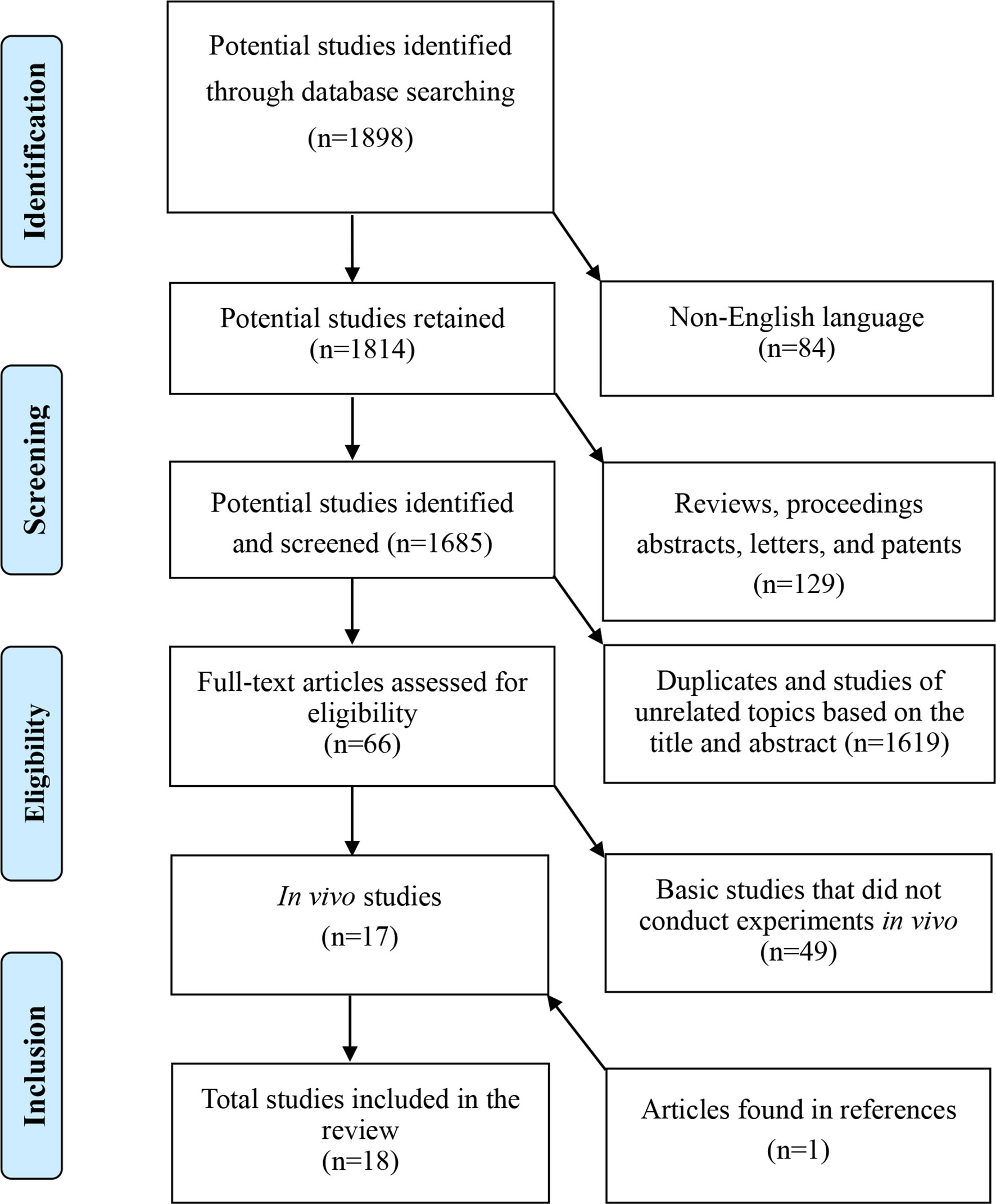
Figure 1 A flowchart of the literature search.
Overall, 1,898 publications were retrieved from the initial search. A total of 1,685 potential studies were retained for further identification after 84 non-English language studies and 129 review articles, letters, editorials, conference, and meeting abstracts were excluded. Furthermore, 1,619 duplicates and studies of unrelated topics based on the title and abstract, and 49 basic studies that did not conduct experiments in vivo were excluded. We identified 17 in vivo studies consisting of 7 human trials and 10 animal studies published between 1990 and 2019 using this retrieval strategy. All reference lists of the 17 included studies were investigated, and an additional human trial (Jancewicz et al., 2004) was identified and included in this review. Finally, data from the 18 studies [8 human studies (Jancewicz et al., 2004; Saw et al., 2011; Skowroński et al., 2012; Saw et al., 2013; Skowroński and Rutka, 2013; Turajane et al., 2013; Fu et al., 2014a; Saw et al., 2015) and 10 animal studies (Spaas et al., 2012; Broeckx et al., 2014a; Broeckx et al., 2014b; Fu et al., 2014a; Deng et al., 2015; Hopper et al., 2015b; Zhao et al., 2018; Daems et al., 2019; Broeckx et al., 2019a; Broeckx et al., 2019b) published by investigators from seven countries or regions] were analyzed.
Among the 18 studies, 7 were case reports [6 in humans (Jancewicz et al., 2004; Saw et al., 2011; Skowroński et al., 2012; Turajane et al., 2013; Fu et al., 2014a; Saw et al., 2015) and 1 in horses (Spaas et al., 2012)], 1 was a human comparative study (Skowroński and Rutka, 2013), 1 was a human randomized controlled trial (RCT) (Saw et al., 2013), 1 was a preliminary study (in horses) (Broeckx et al., 2014a), 1 was a pilot study (in horses) (Broeckx et al., 2014b), 4 involved animal models [rabbits (Fu et al., 2014b), sheep (Hopper et al., 2015b), rats (Deng et al., 2015), and pigs (Zhao et al., 2018), 1 was a prospective placebo-controlled study (in dog) (Daems et al., 2019), and 2 were randomized, double-blinded, placebo-controlled proof-of-concept study (in horses) (Broeckx et al., 2019a; Broeckx et al., 2019b) (Table 1).
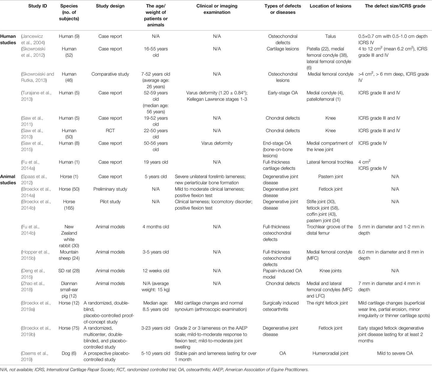
Table 1 Preoperative characteristics of patients and animals.
The age of the patients ranged from 7 to 59 years in the 8 human studies (Jancewicz et al., 2004; Saw et al., 2011; Skowroński et al., 2012; Saw et al., 2013; Skowroński and Rutka, 2013; Turajane et al., 2013; Fu et al., 2014a; Saw et al., 2015). Lesions were mainly located in the tibial plateaus (Saw et al., 2011; Saw et al., 2013), patella (Saw et al., 2011; Skowroński et al., 2012; Saw et al., 2013), femoral condyles (Saw et al., 2011; Skowroński et al., 2012; Saw et al., 2013; Skowroński and Rutka, 2013; Turajane et al., 2013; Saw et al., 2015), femoral trochlea (Saw et al., 2011; Saw et al., 2013; Turajane et al., 2013; Fu et al., 2014a), intercondylar notch (Saw et al., 2011), and talus of the ankle joint (Jancewicz et al., 2004). The types of lesions included cartilage defects (Saw et al., 2011; Skowroński et al., 2012; Saw et al., 2013; Fu et al., 2014a), osteochondral defects (Jancewicz et al., 2004; Skowroński and Rutka, 2013), and early- and late-stage osteoarthritis (Turajane et al., 2013; Saw et al., 2015). The International Cartilage Repair Society (ICRS) scores were all grade III–IV (Jancewicz et al., 2004; Saw et al., 2011; Skowroński et al., 2012; Saw et al., 2013; Skowroński and Rutka, 2013; Turajane et al., 2013; Fu et al., 2014a; Saw et al., 2015).
Types of lesions included spontaneous and induced osteoarthritis (Spaas et al., 2012; Broeckx et al., 2014a; Broeckx et al., 2014b; Deng et al., 2015; Daems et al., 2019; Broeckx et al., 2019a; Broeckx et al., 2019b), cartilage defects (Zhao et al., 2018), and osteochondral defects (Fu et al., 2014b; Hopper et al., 2015b) in the 10 animal studies. The lesions were in the knee joint (Broeckx et al., 2014a; Fu et al., 2014b; Deng et al., 2015; Hopper et al., 2015b; Zhao et al., 2018), fetlock joint (Broeckx et al., 2014a; Broeckx et al., 2014b; Broeckx et al., 2019a; Broeckx et al., 2019b), pastern joint (Broeckx et al., 2014a), coffin joint (Broeckx et al., 2014b), and humeroradial joint (Daems et al., 2019). The preoperative characteristics of the patients and animals, such as the age, clinical and imaging examination, types of defects and diseases, location of lesions, and defect size/ICRS grade are shown in Table 1.
Eight human studies (Jancewicz et al., 2004; Saw et al., 2011; Skowroński et al., 2012; Saw et al., 2013; Skowroński and Rutka, 2013; Turajane et al., 2013; Fu et al., 2014a; Saw et al., 2015) and 1 animal (Hopper et al., 2015b) study used autologous nonculture-expanded condensed PBSCs, 1 animal study (Deng et al., 2015) used allogenic condensed PBSCs, 1 animal study (Spaas et al., 2012) used autologous culture-expanded PB-MSCs, 6 animal studies (Broeckx et al., 2014a; Broeckx et al., 2014b; Fu et al., 2014b; Zhao et al., 2018; Broeckx et al., 2019a; Broeckx et al., 2019b) used allogenic culture-expanded PB-MSCs, and 1 animal study (Daems et al., 2019) used xenogeneic culture-expanded PB-MSCs as seed cells for cartilage repair and regeneration.
All 8 human studies (Jancewicz et al., 2004; Saw et al., 2011; Skowroński et al., 2012; Saw et al., 2013; Skowroński and Rutka, 2013; Turajane et al., 2013; Fu et al., 2014a; Saw et al., 2015) with 130 patients used a blood cell separator to collect PBSCs. One animal study (Deng et al., 2015) with 28 Sprague-Dawley (SD) rats used the density gradient centrifugation (DGC) method to isolate PBSCs. Eight animal studies (Spaas et al., 2012; Broeckx et al., 2014a; Broeckx et al., 2014b; Hopper et al., 2015b; Zhao et al., 2018; Daems et al., 2019; Broeckx et al., 2019a; Broeckx et al., 2019b) with 272 horses, 24 mountain sheep, 12 Diannan small-ear pigs, and 6 dogs used the DGC and plastic adherence (PA) methods to isolate PB-MSCs and PBSCs. Furthermore, one animal study (Fu et al., 2014b) with 30 New Zealand White rabbits used the erythrocyte lysis and PA methods to isolate PB-MSCs.
The seeding dosage in 5 human studies (Saw et al., 2011; Saw et al., 2013; Skowroński and Rutka, 2013; Fu et al., 2014a; Saw et al., 2015) and 1 animal study (Deng et al., 2015) using nonculture-expanded PBSCs as seed cells ranged from 5.0×10⁶ to 3.5×10⁷ cells/ml (or cells/injection), and the seeding dosage in 2 human studies (Skowroński et al., 2012; Turajane et al., 2013) and 1 animal study (Hopper et al., 2015b) was less than 5.0×10⁶ cells/ml (or cells/injection). In 5 animal studies using PB-MSCs as seed cells, the seeding dosage in 3 studies ranged from 1×10⁶ to 5.0×10⁶ cells/ml (or cells/injection) (Spaas et al., 2012; Fu et al., 2014b; Daems et al., 2019; Broeckx et al., 2019a; Broeckx et al., 2019b). One human study (Jancewicz et al., 2004) and 3 animal studies(Broeckx et al., 2014a; Broeckx et al., 2014b; Zhao et al., 2018) did not mention the cell seeding dosage.
Four human studies (Jancewicz et al., 2004; Skowroński et al., 2012; Skowroński and Rutka, 2013; Fu et al., 2014a) and 3 animal studies (Fu et al., 2014b; Hopper et al., 2015b; Zhao et al., 2018) implanted cells by surgery, while the remaining 4 human studies (Saw et al., 2011; Saw et al., 2013; Turajane et al., 2013; Saw et al., 2015) and 7 animal studies (Spaas et al., 2012; Broeckx et al., 2014a; Broeckx et al., 2014b; Deng et al., 2015; Daems et al., 2019; Broeckx et al., 2019a; Broeckx et al., 2019b) implanted cells by single or repeated intra-articular injections.
All human studies used a variety of other treatments, such as intra-articular debridement (Jancewicz et al., 2004; Saw et al., 2011; Skowroński et al., 2012; Saw et al., 2013; Skowroński and Rutka, 2013; Turajane et al., 2013; Fu et al., 2014a; Saw et al., 2015), the modified sandwich technique (Jancewicz et al., 2004; Skowroński and Rutka, 2013), BMS (Saw et al., 2011; Skowroński et al., 2012; Saw et al., 2013; Saw et al., 2015), high tibial osteotomy (HTO) (Saw et al., 2011; Saw et al., 2015), and patellofemoral realignment (Fu et al., 2014a), to promote cartilage repair and regeneration while implanting cells. Strict rehabilitation programmes and passive or active exercises (Saw et al., 2011; Saw et al., 2013; Skowroński and Rutka, 2013; Turajane et al., 2013; Fu et al., 2014a; Saw et al., 2015) were followed to avoid early weight bearing, joint stiffness, and adhesion.
The animal studies used other treatments, such as decalcified bone matrix (DBM) scaffolds (Fu et al., 2014a; Zhao et al., 2018), collagen-glycosaminoglycan (GAG) scaffolds (Hopper et al., 2015b), platelet-rich plasma (PRP) injections (Broeckx et al., 2014a; Broeckx et al., 2014b), and equine allogeneic plasma (EAP) (Broeckx et al., 2019a; Broeckx et al., 2019b), while implanting cells. Except for 2 studies (Broeckx et al., 2019a; Broeckx et al., 2019b), there were no strict rehabilitation plans in the other animal studies.
Table 2 summarizes the details of the application of PBSCs to cartilage repair and regeneration in humans and animals.
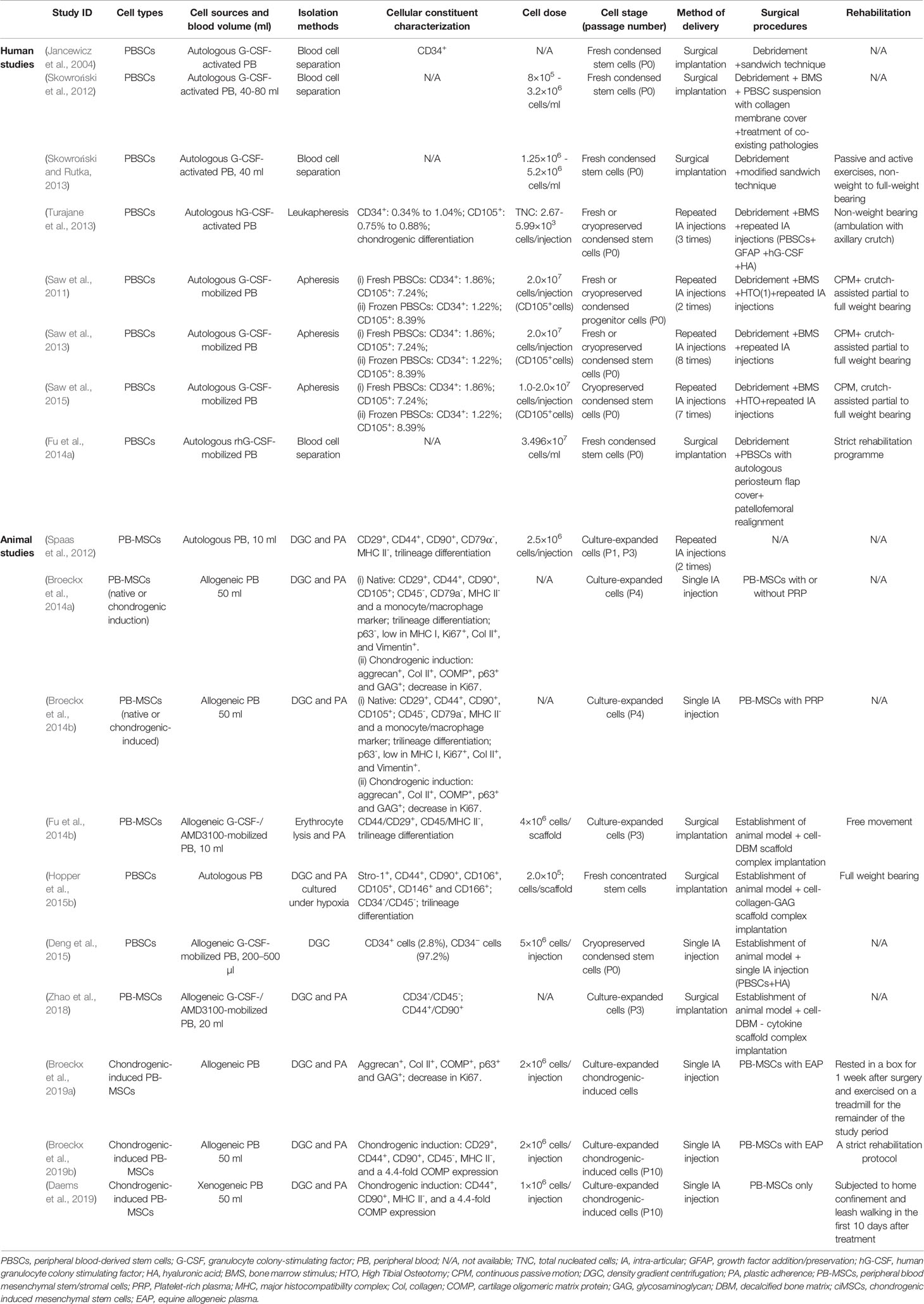
Table 2 Treatment Details of PBSCs for Cartilage Repair and Regeneration in Humans and Animals.
We assessed the adverse events and the clinical, radiographic, and histologic results to determine the treatment efficacy and safety (Table 3).
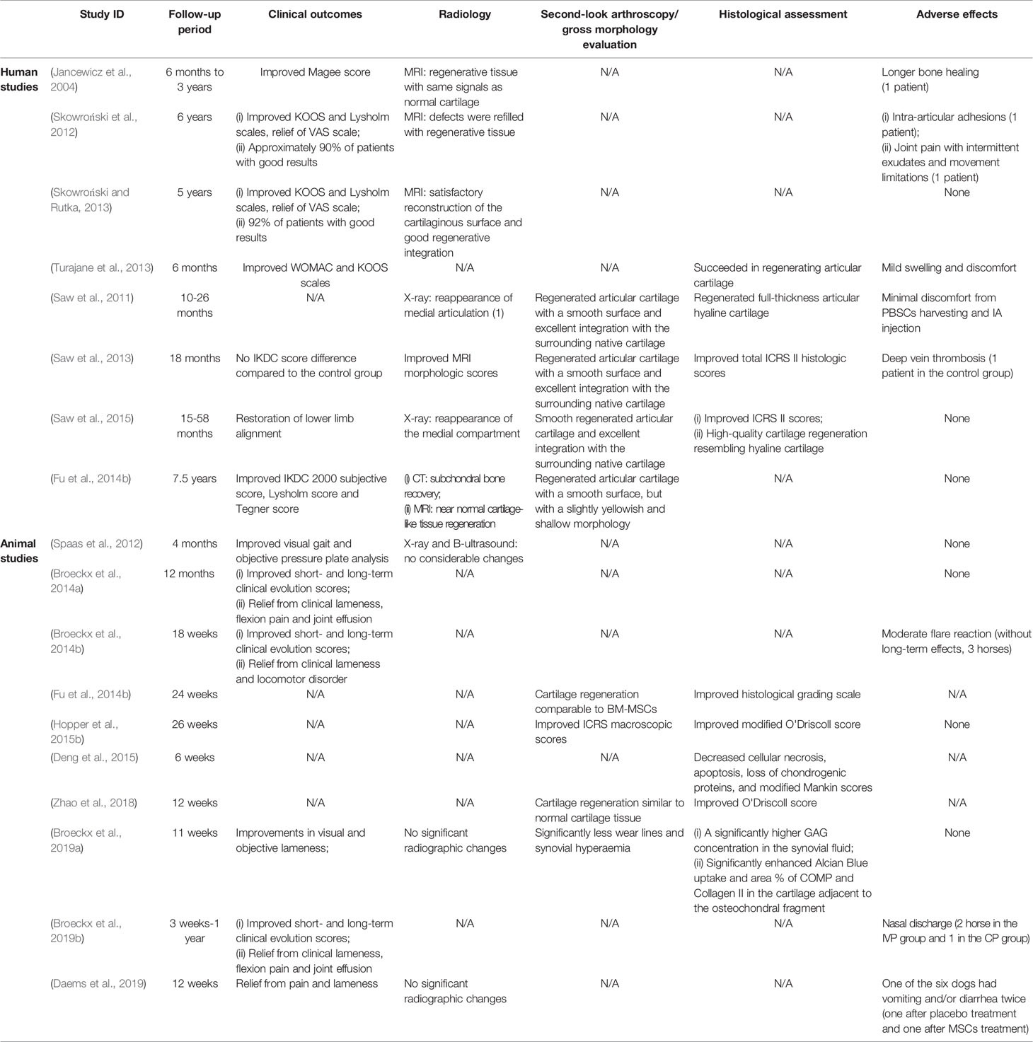
Table 3 Efficacy and safety of treatment.
The follow-up time of the 8 human trials ranged from 6 months to 7.5 years. The clinical evaluation results of 5 studies showed that Magee score (Jancewicz et al., 2004), KOOS scales (Skowroński et al., 2012; Skowroński and Rutka, 2013; Turajane et al., 2013), Lysholm scales (Skowroński et al., 2012; Skowroński and Rutka, 2013), WOMAC scales (Turajane et al., 2013), IKDC 2000 subjective score (Fu et al., 2014a) or Tegner score (Fu et al., 2014a) were improved, VAS scales (Skowroński et al., 2012; Skowroński and Rutka, 2013) were relieved, and Skowroski et al. (Skowroński et al., 2012; Skowroński and Rutka, 2013) reported 90 and 92% of patients with good results in 2012 and 2013, respectively. One study (Saw et al., 2013) reported that there was no IKDC score difference compared to the control group. One study (Saw et al., 2015) reported lower limb line recovery, and one study (Saw et al., 2011) did not report clinical evaluation results.
Five animal studies (Spaas et al., 2012; Broeckx et al., 2014a; Broeckx et al., 2014b; Broeckx et al., 2019a; Broeckx et al., 2019b) on horses reported improved visual gait, objective pressure plate analysis, short- and long-term clinical evolution scores, and relief of visual and objective lameness, flexion pain, and joint effusion.
Radiological examination, which is a non-invasive examination method, was widely used to evaluate the efficacy of cartilage repair and regeneration. Seven human studies used MRI (Jancewicz et al., 2004; Skowroński et al., 2012; Saw et al., 2013; Skowroński and Rutka, 2013; Fu et al., 2014a), X-ray (Saw et al., 2011; Saw et al., 2015) or CT (Fu et al., 2014a) to evaluate the repair effect and reported improved MRI morphologic scores, regenerative tissue with the same signal as normal cartilage, subchondral bone recovery, or reappearance of the medial compartment. However, radiological examination was rarely used in the animal studies. Three animal studies (Spaas et al., 2012; Daems et al., 2019; Broeckx et al., 2019a) reported no significant radiographic changes.
Four human studies (Saw et al., 2011; Saw et al., 2013; Fu et al., 2014a; Saw et al., 2015) evaluated cartilage repair with the method of second-look arthroscopy and suggested that cartilage regeneration was comparable to BM-MSCs with improved ICRS macroscopic scores, cartilage regeneration similar to normal cartilage tissue, or significantly fewer wear lines and synovial hyperaemia.
Histopathological examination is the gold standard for evaluation. Four human studies (Saw et al., 2011; Saw et al., 2013; Turajane et al., 2013; Saw et al., 2015) reported high-quality cartilage regeneration resembling hyaline cartilage and/or improved total ICRS II histologic scores. Five animal studies (Fu et al., 2014b; Deng et al., 2015; Hopper et al., 2015b; Zhao et al., 2018; Broeckx et al., 2019a) reported an improved histological grading scale, modified O'Driscoll score, modified Mankin scores, O'Driscoll score, or significantly higher Alcian blue uptake.
In eight human studies, the major adverse events included longer bone healing (1 patient) (Jancewicz et al., 2004), intra-articular adhesions (1 patient) (Skowroński et al., 2012), joint pain with intermittent exudates and motion limitation (1 patient) (Skowroński et al., 2012), mild swelling (Turajane et al., 2013), and minimal discomfort during PBSCs harvesting and intra-articular (IA) injection (Saw et al., 2011). No serious adverse events occurred during the isolation and treatment of PBSCs. In one human study (Saw et al., 2013), a case of deep vein thrombosis occurred in the control group. In animal studies, moderate flare reactions (3 in 165 horses) (Broeckx et al., 2014a), nasal discharge (3 in 75 horses) (Broeckx et al., 2019), and vomiting and diarrhea (1 in 6 dogs) (Daems et al., 2019) occurred without long-term effects.
Researchers have conducted investigations of PBSCs in cartilage repair and regeneration because of the advantages of PBSCs and limitations of chondrogenic progenitor cells from other sources, such as bone marrow (Bain, 2003), synovial membranes (Murata et al., 2018), and adipose tissue (Kuroda et al., 2015). Increasing evidence has shown that PB-MSCs have a similar potential for proliferation and trilineage differentiation as BM-MSCs and might be a promising source of seed cells for cartilage repair (Wang et al., 2016b) since Fernández et al. (Fernandez et al., 1997) reported the presence of stromal cells in hG-CSF-mobilized PB from patients with breast cancer for the first time in 1997. However, PBSCs were not used to treat chondral defects and promote cartilage regeneration in vivo until 2004, as reported by investigators in Poland (Jancewicz et al., 2004).
For the first time, this review comprehensively evaluated the feasibility, efficacy, and safety of using PBSCs for cartilage repair and regeneration in vivo by analyzing the preoperative characteristics, therapeutic details, outcomes, and adverse events reported in currently published literature. This review might provide new insights and strategies for further foundational research and clinical applications of PBSCs.
Autologous nonculture-expanded PBSCs are easy to harvest and manipulate from G-CSF-activated PB without the concerns of disease transmission, immune rejection, and ethical issues (Fu et al., 2014a; Saw et al., 2015). PBSCs are currently the most commonly used cell type for cartilage repair in all stem cell types derived from PB. It has been demonstrated that nonculture-expanded PBSCs comprise haematopoietic stem cells (HSCs), fibrocytes, a population of MSCs/mesenchymal progenitor cells (MSCs/MPCs), white blood cells, platelets, growth factors, and a small percentage of red blood cells (Stroncek et al., 1997; Cesselli et al., 2009). When PBSCs are being cultured, other impure cell types except MSCs/MPCs are not present anymore. To a certain degree, the cell composition of nonculture-expanded PBSCs is similar to that of the bone marrow-derived buffy coat (BMBC), which is separated from bone marrow using a Ficoll gradient centrifugation system. The bone marrow-derived buffy coat has been widely used as a source of MSCs for cartilage repair and regeneration and has achieved good to excellent results (Fortier et al., 2010; Jin et al., 2011). Several possible mechanisms of action of PBSCs might contribute to cartilage repair. Hopper et al. (Hopper et al., 2015a; Hopper et al., 2015c) found that PBSCs stimulate the upregulation of eight genes associated with chondrogenic differentiation of knee infrapatellar fat pad-derived MSCs, increase the total number of MSCs, increase native chondrocyte migration, and accelerate the rate of cell movement. Exogenous MSCs, HSCs, and growth factors in PBSCs initiate cartilage regeneration and augment endogenous MSC recruitment from bone marrow to subchondral drilling sites (Khaldoyanidi, 2008; Onuora, 2015; Saw et al., 2015). Deng et al. (2015) suggested that PBSCs prevent the progression of papain-induced knee OA in a rat model by reducing articular surface fibrillation, irregularity, and erosion, and by inhibiting chondrocyte necrosis and loss of chondrogenic proteins. HSCs and non-HSCs, such as MSCs, endothelial progenitor cells, and very small embryonic-like (VSEL) cells, contained in PBSCs might play an important role through a paracrine mechanism (Kucia et al., 2007; Onuora, 2015). Although the term “PBSCs” had different expressions in different studies, such as PB progenitor cells (PBPCs) (Saw et al., 2011) and PB mononuclear cells (PBMCs) (Hopper et al., 2015b), we found that the cell acquisition method and cell composition were basically the same. For the convenience of expression, “PBSCs” was used uniformly in this paper.
The transplantation of autologous culture-expanded PB-MSCs requires two procedures for obtaining patient cells and transplanting the cells after cultivation, which prolongs hospital stays, increases costs and risks contamination related to in vitro culture, possibly limiting the clinical application of autologous PB-MSCs (Saw et al., 2013; Fu et al., 2014b). Moreover, an age-related decline in MSC numbers, proliferation, and clonogenicity, which lead to more difficult culture in vitro and a longer culture cycle than MSCs from other tissue sources, might be another significant cause for the lack of clinical applications of autologous culture-expanded PB-MSCs (Kassis et al., 2006; Bourzac et al., 2010; Chong et al., 2012; Spaas et al., 2013; Wang et al., 2016b). For example, MSCs derived from bone marrow, synovium or adipose tissue reached 80–90% confluence within 7 to 14 days (Zhang et al., 2014; Jin et al., 2016; Shimomura et al., 2016). However, MSCs derived from PB did not achieve the same confluence until about 21 days after primary culture (Chen et al., 2019). It takes longer to obtain the culture-expanded PB-MSCs than other tissue-derived MSCs. The presence of MSCs in human PB is debatable and their identification may be hampered, among others, by: (i) their low frequency in PB of healthy individuals, and (ii) the large biological variations related to donor age, pathology, disease status, and corresponding treatment regimens (Fox et al., 2007; Moll et al., 2019). Most investigators agree that their frequency in blood is low in healthy individuals, but that the amounts of circulating MSCs may increase under special mobilization conditions, thus supporting the notion that MSCs can be transiently found circulating in blood (Moll et al., 2020). Jain et al. provide evidence that MSCs can be found in PB and apheresis product of patients treated with a typical G-CSF-based HSCs mobilization regimen by using flow cytometry (Jain et al., 2020). However, a systematic review strongly indicated the existence of MSCs in the PB of animals (Wang et al., 2016b), this might be because researchers could improve the success rate of PB-MSCs in animal studies by optimizing mobilization and culture procedures, prolonging the culture time, and increasing the number of animals and the frequency of blood drawn (Pitchford et al., 2009; To et al., 2011; Spaas et al., 2013). To et al. noted in a baboon model that MSC mobilization and colony-forming unit fibroblast (CFUF) in PB in response to G-CSF did only occur when adding stem cell factor (To et al., 2011). Pitchford et al. found, that MSCs/CFU-F were not found in mice PB post-mobilization with G-CSF, but when adding vascular endothelial growth factor and CXCR4-antagonist (Pitchford et al., 2009). Spaas et al. systematically studied the isolation and culture methods, cell characteristics, and clinical safety of equine PB-MSCs, and applied them to many veterinary clinical studies, such as promoting cartilage repair, cutaneous wound healing, and healing of tendon and ligament lesions (Spaas et al., 2013; Beerts et al., 2017; Martinello et al., 2018; Broeckx et al., 2019a). Allogenic or xenogeneic MSCs banks, improving the mobilization and purification techniques, and shortening the culture cycle might effectively account for deficiencies in autologous MSCs, reduce the burden on both patients and treatment providers, and promote the development of single-stage procedures (Moroni and Fornasari, 2013; Pescador et al., 2017).
MSCs inhibit immune responses and are not restricted by the HLA system through immune evasion and immune privilege mechanisms (Paterson et al., 2014; Vega et al., 2015). Moreover, the strong immunomodulatory and immunosuppressive properties of MSCs may play an important role in modifying graft-versus-host reactions during allogenic transplantations (Le Blanc and Ringden, 2007). Two animal studies used allogenic native and chondrogenic-induced PB-MSCs as a treatment for degenerative joint disease in horses and significantly improved the short- and long-term effects without serious adverse events (Broeckx et al., 2014a; Broeckx et al., 2014b). Vega et al. (2015) performed an RCT to assess the feasibility and safety of treating osteoarthritis with allogeneic MSCs in humans, and they concluded that allogeneic MSCs might be a convenient and effective alternative to autologous MSCs for the treatment of OA in the knee without serious transplantation-related adverse events. A number of published papers have indicated that transplanted MSCs influence the local microenvironment of cartilage by paracrine actions, such as the secretion of various growth factors, cytokines, and chemokines, to exert anti-inflammatory, anti-apoptotic, and anti-fibrotic effects on chondrocytes(Kuroda et al., 2015; Mancuso et al., 2019). Another possible mechanism of action of MSCs in cartilage repair and regeneration is that transplanted progenitor cells migrate to damaged cartilage areas and differentiate into chondrocytes and osteocytes (Cesselli et al., 2009). The fate of MSCs injected into the articular cavity can be monitored by labelling with green fluorescent protein (GFP) or carboxyfluorescein diacetate succinimidyl ester (CFDA-SE) (Guest et al., 2008; Sato et al., 2012). Murphy et al. found that the implantation of MSCs into the knee joints of goats with OA showed a strong and sustained effect in promoting cartilage repair. However, further tracing the labelled MSCs showed that the cell retention rate was very low, usually about 3%, and most cells disappeared within a few days (Murphy et al., 2003). This suggests that MSCs may not directly differentiate into chondrocytes to participate in tissue repair in vivo, but promote cartilage regeneration through other mechanisms. In recent years, more and more researchers believed that exosomes secreted by MSCs played an important role in cartilage repair and regeneration (Marote et al., 2016; Yan and Wu, 2019; Jin et al., 2020; Liu et al., 2020). Exosomes are generally considered as communication vectors between cells, and carry a large number of complex nucleic acids (mRNA and miRNA lncRNA), proteins and lipids that can regulate and restore extracellular matrix (ECM) homeostasis (Colombo et al., 2014). For example, MSCs exosomes with overexpressing of miR-140-5p blocked other Wnt signals in vitro by inhibiting v-ral simian leukemia viral oncogene homolog A (RalA) and activating sex determining region Y-box 9 (SOX9), and regulate the expression of Col II and aggrecan (ACAN) in vivo to promote cartilage regeneration (Tao et al., 2017). It may also be an important mechanism for PBSCs to promote cartilage repair.
Blood cell separation is the most commonly used method for collecting PBSCs. It is a developed and simple technique that has been widely used in the treatment of systemic blood diseases. In a monocyte suspension isolated by blood cell separation, CD105+ cells have been shown to be more abundance than CD34+ cells, and the proportion of CD105+ cells increased after cryopreservation (Saw et al., 2011). However, there is no study on the subsequent isolation and culture of PB-MSCs from PBSCs collected by blood cell separation. The current standard methods of PB-MSC isolation are DGC (such as Ficoll, Lymphoprep, and Percoll) and PA (Bourzac et al., 2010).
As one of the most fundamental parameters that might influence the outcome of cartilage repair (Gupta et al., 2016), the optimal density or dosage of PBSCs used for cartilage regeneration in different methods and species has not been fully investigated. Skowroński et al. (Skowroński and Rutka, 2013) reported a slightly poorer outcome of cartilage repair in a group treated with a bone marrow concentrate than a group treated with fresh condensed PBSCs, and they attributed this result to the lower cell count in the suspension obtained from bone marrow. The main concern of using nonculture-expanded PBSCs to promote tissue regeneration is the low content of MSCs within harvests. The number of HSCs (with a CD34+ surface marker) and MSCs (with a CD105+ surface marker) were quantified by flow cytometry in a study carried out by Saw et al. (Saw et al., 2011). The flow cytometry result showed that the proportion of CD105+ cells in fresh PBSC suspension was 7.24% (2.32×10⁶ cells/ml). Interestingly, the proportion of CD105+ cells reached 8.39% (2.69×10⁶ cells/ml) after cryopreservation. However, the CD105+ cell counts vary between different studies. Turajane et al. (Turajane et al., 2013) reported that a proportion of CD105+ cells ranging from 0.75 to 0.88%. The difference of the proportion of CD105+ cells in the two studies was probably due to the younger patients in the previous study and the older patients in the latter.
To increase the yield of MSCs from autologous PB, repeated intra-articular injections were implemented in some studies (Saw et al., 2011; Saw et al., 2013; Turajane et al., 2013; Saw et al., 2015), and they speculated that this method is more efficacious than a single injection for the enhancement of cartilage repair on the basis of a suggestion from an animal study (Saw et al., 2009). However, repeated IA injections of culture-expanded allogeneic MSCs is not recommended due to a significant adverse response that might be initiated by immune recognition of allogeneic MSCs after a second exposure (Joswig et al., 2017).
Currently, the optimal seeding density of MSCs also remains unknown. A systematic review showed that the dose of MSCs for cartilage repair varies from 2×10⁶–7.7×10⁷ cells in human clinical studies (Goldberg et al., 2017). Gupta et al. (Gupta et al., 2016) found that an MSC dose of 2.5×10⁷ with the IA injection method for treating OA showed the best improvement for relieving pain and the lowest adverse events compared with other higher dose groups. They hypothesized that a higher cell dosage causes cell aggregation and subsequent cell death due to limited space in the knee joint. A prospective RCT demonstrated that an intra-articular injection of cultured MSCs with a mean dose of 1.46×10⁷ cells for treating OA is effective in improving clinical and magnetic resonance observation of cartilage repair tissue (MOCART) scores after a 2-year follow-up (Wong et al., 2013). Given the limited evidence of clinical application of PB-MSCs in cartilage repair and regeneration, the optimal therapeutic dose of PB-MSCs remains to be further studied.
Moreover, a number of studies have reported concomitant procedures, such as abrasion arthroplasty (Beckmann et al., 2015), autologous bone grafting to restore bone mass (Sadlik et al., 2017), treatment of co-existing pathologies (Wong et al., 2013), and BMS (Jin et al., 2011), PRP (Broeckx et al., 2019a) and HA (Charlesworth et al., 2019) to repair cartilage defects. Thus, the abovementioned methods are recommended to supplement PBSCs for cartilage repair and regeneration. A rigorous postoperative rehabilitation programme is required to protect grafts and avoid the effusion of PBSC suspensions (Skowroński and Rutka, 2013; Fu et al., 2014a).
Compared with other tissue-derived MSCs, the culture of PB-MSCs was relatively difficult, which resulted in less reports of its application in vivo, but it does not affect its application prospects. On the contrary, it is ethically more suitable for clinical application due to its unique advantages, such as minimally invasive sample acquisition procedure, repeatable sampling, and high recognition of patients (Fu et al., 2014a; Fu et al., 2014b; Wang et al., 2016a; Chen et al., 2019). In this review, we have summarized all the currently published researches on the use of PBSCs for cartilage repair and regeneration in vivo. Although only 5 human and veterinary clinical studies (Saw et al., 2013; Skowroński and Rutka, 2013; Daems et al., 2019; Broeckx et al., 2019a; Broeckx et al., 2019b) had a control group, the results were still very useful for readers, and can reflect the progress and problems in this field to a certain extent.
This review evaluated the use of PBSCs in cartilage repair and regeneration in vivo for the first time. Autologous PBSCs are easy to obtain and are free of transmittable diseases, infection risks, and medical ethical restrictions. They are currently the most commonly used cell type for cartilage repair among all stem cell types derived from PB. Blood cell separation technology is developed, simple, and convenient, making it the most commonly used method to obtain PBSC suspensions. Allogeneic culture-expanded PB-MSCs are more widely used in animal research and are potential seed cell types for cartilage repair and regeneration in the future. DGC and PA are the most commonly used methods for PB-MSC isolation. Improving the purification technology and shortening the culture cycle of culture-expanded PB-MSCs will obviously promote the researchers' interest. PBSCs are safe in cartilage repair and regeneration. Although all reviewed articles indicated that using PB as a cell source enhances cartilage repair and regeneration in vivo by the IA injection and surgery implantation methods, we should maintain a prudent attitude towards the positive therapeutic effect of PBSCs considering the deficiency of studies with a high level of evidence, incomplete assessment system of outcomes, and combined use of multiple other treatments. In summary, the use of PBSCs in cartilage repair and regeneration warrants considerable efforts for further investigations due to its superiorities and safety in clinical settings and positive effects despite limited evidence in human.
Conception and design: J-KY and DJ. Analysis and interpretation of the data: Y-RC, XY, and F-ZY. Drafting of the article: Y-RC, XY, and F-ZY. Information collection and sorting: JY, B-BX, Z-XZ, Z-MM, and JG. Manuscript editing and proofreading: Y-FS, Z-WS, X-JW, and Z-YC. D-YW, B-SF, MY, and S-TS provided oversight. Critical revision of the article for important intellectual content: J-KY and DJ. All authors read and approved the final manuscript.
This work was supported by the National Natural Science Foundation of China (Grant Nos. 51773004, 81630056, 51920105006, 31670982) and National Key Research and Development Program (Grant No. 2016YFC1100704).
The authors declare that the research was conducted in the absence of any commercial or financial relationships that could be construed as a potential conflict of interest.
The handling editor declared a past co-authorship with one of the authors JK-Y.
PBSCs, Peripheral blood-derived stem cells; PB-MSCs, Peripheral blood-derived mesenchymal stem cells; DGC, Density gradient centrifugation; PA, Plastic adherence; BMS, Bone marrow stimulation; ACI, Autologous chondrocyte implantation; BM-MSCs, Bone marrow-derived mesenchymal stem cells; PRISMA, Preferred Reporting Items for Systematic Reviews and Meta-analyses; RCT, Randomized controlled trial; HTO, High tibial osteotomy; DBM, Decalcified bone matrix; ICRS, International Cartilage Repair Society; OA, Osteoarthritis; IA, Intra-articular; hG-CSF, Human granulocyte colony stimulating factor; HA, Hyaluronic acid; CPM, Continuous passive motion; PRP, Platelet-rich plasma; MHC, Major histocompatibility complex; COMP, Cartilage oligomeric matrix protein; GAG, Glycosaminoglycan; MRI, Magnetic Resonance Imaging; CT, Computed Tomography; KOOS, Knee injury and osteoarthritis outcome score; VAS, Visual Analogue Scale; WOMAC, The Western Ontario and McMaster Universities Osteoarthritis Index; IKDC, The International Knee Documentation Committee Knee Uation Form; ECM, extracellular matrix; GFP, green fluorescent protein; CFDA-SE, carboxyfluorescein diacetate succinimidyl ester; RalA, v-ral simian leukemia viral oncogene homolog A; SOX9, sex determining region Y-box 9; ACAN, aggrecan; CFUF, colony-forming unit fibroblast.
Andriolo, L., Merli, G., Filardo, G., Marcacci, M., Kon, E. (2017). Failure of Autologous Chondrocyte Implantation. Sports Med. Arthrosc Rev. 25, 10–18. doi: 10.1097/JSA.0000000000000137
Bain, B. J. (2003). Bone marrow biopsy morbidity and mortality. Br. J. Haematol. 121, 949–951. doi: 10.1111/j.1365-2257.2004.00630.x
Beckmann, R., Lippross, S., Hartz, C., Tohidnezhad, M., Ferreira, M. S., Neuss-Stein, S., et al. (2015). Abrasion arthroplasty increases mesenchymal stem cell content of postoperative joint effusions. BMC Musculoskelet Disord. 16, 250. doi: 10.1186/s12891-015-0705-0
Beerts, C., Suls, M., Broeckx, S. Y., Seys, B., Vandenberghe, A., Declercq, J., et al. (2017). Tenogenically Induced Allogeneic Peripheral Blood Mesenchymal Stem Cells in Allogeneic Platelet-Rich Plasma: 2-Year Follow-up after Tendon or Ligament Treatment in Horses. Front. Vet. Sci. 4, 158. doi: 10.3389/fvets.2017.00158
Bourzac, C., Smith, L. C., Vincent, P., Beauchamp, G., Lavoie, J. P., Laverty, S. (2010). Isolation of equine bone marrow-derived mesenchymal stem cells: a comparison between three protocols. Equine Vet. J. 42, 519–527. doi: 10.1111/j.2042-3306.2010.00098.x
Broeckx, S., Suls, M., Beerts, C., Vandenberghe, A., Seys, B., Wuertz-Kozak, K., et al. (2014a). Allogenic mesenchymal stem cells as a treatment for equine degenerative joint disease: a pilot study. Curr. Stem Cell Res. T. 9, 497. doi: 10.2174/1574888x09666140826110601
Broeckx, S., Zimmerman, M., Crocetti, S., Suls, M., Mariën, T., Ferguson, S. J., et al. (2014b). Regenerative Therapies for Equine Degenerative Joint Disease: A Preliminary Study. PLoS One 9, e85917. doi: 10.1371/journal.pone.0085917
Broeckx, S. Y., Martens, A. M., Bertone, A. L., Van Brantegem, L., Duchateau, L., Van Hecke, L., et al. (2019a). The use of equine chondrogenic-induced mesenchymal stem cells as a treatment for osteoarthritis: A randomised, double-blinded, placebo-controlled proof-of-concept study. Equine Vet. J. 51, 787–794. doi: 10.1111/evj.13089
Broeckx, S. Y., Seys, B., Suls, M., Vandenberghe, A., Mariën, T., Adriaensen, E., et al. (2019b). Equine Allogeneic Chondrogenic Induced Mesenchymal Stem Cells Are an Effective Treatment for Degenerative Joint Disease in Horses. Stem Cells Dev. 28, 410–422. doi: 10.1089/scd.2018.0061
Cesselli, D., Beltrami, A. P., Rigo, S., Bergamin, N., D'Aurizio, F., Verardo, R., et al. (2009). Multipotent progenitor cells are present in human peripheral blood. Circ. Res. 104, 1225–1234. doi: 10.1161/CIRCRESAHA.109.195859
Charlesworth, J., Fitzpatrick, J., Perera, N., Orchard, J. (2019). Osteoarthritis- a systematic review of long-term safety implications for osteoarthritis of the knee. BMC Musculoskelet Disord. 20, 151. doi: 10.1186/s12891-019-2525-0
Chen, S., Fu, P., Wu, H., Pei, M. (2017). Meniscus, articular cartilage and nucleus pulposus: a comparative review of cartilage-like tissues in anatomy, development and function. Cell Tissue Res. 370, 53–70. doi: 10.1007/s00441-017-2613-0
Chen, Y. R., Zhou, Z. X., Zhang, J. Y., Yuan, F. Z., Xu, B. B., Guan, J., et al. (2019). Low-Molecular-Weight Heparin-Functionalized Chitosan-Chondroitin Sulfate Hydrogels for Controlled Release of TGF-beta3 and in vitro Neocartilage Formation. Front. Chem. 7, 745. doi: 10.3389/fchem.2019.00745
Chong, P. P., Selvaratnam, L., Abbas, A. A., Kamarul, T. (2012). Human peripheral blood derived mesenchymal stem cells demonstrate similar characteristics and chondrogenic differentiation potential to bone marrow derived mesenchymal stem cells. J. Orthop. Res. 30, 634–642. doi: 10.1002/jor.21556
Colombo, M., Raposo, G., Thery, C. (2014). Biogenesis, secretion, and intercellular interactions of exosomes and other extracellular vesicles. Annu. Rev. Cell Dev. Biol. 30, 255–289. doi: 10.1146/annurev-cellbio-101512-122326
Daems, R., Van Hecke, L., Schwarzkopf, I., Depuydt, E., Broeckx, S. Y., David, M., et al. (2019). A Feasibility Study on the Use of Equine Chondrogenic Induced Mesenchymal Stem Cells as a Treatment for Natural Occurring Osteoarthritis in Dogs. Stem Cells Int. 2019, 1–11. doi: 10.1155/2019/4587594
Deng, M., Wei, S., Yew, T., Lee, P., Yang, T., Chu, H., et al. (2015). Cell Therapy with G-CSF-Mobilized Stem Cells in a Rat Osteoarthritis Model. Cell Transplant 24, 1085–1096. doi: 10.3727/096368914X680091
Ding, J., Zhang, J., Li, J., Li, D., Xiao, C., Xiao, H., et al. (2019). Electrospun polymer biomaterials. Prog. Polym. Sci. 90, 1–34. doi: 10.1016/j.progpolymsci.2019.01.002
Fernandez, M., Simon, V., Herrera, G., Cao, C., Del, F. H., Minguell, J. J. (1997). Detection of stromal cells in peripheral blood progenitor cell collections from breast cancer patients. Bone Marrow Transplant. 20, 265–271. doi: 10.1038/sj.bmt.1700890
Fortier, L. A., Potter, H. G., Rickey, E. J., Schnabel, L. V., Foo, L. F., Chong, L. R., et al. (2010). Concentrated bone marrow aspirate improves full-thickness cartilage repair compared with microfracture in the equine model. J. Bone Joint Surg. Am. 92, 1927–1937. doi: 10.2106/JBJS.I.01284
Fox, J. M., Chamberlain, G., Ashton, B. A., Middleton, J. (2007). Recent advances into the understanding of mesenchymal stem cell trafficking. Br. J. Haematol. 137, 491–502. doi: 10.1111/j.1365-2141.2007.06610.x
Fu, W. L., Ao, Y., Ke, X., Zheng, Z., Gong, X., Jiang, D., et al. (2014a). Repair of large full-thickness cartilage defect by activating endogenous peripheral blood stem cells and autologous periosteum flap transplantation combined with patellofemoral realignment. Knee 21, 609–612. doi: 10.1016/j.knee.2013.10.010
Fu, W. L., Zhou, C. Y., Yu, J. K. (2014b). A new source of mesenchymal stem cells for articular cartilage repair: MSCs derived from mobilized peripheral blood share similar biological characteristics in vitro and chondrogenesis in vivo as MSCs from bone marrow in a rabbit model. Am. J. Sports Med. 42, 592–601. doi: 10.1177/0363546513512778
Goldberg, A., Mitchell, K., Soans, J., Kim, L., Zaidi, R. (2017). The use of mesenchymal stem cells for cartilage repair and regeneration: a systematic review. J. Orthop. Surg. Res. 12, 39. doi: 10.1186/s13018-017-0534-y
Guadix, J. A., Zugaza, J. L., Galvez-Martin, P. (2017). Characteristics, applications and prospects of mesenchymal stem cells in cell therapy. Med. Clin. (Barc) 148, 408–414. doi: 10.1016/j.medcli.2016.11.033
Guest, D. J., Smith, M. R., Allen, W. R. (2008). Monitoring the fate of autologous and allogeneic mesenchymal progenitor cells injected into the superficial digital flexor tendon of horses: preliminary study. Equine Vet. J. 40, 178–181. doi: 10.2746/042516408X276942
Gupta, P. K., Chullikana, A., Rengasamy, M., Shetty, N., Pandey, V., Agarwal, V., et al. (2016). Efficacy and safety of adult human bone marrow-derived, cultured, pooled, allogeneic mesenchymal stromal cells (Stempeucel(R)): preclinical and clinical trial in osteoarthritis of the knee joint. Arthritis Res. Ther. 18, 301. doi: 10.1186/s13075-016-1195-7
Han, Y., Li, X., Zhang, Y., Han, Y., Chang, F., Ding, J. (2019). Mesenchymal Stem Cells for Regenerative Medicine. Cells 8, 886. doi: 10.3390/cells8080886
Hernigou, P., Homma, Y., Flouzat-Lachaniette, C. H., Poignard, A., Chevallier, N., Rouard, H. (2013). Cancer risk is not increased in patients treated for orthopaedic diseases with autologous bone marrow cell concentrate. J. Bone Joint Surg. Am. 95, 2215–2221. doi: 10.2106/JBJS.M.00261
Hopper, N., Henson, F., Brooks, R., Ali, E., Rushton, N., Wardale, J. (2015a). Peripheral blood derived mononuclear cells enhance osteoarthritic human chondrocyte migration. Arthritis Res. Ther. 17, 199. doi: 10.1186/s13075-015-0709-z
Hopper, N., Wardale, J., Brooks, R., Power, J., Rushton, N., Henson, F. (2015b). Peripheral Blood Mononuclear Cells Enhance Cartilage Repair in in vivo Osteochondral Defect Model. PLoS One 10, e0133937. doi: 10.1371/journal.pone.0133937
Hopper, N., Wardale, J., Howard, D., Brooks, R., Rushton, N., Henson, F. (2015c). Peripheral blood derived mononuclear cells enhance the migration and chondrogenic differentiation of multipotent mesenchymal stromal cells. Stem Cells Int. 2015, 323454. doi: 10.1155/2015/323454
Jain, A., Khadwal, A., Sachdeva, M., Bose, P., Lad, D., Bhattacharya, S., et al. (2020). Variables affecting the presence of mesenchymal stromal cells in peripheral blood and their relationship with apheresis products. Br. J. Haematol. doi: 10.1111/bjh.16412
Jancewicz, P., Dzienis, W., Pietruczuk, M., Skowroński, J., Bielecki, M. (2004). Osteochondral defects of the talus treated by mesenchymal stem cell implantation–early results. Roczniki Akademii Medycznej w Bialymstoku (1995) 49 (Suppl 1), 25.
Jin, L. H., Choi, B. H., Kim, Y. J., Park, S. R., Jin, C. Z., Min, B. H. (2011). Implantation of bone marrow-derived buffy coat can supplement bone marrow stimulation for articular cartilage repair. Osteoarthritis Cartilage 19, 1440–1448. doi: 10.1016/j.joca.2011.07.012
Jin, G., Park, J., Wall, I., Kim, H. (2016). Isolation and culture of primary rat adipose derived stem cells using porous biopolymer microcarriers. Tissue Eng. Regen. Med. 13, 242–250. doi: 10.1007/s13770-016-0040-z
Jin, Z., Ren, J., Qi, S. (2020). Human bone mesenchymal stem cells-derived exosomes overexpressing microRNA-26a-5p alleviate osteoarthritis via down-regulation of PTGS2. Int. Immunopharmacol. 78, 105946. doi: 10.1016/j.intimp.2019.105946
Joswig, A. J., Mitchell, A., Cummings, K. J., Levine, G. J., Gregory, C. A., Smith, R. R., et al. (2017). Repeated intra-articular injection of allogeneic mesenchymal stem cells causes an adverse response compared to autologous cells in the equine model. Stem Cell Res. Ther. 8, 42. doi: 10.1186/s13287-017-0503-8
Kassis, I., Zangi, L., Rivkin, R., Levdansky, L., Samuel, S., Marx, G., et al. (2006). Isolation of mesenchymal stem cells from G-CSF-mobilized human peripheral blood using fibrin microbeads. Bone Marrow Transplant. 37, 967–976. doi: 10.1038/sj.bmt.1705358
Khaldoyanidi, S. (2008). Directing stem cell homing. Cell Stem Cell 2, 198–200. doi: 10.1016/j.stem.2008.02.012
Kucia, M., Halasa, M., Wysoczynski, M., Baskiewicz-Masiuk, M., Moldenhawer, S., Zuba-Surma, E., et al. (2007). Morphological and molecular characterization of novel population of CXCR4+ SSEA-4+ Oct-4+ very small embryonic-like cells purified from human cord blood: preliminary report. Leukemia 21, 297–303. doi: 10.1038/sj.leu.2404470
Kuroda, K., Kabata, T., Hayashi, K., Maeda, T., Kajino, Y., Iwai, S., et al. (2015). The paracrine effect of adipose-derived stem cells inhibits osteoarthritis progression. BMC Musculoskelet Disord. 16, 236. doi: 10.1186/s12891-015-0701-4
Le Blanc, K., Ringden, O. (2007). Immunomodulation by mesenchymal stem cells and clinical experience. J. Intern. Med. 262, 509–525. doi: 10.1111/j.1365-2796.2007.01844.x
Li, X., Ding, J., Zhang, Z., Yang, M., Yu, J., Wang, J., et al. (2016). Kartogenin-Incorporated Thermogel Supports Stem Cells for Significant Cartilage Regeneration. ACS Appl. Mater. Interfaces 8, 5148–5159. doi: 10.1021/acsami.5b12212
Liu, H., Cheng, Y., Chen, J., Chang, F., Wang, J., Ding, J., et al. (2018). Component effect of stem cell-loaded thermosensitive polypeptide hydrogels on cartilage repair. Acta Biomater. 73, 103–111. doi: 10.1016/j.actbio.2018.04.035
Liu, C., Li, Y., Yang, Z., Zhou, Z., Lou, Z., Zhang, Q. (2020). Kartogenin enhances the therapeutic effect of bone marrow mesenchymal stem cells derived exosomes in cartilage repair. Nanomedicine (Lond) 15, 273–288. doi: 10.2217/nnm-2019-0208
Makris, E. A., Gomoll, A. H., Malizos, K. N., Hu, J. C., Athanasiou, K. A. (2015). Repair and tissue engineering techniques for articular cartilage. Nat. Rev. Rheumatol. 11, 21–34. doi: 10.1038/nrrheum.2014.157
Mancuso, P., Raman, S., Glynn, A., Barry, F., Murphy, J. M. (2019). Mesenchymal Stem Cell Therapy for Osteoarthritis: The Critical Role of the Cell Secretome. Front. Bioeng. Biotechnol. 7, 9. doi: 10.3389/fbioe.2019.00009
Marote, A., Teixeira, F. G., Mendes-Pinheiro, B., Salgado, A. J. (2016). MSCs-Derived Exosomes: Cell-Secreted Nanovesicles with Regenerative Potential. Front. Pharmacol. 7, 231. doi: 10.3389/fphar.2016.00231
Martinello, T., Gomiero, C., Perazzi, A., Iacopetti, I., Gemignani, F., DeBenedictis, G. M., et al. (2018). Allogeneic mesenchymal stem cells improve the wound healing process of sheep skin. BMC Vet. Res. 14, 202. doi: 10.1186/s12917-018-1527-8
Moll, G., Ankrum, J. A., Kamhieh-Milz, J., Bieback, K., Ringden, O., Volk, H. D., et al. (2019). Intravascular Mesenchymal Stromal/Stem Cell Therapy Product Diversification: Time for New Clinical Guidelines. Trends Mol. Med. 25, 149–163. doi: 10.1016/j.molmed.2018.12.006
Moll, G., Drzeniek, N., Kamhieh-Milz, J., Geissler, S., Reinke, P. (2020). Editorial comment: variables affecting the presence of mesenchymal stromal cells in the peripheral blood and their relationship with apheresis product. Br. J. Haematol. doi: 10.1111/bjh.16389
Moroni, L., Fornasari, P. M. (2013). Human mesenchymal stem cells: a bank perspective on the isolation, characterization and potential of alternative sources for the regeneration of musculoskeletal tissues. J. Cell. Physiol. 228, 680–687. doi: 10.1002/jcp.24223
Murata, Y., Uchida, S., Utsunomiya, H., Hatakeyama, A., Nakashima, H., Chang, A., et al. (2018). Synovial Mesenchymal Stem Cells Derived From the Cotyloid Fossa Synovium Have Higher Self-renewal and Differentiation Potential Than Those From the Paralabral Synovium in the Hip Joint. Am. J. Sports Med. 46, 2942–2953. doi: 10.1177/0363546518794664
Murphy, J. M., Fink, D. J., Hunziker, E. B., Barry, F. P. (2003). Stem cell therapy in a caprine model of osteoarthritis. Arthritis Rheum. 48, 3464–3474. doi: 10.1002/art.11365
Onuora, S. (2015). PBMCs stimulate chondrocyte migration and cartilage repair. Nat. Rev. Rheumatol. 11, 563–563. doi: 10.1038/nrrheum.2015.118
Paterson, Y. Z., Rash, N., Garvican, E. R., Paillot, R., Guest, D. J. (2014). Equine mesenchymal stromal cells and embryo-derived stem cells are immune privileged in vitro. Stem Cell Res. Ther. 5, 90. doi: 10.1186/scrt479
Pescador, D., Ibanez-Fonseca, A., Sanchez-Guijo, F., Brinon, J. G., Arias, F. J., Muntion, S., et al. (2017). Regeneration of hyaline cartilage promoted by xenogeneic mesenchymal stromal cells embedded within elastin-like recombinamer-based bioactive hydrogels. J. Mater. Sci. Mater. Med. 28, 115. doi: 10.1007/s10856-017-5928-1
Pitchford, S. C., Furze, R. C., Jones, C. P., Wengner, A. M., Rankin, S. M. (2009). Differential mobilization of subsets of progenitor cells from the bone marrow. Cell Stem Cell 4, 62–72. doi: 10.1016/j.stem.2008.10.017
Redondo, M. L., Naveen, N. B., Liu, J. N., Tauro, T. M., Southworth, T. M., Cole, B. J. (2018). Preservation of Knee Articular Cartilage. Sports Med. Arthrosc. Rev. 26, e23–e30. doi: 10.1097/JSA.0000000000000226
Riboh, J. C., Cvetanovich, G. L., Cole, B. J., Yanke, A. B. (2017). Comparative efficacy of cartilage repair procedures in the knee: a network meta-analysis. Knee Surg. Sports Traumatol. Arthrosc. 25, 3786–3799. doi: 10.1007/s00167-016-4300-1
Sadlik, B., Gobbi, A., Puszkarz, M., Klon, W., Whyte, G. P. (2017). Biologic Inlay Osteochondral Reconstruction: Arthroscopic One-Step Osteochondral Lesion Repair in the Knee Using Morselized Bone Grafting and Hyaluronic Acid-Based Scaffold Embedded With Bone Marrow Aspirate Concentrate. Arthrosc. Tech. 6, e383–e389. doi: 10.1016/j.eats.2016.10.023
Sato, M., Uchida, K., Nakajima, H., Miyazaki, T., Guerrero, A. R., Watanabe, S., et al. (2012). Direct transplantation of mesenchymal stem cells into the knee joints of Hartley strain guinea pigs with spontaneous osteoarthritis. Arthritis Res. Ther. 14, R31. doi: 10.1186/ar3735
Saw, K. Y., Hussin, P., Loke, S. C., Azam, M., Chen, H. C., Tay, Y. G., et al. (2009). Articular cartilage regeneration with autologous marrow aspirate and hyaluronic Acid: an experimental study in a goat model. Arthroscopy 25, 1391–1400. doi: 10.1016/j.arthro.2009.07.011
Saw, K., Anz, A., Merican, S., Tay, Y., Ragavanaidu, K., Jee, C. S. Y., et al. (2011). Articular Cartilage Regeneration With Autologous Peripheral Blood Progenitor Cells and Hyaluronic Acid After Arthroscopic Subchondral Drilling: A Report of 5 Cases With Histology. Arthroscopy: J. Arthroscopic Related Surg. 27, 493–506. doi: 10.1016/j.arthro.2010.11.054
Saw, K., Anz, A., Siew-Yoke Jee, C., Merican, S., Ching-Soong Ng, R., Roohi, S. A., et al. (2013). Articular Cartilage Regeneration With Autologous Peripheral Blood Stem Cells Versus Hyaluronic Acid: A Randomized Controlled Trial. Arthroscopy: J. Arthroscopic Related Surg. 29, 684–694. doi: 10.1016/j.arthro.2012.12.008
Saw, K., Anz, A., Jee, C. S., Ng, R. C., Mohtarrudin, N., Ragavanaidu, K. (2015). High Tibial Osteotomy in Combination With Chondrogenesis After Stem Cell Therapy: A Histologic Report of 8 Cases. Arthroscopy: J. Arthroscopic Related Surg. 31, 1909–1920. doi: 10.1016/j.arthro.2015.03.038
Shimomura, K., Moriguchi, Y., Nansai, R., Fujie, H., Ando, W., Horibe, S., et al. (2016). Comparison of 2 Different Formulations of Artificial Bone for a Hybrid Implant With a Tissue-Engineered Construct Derived From Synovial Mesenchymal Stem Cells: A Study Using a Rabbit Osteochondral Defect Model. Am. J. Sports Med. 45, 666–675. doi: 10.1177/0363546516668835
Skowroński, J., Rutka, M. (2013). Osteochondral lesions of the knee reconstructed with mesenchymal stem cells - results. Ortopedia Traumatologia Rehabilitacja 15, 1–1. doi: 10.5604/15093492.1058409
Skowroński, J., Rutka, M., Skowroński, R. (2012). Cartilage lesions of the knee treated with blood mesenchymal stem cells - results. Ortopedia Traumatologia Rehabilitacja 14, 1–10. doi: 10.5604/15093492.1012404
Spaas, J. H., Oosterlinck, M., Broeckx, S., Dumoulin, M., Saunders, J., Van Soom, A., et al. (2012). Treatment of equine degenerative joint disease with autologous peripheral blood-derived mesenchymal stem cells: A case report. Vlaams Diergen. Tijds. 81, 11–15. doi: 10.1080/00480169.2012.658737
Spaas, J. H., De Schauwer, C., Cornillie, P., Meyer, E., Van Soom, A., Van de Walle, G. R. (2013). Culture and characterisation of equine peripheral blood mesenchymal stromal cells. Vet. J. 195, 107–113. doi: 10.1016/j.tvjl.2012.05.006
Stroncek, D. F., Clay, M. E., Smith, J., Herr, G., Ilstrup, S., Kunkel, L. A., et al. (1997). Composition of peripheral blood progenitor cell components collected from healthy donors. Transfusion 37, 411–417. doi: 10.1046/j.1537-2995.1997.37497265342.x
Tao, S. C., Yuan, T., Zhang, Y. L., Yin, W. J., Guo, S. C., Zhang, C. Q. (2017). Exosomes derived from miR-140-5p-overexpressing human synovial mesenchymal stem cells enhance cartilage tissue regeneration and prevent osteoarthritis of the knee in a rat model. Theranostics 7, 180–195. doi: 10.7150/thno.17133
To, L. B., Levesque, J. P., Herbert, K. E., Winkler, I. G., Bendall, L. J., Hiwase, D. K., et al. (2011). Mobilisation strategies for normal and malignant cells. Pathology 43, 547–565. doi: 10.1097/PAT.0b013e32834a9eb8
Turajane, T., Chaweewannakorn, U., Larbpaiboonpong, V., Aojanepong, J., Thitiset, T., Honsawek, S., et al. (2013). Combination of intra-articular autologous activated peripheral blood stem cells with growth factor addition/ preservation and hyaluronic acid in conjunction with arthroscopic microdrilling mesenchymal cell stimulation Improves quality of life and regenerates articular cartilage in early osteoarthritic knee disease. J. Med. Assoc. Thailand 96, 580. doi: 10.1109/JPROC.2015.2471838
Vega, A., Martin-Ferrero, M. A., Del, C. F., Alberca, M., Garcia, V., Munar, A., et al. (2015). Treatment of Knee Osteoarthritis With Allogeneic Bone Marrow Mesenchymal Stem Cells: A Randomized Controlled Trial. Transplantation 99, 1681–1690. doi: 10.1097/TP.0000000000000678
Wang, S. J., Jiang, D., Zhang, Z. Z., Huang, A. B., Qi, Y. S., Wang, H. J., et al. (2016a). Chondrogenic Potential of Peripheral Blood Derived Mesenchymal Stem Cells Seeded on Demineralized Cancellous Bone Scaffolds. Sci. Rep. 6, 36400. doi: 10.1038/srep36400
Wang, S. J., Yin, M. H., Jiang, D., Zhang, Z. Z., Qi, Y. S., Wang, H. J., et al. (2016b). The Chondrogenic Potential of Progenitor Cells Derived from Peripheral Blood: A Systematic Review. Stem Cells Dev. 25, 1195–1207. doi: 10.1089/scd.2016.0055
Wang, C., Feng, N., Chang, F., Wang, J., Yuan, B., Cheng, Y., et al. (2019). Injectable Cholesterol-Enhanced Stereocomplex Polylactide Thermogel Loading Chondrocytes for Optimized Cartilage Regeneration. Adv. Healthc. Mater. 8, e1900312. doi: 10.1002/adhm.201900312
Wong, K. L., Lee, K. B., Tai, B. C., Law, P., Lee, E. H., Hui, J. H. (2013). Injectable cultured bone marrow-derived mesenchymal stem cells in varus knees with cartilage defects undergoing high tibial osteotomy: a prospective, randomized controlled clinical trial with 2 years' follow-up. Arthroscopy 29, 2020–2028. doi: 10.1016/j.arthro.2013.09.074
Yan, L., Wu, X. (2019). Exosomes produced from 3D cultures of umbilical cord mesenchymal stem cells in a hollow-fiber bioreactor show improved osteochondral regeneration activity. Cell Biol. Toxicol. 35, 471–484. doi: 10.1007/s10565-019-09504-5
Zhang, W., Zhang, F., Shi, H., Tan, R., Han, S., Ye, G., et al. (2014). Comparisons of rabbit bone marrow mesenchymal stem cell isolation and culture methods in vitro. PloS One 9, e88794. doi: 10.1371/journal.pone.0088794
Zhang, Y., Yu, J., Ren, K., Zuo, J., Ding, J., Chen, X. (2019). Thermosensitive Hydrogels as Scaffolds for Cartilage Tissue Engineering. Biomacromolecules 20, 1478–1492. doi: 10.1021/acs.biomac.9b00043
Keywords: peripheral blood, stem cell, cartilage, in vivo, review
Citation: Chen Y-R, Yan X, Yuan F-Z, Ye J, Xu B-B, Zhou Z-X, Mao Z-M, Guan J, Song Y-F, Sun Z-W, Wang X-J, Chen Z-Y, Wang D-Y, Fan B-S, Yang M, Song S-T, Jiang D and Yu J-K (2020) The Use of Peripheral Blood-Derived Stem Cells for Cartilage Repair and Regeneration In Vivo: A Review. Front. Pharmacol. 11:404. doi: 10.3389/fphar.2020.00404
Received: 05 January 2020; Accepted: 17 March 2020;
Published: 03 April 2020.
Edited by:
Jianxun Ding, Chinese Academy of Sciences, ChinaReviewed by:
Jiashu Sun, National Center for Nanoscience and Technology (CAS), ChinaCopyright © 2020 Chen, Yan, Yuan, Ye, Xu, Zhou, Mao, Guan, Song, Sun, Wang, Chen, Wang, Fan, Yang, Song, Jiang and Yu. This is an open-access article distributed under the terms of the Creative Commons Attribution License (CC BY). The use, distribution or reproduction in other forums is permitted, provided the original author(s) and the copyright owner(s) are credited and that the original publication in this journal is cited, in accordance with accepted academic practice. No use, distribution or reproduction is permitted which does not comply with these terms.
*Correspondence: Jia-Kuo Yu, eXVqaWFrdW9AMTI2LmNvbQ==; Dong Jiang, YnlzeWppYW5nZG9uZ0AxMjYuY29t
†These authors have contributed equally to this work
Disclaimer: All claims expressed in this article are solely those of the authors and do not necessarily represent those of their affiliated organizations, or those of the publisher, the editors and the reviewers. Any product that may be evaluated in this article or claim that may be made by its manufacturer is not guaranteed or endorsed by the publisher.
Research integrity at Frontiers
Learn more about the work of our research integrity team to safeguard the quality of each article we publish.