 Qin Huo
 Zhenwei Li2†
 Lixin Cheng
95% of researchers rate our articles as excellent or good
Learn more about the work of our research integrity team to safeguard the quality of each article we publish.
Find out more
ORIGINAL RESEARCH article
Front. Oncol. , 12 May 2020
Sec. Cancer Immunity and Immunotherapy
Volume 10 - 2020 | https://doi.org/10.3389/fonc.2020.00621
Background: Sirtuin 7 (SIRT7), a protein-coding gene whose abnormal expression and function are associated with carcinogenesis. However, the prognosis of SIRT7 in different breast cancer subtypes and its correlation with tumor-infiltrating lymphocytes remain unclear.
Methods: The expression and survival data of SIRT7 in patients with breast cancer were analyzed using Tumor Immune Estimation Resource (TIMER), Gene Expression Profiling Interaction Analysis (GEPIA), The Human Protein Atlas (HPA), UALCAN, Breast Cancer Gene-Expression Miner (BC-GenExMiner), and Kaplan-Meier plotter databases. Also, the expression correlations between SIRT7 and immune infiltration gene markers were analyzed using TIMER and further verified the results using immunohistochemistry.
Results: SIRT7 exhibited higher expression levels in breast cancer tissues than the adjacent normal tissues. SIRT7 expression was significantly correlated with sample type, subclass, cancer stage, menopause status, age, nodal status, estrogen receptor (ER), progesterone receptor (PR), and triple-negative status. High SIRT7 expression was associated with poor prognosis in breast cancer-luminal A [overall survival (OS): hazard ratio (HR) = 1.54, p = 1.70e-02; distant metastasis-free survival (DMFS): HR = 1.56, p = 2.60e-03]. Moreover, the expression of SIRT7 was positively correlated with the expression of IRF5 (M1 macrophages marker, r = 0.165, p = 1.13e-04) and PD1 (T cell exhaustion marker, r = 0.134, p = 1.74e-03). These results suggested that the expression of SIRT7 was related to M1 macrophages and T cell exhaustion infiltration in breast cancer-luminal.
Conclusions: These findings demonstrate that the high expression of SIRT7 indicates poor prognosis in breast cancer as well as increased immune infiltration levels of M1 macrophages and T cell exhaustion in breast cancer-luminal. Thus, SIRT7 may serve as a candidate prognostic biomarker for determining prognosis associated with immune infiltration in breast cancer-luminal.
Breast cancer is the most common malignant disease affecting women worldwide (1). Most breast cancer-related deaths are caused by metastases (2). Based on gene expression profiles, breast cancer is classified into three main subtypes: luminal (luminal A and luminal B), human epidermal growth factor receptor (HER2)-positive, and triple-negative breast cancer (TNBC) (3). A large number of patients experienced relapse due to organ metastases, especially those with the worst TNBC prognosis (4). In addition, a study reported that patients with distant metastatic breast cancer faced a 5-year survival rate of less than 25% (5). Currently, surgery, radiotherapy, chemotherapy, endocrine therapy, and other combined therapies are commonly used in the clinic with certain effects (6). However, these conventional therapies work only at the early stages but not that effective for patients at advanced stages or with distant metastasis.
Over the past decade, several studies demonstrated the role of adaptive immune response in regulating cancer growth. Recently, a study published in the Cancer Cell identified key changes in immune cells in cancerous tumors that may help predict the prognosis of cancer patients (7). The results may help researchers develop new technologies to diagnose and predict the survival status of breast cancer patients and to study the behavioral characteristics of other common cancer lesions. The absolute number of tumor-infiltrating lymphocytes (TILs) is essential to confer potential protective immunity against tumor (8) and may be an independent prognostic factor for some tumors (9). Murray et al. (10) reported that the patients with high TILs in tumor tissue had a better prognosis than patients with low TILs. Subsequently, several studies have demonstrated this phenomenon in diverse tumor types, including breast cancer (11), colorectal cancer (12), and renal cell cancer (13). Many authors attempted to describe the immune response to breast cancer to evaluate its role and efficacy as a prognostic marker of cancer (14). The prognosis of breast cancer is related to not only the biological characteristics but also the tumor microenvironment (15). CD8+ lymphocyte is a crucial member of the tumor microenvironment that mediates tumor-specific immune responses. In breast cancer patients, high infiltration of CD8+ lymphocytes predicts a significantly higher response to chemotherapy compared to low infiltration (16). Previous studies on the prognostic value of invasive immune cells in breast cancer reported that numerous invasive CD8+ cytotoxic lymphocytes in tumor tissues were significantly associated with the survival and prognosis of patients (17). Macchetti et al. (18) performed a flow cytometric analysis of TILs in 23 patients with T1–T2 breast cancer. They found that the average percentage of tumor-infiltrating CD4+ T cells increased, rather than CD8+ T cells, and that this infiltration was associated with poor patient prognosis. A recent study has demonstrated that stromal TILs can predict the disease-free survival in patients with HER2+ breast cancer to some extent (19). Therefore, there is an urgent need to identify novel immune-related therapeutic targets in breast cancer.
Sirtuins (SIRTs) belong to the nicotinamide adenine dinucleotide (NAD+)-dependent histone deacetylases (HDACs) Class III family, which are highly conserved between bacteria and human (20). SIRTs are implemented in pathways of DNA repair, inflammation, aging, and cell survival through substrate deacetylation (21). SIRT7, a key member of the SIRTs family, is involved in several physiological processes, including rRNA transcription and modification, cellular metabolism, cellular stress, and DNA damage repair. Tang et al. (4) reported that SIRT7 was significantly downregulated in human and mouse breast cancer with lung metastasis. Additional studies have shown that resveratrol-dependent activation of SIRT7 deacetylase activity can antagonize transforming growth factor (TGF)-β signaling, inhibit epithelial-mesenchymal transition, and ultimately inhibit lung metastasis of breast cancer, thereby improving survival rates. It has been reported that certain genes, such as LAYN and BRD4, have multiple functions in TILs (22–24). However, the potential functions of SIRT7 in tumor progression and tumor immune response remain unclear.
In this study, we comprehensively investigated the expression pattern of SIRT7 and its association with the prognosis of breast cancer patients using several bioinformatics web servers including Tumor Immune Estimation Resource (TIMER), Gene Expression Profiling Interaction Analysis (GEPIA), The Human Protein Atlas (HPA), UALCAN, Breast Cancer Gene-Expression Miner (BC-GenExMiner), and Kaplan-Meier plotter. We also evaluated the relationship between SIRT7 expression levels and different clinical pathological parameters of breast cancer, such as sample type, patient's age, cancer stage, and breast cancer subclass. Next, we compared the expression levels of SIRT7 association with prognosis in different breast cancer subtypes. Moreover, we investigated the correlation between SIRT7 expression and immune infiltration levels in breast cancer by TIMER database and immunohistochemistry.
TIMER (https://cistrome.shinyapps.io/timer/) is a comprehensive resource for systematic analysis of immune infiltrates across diverse cancer types (25). It includes 10,897 samples from 32 cancer types to estimate the role of immune infiltration. The infiltration of immune cells in tumor tissues can be detected and quantified from the RNA-seq expression profile data, thereby determining the relationship between tumor and immune cells. Moreover, the database can accurately quantify the purity of tumors and the immune infiltration levels and assess the correlation between infiltration and clinical prognosis. We analyzed the expression of SIRT7 in different cancer types by gene module and the correlation between SIRT7 expression and immune infiltration, including B cells, CD4+ T and CD8+ T cells, neutrophils, macrophages, and dendritic cells. Different cancer types (tumor/normal) were plotted on the x-axis and SIRT7 expression on the y-axis. The levels of gene expression were represented by log2 RSEM.
Also, we explored the correlation between SIRT7 expression and gene markers of immune cells [CD8+ T cells, T cells, B cells, monocytes, tumor-associated macrophages (TAMs), M1 and M2 macrophages, neutrophils, natural killer cells, and dendritic cells] through relevant modules. Relevant modules generated scatter maps of expression between a pair of user-defined genes in a given cancer type, as well as the statistical significance of Spearman's correlation and estimation.
GEPIA (http://gepia.cancer-pku.cn/) is an interactive web application based on the gene expression analysis of 9,736 tumors and 8,587 healthy tissue samples from The Cancer Genome Atlas (TCGA) and The Genotype-Tissue Expression (GTEx) databases. The analysis results included ~20,000 coding genes, ~25,000 non-coding genes, ~14,000 pseudogenes, and ~400 T-cell receptor fragments (26). In the study, we used the GEPIA database to analyze the expression levels of SIRT7 in breast cancer tissues and normal tissues by the “Expression DIY” tab.
HPA (https://www.Proteinatlas.org/) makes use of antibody method for immunostaining on tissues and cell lines as well as for differential expression analysis of proteins in normal and tumor tissues (27). In this study, we checked the expression of SIRT7 in the protein expression module of the HPA database and analyzed the immunohistochemical results of SIRT7 in tumor tissues and normal tissues (Antibody: HPA053669).
UALCAN (http://ualcan.path.uab.edu/) is an effective online analysis and mining website for an in-depth analysis of gene expression data using TCGA levels 3 RNA-seq and clinical data from 31 cancer types (28). It allows the relative expression of genes between tumors and normal samples, as well as in different tumor subgroups based on sample type, individual tumor stage, major subclasses, and other clinical pathological features. We entered the target gene SIRT7 on the home page of the website, selected breast invasive carcinoma, and obtained differential expression of the target gene in breast cancer tissues and normal tissues. This study will analyze the differential expression of SIRT7 from various angles such as sample type (normal/primary tumor), breast cancer subclass (luminal, HER2+, and triple-negative), cancer stage (stages 1, 2, 3, and 4), and menopause status (premenopause, perimenopause, and postmenopause).
BC-GenExMiner (http://bcgenex.centregauducheau.fr/), a statistical mining tool of published annotated breast cancer transcriptomic data [DNA microarrays (n = 10,001) and RNA-seq (n = 4,712)] (29, 30). The prognostic module measures the prognostic effect of candidate genes in breast cancer and identifies potential prognostic markers. The correlation module calculates the correlation between genes located in the same chromosomal region or related coexpression gene clusters (31). First, we logged in to the BC-GenExMiner home page, selected RNA-seq TCGA for gene expression data, and entered the gene “SIRT7.” We then analyzed SIRT7 expression levels based on various classified parameters such as age (≤51 and >51); nodal status (N+/N−); estrogen receptor (ER), progesterone receptor (PR), and HER2 status (ER+/ER−, PR+/PR−, HER2+/HER2−); molecular subtypes; and triple-negative status (TNBC vs. Not TNBC).
The Kaplan-Meier database (http://kmplot.com/analysis/) could assess the effects of 54,675 genes on survival using 10,461 cancer tissue samples, which included 5,143 breast cancer, 1,816 ovarian cancer, 2,437 lung cancer, and 1,065 gastric cancer samples (32). We compared the expression levels of SIRT7 in different breast cancer subtypes (basal, luminal A, luminal B, and HER2+). Overall survival (OS) and distant metastasis-free survival (DMFS) were calculated using the Kaplan-Meier method, and the result was shown in Figure 4. Hazard ratio (HR) for 95% confidence interval and p < 0.05 were considered statistically significant.
This study was performed on archived tissues from 10 diagnosed cases of breast cancer-luminal. These samples were obtained from Shenzhen Second People's Hospital. This study was approved by the Ethics Committee of Shenzhen Second People's Hospital in accordance with the principles of the Declaration of Helsinki. To validate the relationship between SIRT7 expression and tumor-infiltrating immune cells, we performed immunohistochemistry to assess SIRT7, IRF5, and PD1. First, the collected tissue samples were fixed and embedded in paraffin to make paraffin tissue sections and placed on slides. The slides were deparaffinized and rehydrated through graded alcohols and were then subjected to an antigen retrieval procedure (10 mM sodium citrate, 0.05 % Tween-20, pH 6.0 for 25 min). Endogenous peroxidase was blocked with 3% H2O2 (freshly made) for 10 min at room temperature. Then the tissues were incubated by primary antibodies and incubated at 4°C overnight and incubated by secondary antibodies. The concentration of the three antibodies was optimized; the following primary antibodies were used: rabbit anti-SIRT7 (1:100, Affinity Biosciences), rabbit anti-IRF (1:100, Affinity Biosciences), and rabbit anti-PD1 (1:200, Affinity Biosciences). MXB was used to detect secondary antibodies. The color reaction was then carried out using 3′-diaminobenzidine (DAB). The expression density of SIRT7, IRF5, and PD1 in breast cancer tissue was quantitated by scoring staining intensity, including negative (–) and weak (+) staining, moderate (++) and strong (+ + +) staining, respectively (33).
The differences in OS and DMFS between high risk and low risk of SIRT7 were analyzed using Kaplan-Meier method. The results of Kaplan-Meier plots and GEPIA were presented with HRs and p-values from a log-rank test. Spearman correlation coefficient was used to measure the expression correlation among genes. A value of p < 0.05 was considered statistically significant.
We examined the difference in SIRT7 expression between tumor and adjacent normal tissues by using RNA-seq data from multiple malignancies in TCGA. The results were shown in Figure 1A. From the TIMER database, we found that SIRT7 was upregulated in breast cancer, cholangiocarcinoma, kidney renal clear cell carcinoma, kidney renal papillary cell carcinoma, liver hepatocellular carcinoma, lung adenocarcinoma, lung squamous cell carcinoma, prostate adenocarcinoma, thyroid carcinoma, and uterine corpus endometrial carcinoma, while it has lower expression levels in colon adenocarcinoma and kidney chromophobe cancers compared with adjacent healthy tissues.
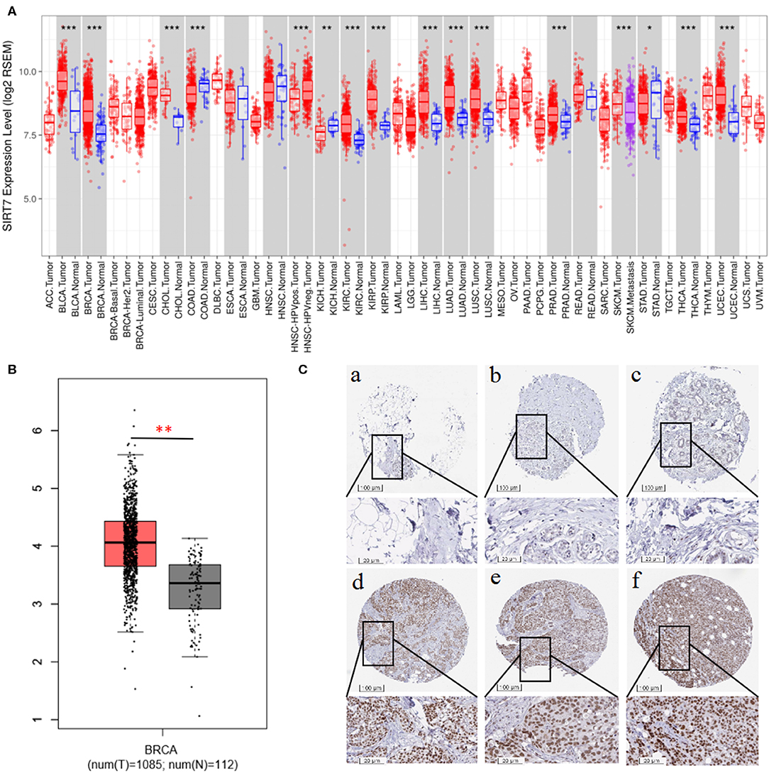
Figure 1. (A) Human sirtuin (SIRT)7 expression levels in different tumor types from The Cancer Genome Atlas (TCGA) database were determined using Tumor Immune Estimation Resource (TIMER). Compared with normal tissues, SIRT7 was upregulated in breast, bladder urothelial carcinoma (BLCA), cholangiocarcinoma (CHOL), kidney renal clear cell carcinoma (KIRC), kidney renal papillary cell carcinoma (KIRP), liver hepatocellular carcinoma (LIHC), lung adenocarcinoma (LUAD), lung squamous cell carcinoma (LUSC), prostate adenocarcinoma (PRAD), thyroid carcinoma (THCA), and uterine corpus endometrial carcinoma (UCEC) and downregulated in colon adenocarcinoma (COAD) and kidney chromophobe (KICH) cancers. (B) Gene Expression Profiling Interaction Analysis (GEPIA) for the expression of SIRT7 in tumor tissues and normal tissues. TCGA data revealed that SIRT7 mRNA was significantly higher in breast cancer tissues (1,085 cases) than that in normal tissues (112 cases). (C) SIRT7 expression in breast cancer tissues and normal tissues from the Human Protein Atlas (HPA) database. Immunohistochemical staining revealed that SIRT7 exhibited low expression in normal tissue samples (a–c) and high expression in tumor tissues (d–f). p-value significant codes: 0 ≤ *** < 0.001 ≤ ** < 0.01 ≤ * < 0.05 ≤. < 0.1.
To validate the results in breast cancer, GEPIA was used to analyze 1,197 samples from the TCGA database. As shown in Figure 1B, TCGA data revealed that SIRT7 mRNA was significantly higher in breast cancer tissues (1,085 cases) than that in normal tissues (112 cases) (p < 0.01), which is consistent with the TIMER database. In addition, we used the antibody HPA053669 of SIRT7 to analyze the immunohistochemical results of normal and tumor tissues using HPA and found that the protein expression and antibody staining level of three cases of breast cancer were moderate (Figure 1C). This further confirmed that the expression of SIRT7 in tumor tissues was significantly higher than that in normal tissues.
UALCAN is used to study gene expression levels based on TCGA data and clinical patient data. It is used not only to compare primary tumors and healthy tissue samples but also to compare clinical pathological parameters of patients based on pathological staging, tumor grade, and other clinical pathology features. We next investigated SIRT7 expression on the basis of patients' different clinical pathological parameters, such as sample type (normal/primary tumor), breast cancer subclass (luminal, HER2+, and triple-negative), cancer stage (stages 1, 2, 3, and 4), and menopause status (premenopause, perimenopause, and postmenopause) using UALCAN database. As shown in Figure 2A, the expression of SIRT7 in breast cancer samples was significantly higher than that in normal breast tissues (p < 10−12). An analysis of subclass showed that SIRT7 was higher expressed in different subclasses than that in normal breast tissues (normal vs. breast cancer-luminal, p < 10−12; normal vs. HER2-positive breast cancer, p < 10−6; normal vs. TNBC, p < 10−12). Compared with subtypes of luminal and HER2, SIRT7 was higher expressed in the three negative subtypes (luminal vs. TNBC, p < 10−4; HER2 vs. TNBC, p = 0.096) (Figure 2B). For cancer stages, the higher expression of SIRT7 in late-stage cancers compared to early stages suggests a possible role of SIRT7 in cancer progression and invasion (Figure 2C). Besides, as shown in Figure 2D, the expression of SIRT7 in postmenopause was higher than that in perimenopause (perimenopause vs. postmenopause, p = 0.04). SIRT7 expression in perimenopause was lower than that in premenopause (premenopause vs. perimenopause, p = 0.04).
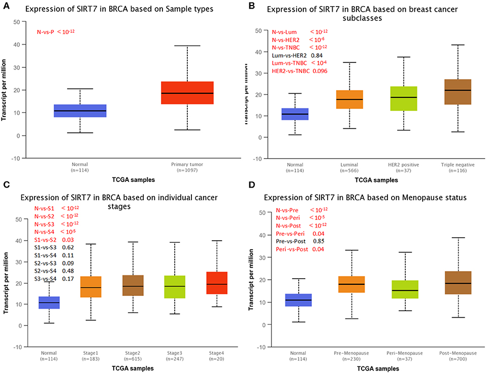
Figure 2. UALCAN analysis for the correlation between sirtuin (SIRT)7 mRNA expression level and clinicopathological parameters of breast cancer. (A) Sample type (normal/primary tumor). (B) Breast cancer subclass (luminal, HER2+, and triple negative). (C) Cancer stage (stages 1, 2, 3, and 4). (D) Menopause status (premenopause, perimenopause, and postmenopause). N, normal; P, primary tumor; Lum, luminal; HER2, HER2+; TNBC, triple negative; S1, stage 1; S2, stage 2; S3, stage 3; S4, stage 4; Pre, premenopause; Peri, perimenopause; Post, postmenopause; BRCA, breast cancer.
BC-GenExMiner 4.4 was used to explore the correlation between SIRT7 mRNA levels and the six common risk factors, namely, age, nodal status, ER status, PR status, HER2 receptor status, and triple-negative status (Table 1). Regarding age, SIRT7 mRNA expression was significantly higher in ≤51 years group than that in >51 years group (p = 5.20e-03; Figure 3A). The results showed that there were remarkably different expression levels of SIRT7 mRNA in nodal status (N+ > N–, p = 0.0004; Figure 3B), ER status (ER– > ER+, p < 0.0001; Figure 3C), PR status (PR– > PR+, p < 0.0001; Figure 3D), triple-negative status (TNBC > not TNBC, p = 0.0073; Figure 3F), respectively. However, no significant expression difference of SIRT7 mRNA was found in HER2 receptor status (Figure 3E). These results suggest that SIRT7 expression may serve as a potential diagnostic indicator in breast cancer.
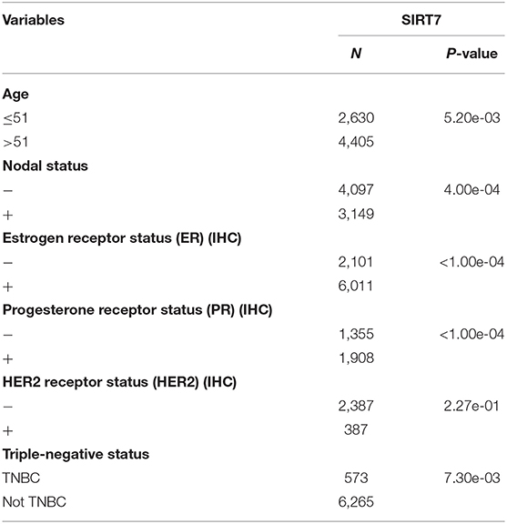
Table 1. Relationship between mRNA expression of SIRT7 and clinicopathological parameters of breast cancer.
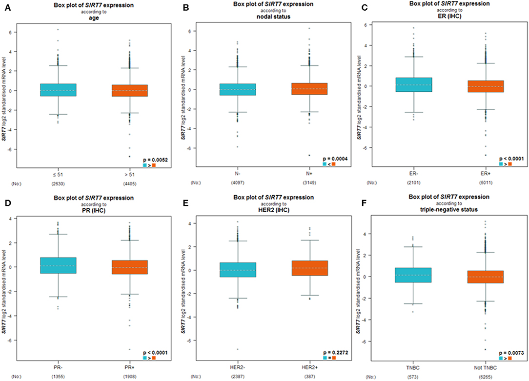
Figure 3. Differential expression levels of sirtuin (SIRT)7 mRNA in patients with breast cancer were performed by bc-GenExMiner v4.4 based on different types of classified parameters: (A) age (≤ 51 and >51 years), (B) nodal status (N+ vs. N−), (C–E) receptor status (ER+ vs. ER−, PR+ vs. PR−, HER2+ vs. HER2−), (F) triple-negative status (TNBC vs. Not TNBC).
The results showed that the expression of SIRT7 mRNA in the breast cancer group was higher than that in the normal group. Therefore, the relationship between SIRT7 transcription level and tumor prognosis warranted exploration. In this study, to determine whether SIRT7 can be used as a prognostic biomarker, we compared the expression levels of SIRT7 association with prognosis in different breast cancer subtypes (luminal A, luminal B, HER2+, and basal) using Kaplan-Meier survival curves. Notably, SIRT7 expression significantly affected the prognosis of different subtypes. In breast cancer-luminal A, high SIRT7 expression was associated with poor prognosis (OS: HR = 1.54, p = 1.70e-02; DMFS: HR = 1.56, p = 2.60e-03) (Figures 4A,B). In contrast, no significant correlation was observed between the expression of SIRT7 and the prognosis in breast cancer-luminal B, breast cancer-HER2+, and basal (Figures 4C–H). These findings indicate that SIRT7 expression may be a prognostic indicator of mortality risk in patients with breast cancer-luminal A.
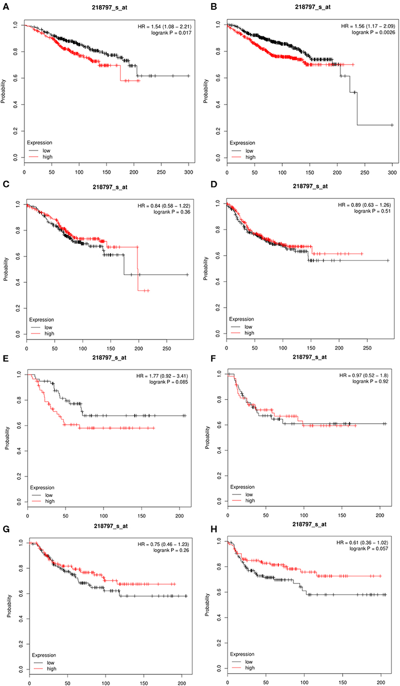
Figure 4. Kaplan-Meier survival curves comparing the high and low expressions of SIRT7 in different breast cancer subtypes (luminal A, luminal B, HER2+, and basal). (A,B) Survival curves of overall survival (OS) and distant metastasis-free survival (DMFS) in breast cancer-luminal A (n = 2,504). (C,D) Survival curves of OS and DMFS in breast cancer-luminal B (n = 1,425). (E,F) Survival curves of OS and DMFS in breast cancer-HER2+ (n = 335). OS and DMFS survival curves of breast cancer (n = 1,402, n = 1,747). (G,H) Survival curves of OS and DMFS in breast cancer-basal (n = 879).
TILs are independent predictors of sentinel lymph node status and survival in patients with cancer (34, 35). Studies showed that the levels of stroma and immune cells in tumor tissue may be related to the clinical features of the disease. Moreover, the infiltration of tumor-associated normal cells may affect the genomic analysis of clinical tumor samples (36). Therefore, we investigated whether the expression of SIRT7 was correlated with the immune infiltration levels in breast cancer using TIMER. Most of the homologous data in TIMER are from TCGA (25), which can be used to detect and quantify the immune infiltration levels in tumor tissues from the expression profile data of RNA-seq.
We used the TIMER database to study the correlation between SIRT7 and various immunocytochemical markers of immune cells (CD8+ T cells, T cells, B cells, monocytes, TAMs, M1 and M2 macrophages, neutrophils, natural killer cells, and dendritic cells) in different breast cancer subtypes (luminal, basal, and HER2+). Tumor purity is an important factor affecting immune infiltration analysis of clinical tumor samples (36). Therefore, we showed that SIRT7 expression is significantly related to tumor purity in different breast cancer subtypes. The results showed that the expression levels of SIRT7 were correlated with most markers of different immune cells in breast cancer subtypes (breast cancer-luminal, n = 611; breast cancer-basal, n = 139; breast cancer-HER2+, n = 67). Particularly, the expression levels of SIRT7 were significantly positively correlated with seven gene markers in breast cancer-luminal (Table 2), such as CD19 (r = 0.105, p = 1.46e-02), IRF5 (r = 0.165, p = 1.13e-04), KIR2DL1 (r = 0.106, p = 1.31e-02), T-bet (r = 0.103, p = 1.00e-02), FOXP3 (r = 0.12, p = 5.06e-03), PD1 (r = 0.134, p =1.74e-03), and GZMB (r = 0.108, p = 1.14e-02). However, the expression levels of SIRT7 were only significantly negatively correlated with two gene markers in breast cancer-luminal, such as VSIG4 (r = −0.108, p = 1.16e-02) and BDCA-1 (r = −0.114, p = 7.69e-03). These findings implicate that SIRT7 plays a specific role in immune cell infiltration, such as B cells, M1 macrophages, M2 macrophages, natural killer cells, dendritic cells, Th1 marker neutrophils, regulatory T cells, and T cell exhaustion, especially M1 macrophages and T cell exhaustion.
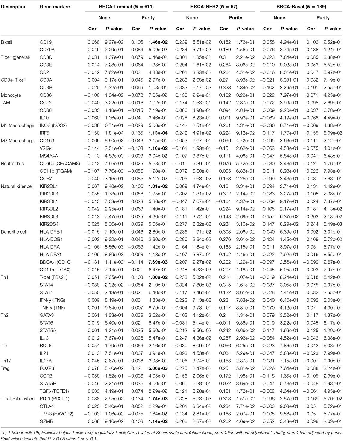
Table 2. Correlation analysis between SIRT7 and relate genes and markers of immune cells in breast cancer.
We investigated the relationship between SIRT7 expression and tumor-infiltrating of immune cells (M1 macrophages and T cell exhaustion) based on the expression level of immune marker genes in the TIMER databases (Figure 5A). We found that the expression of SIRT7 was positively correlated with the expression of IRF5 (M1 macrophages marker) and PD1 (PDCD1) (T cell exhaustion marker) in breast cancer-luminal. These results suggested that the expression of SIRT7 was related to M1 macrophages and T cell exhaustion infiltration.
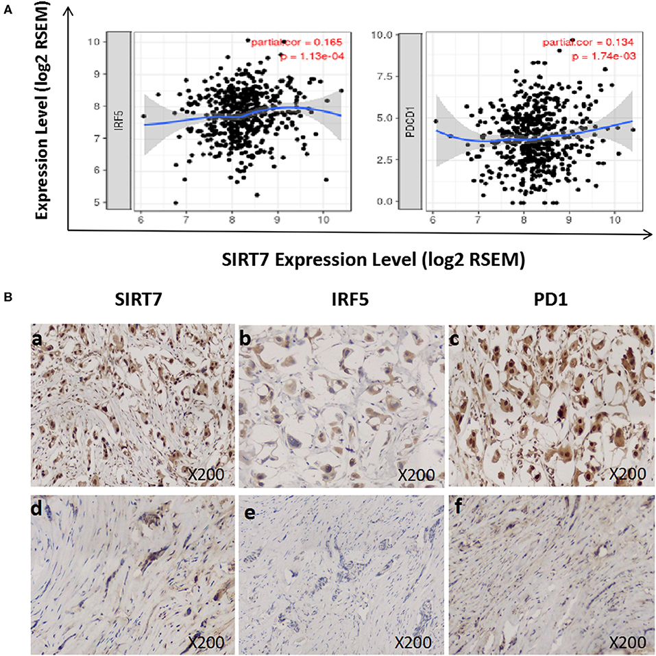
Figure 5. Correlation between sirtuin (SIRT)7 expression and immune infiltration levels of M1 macrophages and T cell exhaustion in breast cancer-luminal. (A) SIRT7 expression is significantly positively correlated with M1 macrophages (r = 0.165, p = 1.13e-04) and T cell exhaustion (r = 0.134, p = 1.74e-03). (B) Tumor infiltration of M1 macrophages and T cell exhaustion in breast cancer-luminal. The expression of SIRT7 (a: + + +, d: +). The expression of IRF5 (b: ++, e: –). The expression of PD1 (PDCD1) (c: + + +, f: +). The expression density of SIRT7, PD1, and IRF5 in breast cancer tissue was quantitated by scoring staining intensity, including negative (–) and weak (+) staining, moderate (++) and strong (+ + +) staining, respectively.
Next, we further analyzed the correlation between SIRT7 expression and these markers by immunohistochemistry, and tumor infiltration of M1 macrophages and T cell exhaustion were shown in Figure 5B. The levels of the expression were quantitated by scoring staining intensity, including negative (–) and weak (+) staining, moderate (++) and strong (+ + +) staining, respectively. We found that SIRT7 mainly localized in the nucleolus and nucleoplasm. SIRT7 showed strong expression in most breast cancer-luminal subtype samples, and few samples showed moderate expression. Interestingly, the expression levels of IRF5 and PD1 were high in SIRT7 strong expression samples, while the expression levels of IRF5 and PD1 were relatively low in SIRT7 weak expression samples. The results further revealed that IRF5 and PD1 tend to express in a positive correlation way, suggesting that the high expression of SIRT7 relates to high infiltration levels of M1 macrophages and T cell exhaustion.
In this study, an extensive bioinformatics examination was used to comprehensively analyze the SIRT7 expression pattern, investigate its association with breast cancer prognosis, and predict the correlation between SIRT7 expression and the immune infiltration levels of different immune cells in breast cancer-luminal.
SIRT7, a member of the SIRT family of NAD+-dependent protein deacetylases, is a key mediator of many cellular activities (37). The SIRT family is associated with malignant progression of tumors, and SIRT7 is supposed to be involved in lymph node metastasis of breast cancer (38). The expression of SIRT7 is related to cell growth. It is highly expressed in metabolically active cells, while it is lowly expressed or not expressed in non-proliferative cells (20, 39, 40). Some studies suggested the carcinogenic potential of SIRT7, whose expression levels were associated with several cancers, including breast, ovarian, and lung cancers, thereby classifying SIRT7 as a carcinogenic gene (41–44). SIRT7 is also a potential prognostic factor for breast cancer (45). However, the potential functions of SIRT7 in tumor progression and tumor immunology remain unclear.
We examined the expression levels of SIRT7 in breast cancer using TIMER, GEPIA, and HPA databases. We found that SIRT7 was highly expressed in breast cancer, which is consistent with Geng et al. (46). Some studies reported that age at diagnosis was an independent prognostic factor for breast cancer (47, 48), especially for breast cancer-luminal A. However, the correlation between age and SIRT7 expression remains unclear (49). In multivariate analysis, after adjusting for age, nodal status, ER, PR, HER2, and triple-negative status, we found that SIRT7 expression was significantly associated with age, nodal status, ER, PR, and triple-negative status. These results suggest that SIRT7 expression may be a potential diagnostic indicator of breast cancer. Different molecular types of breast cancer are associated with different prognostic survival of patients (50). Although SIRT7 expression is associated with poor prognosis in some cancers, such as colorectal cancer (51), the effect of SIRT7 expression on the prognosis of breast cancer subtypes is unclear. To definitively address this question, we classified breast cancer into three main subtypes, refer to the study of Polyak and Filho. (3). We believe that this classification is practical, straightforward, informative, and clinically useful, exhibiting considerable differences between subtypes. To determine whether SIRT7 can be used as a prognostic biomarker, we analyzed the expression of SIRT7 in different subtypes from the Kaplan-Meier plotter. There were significant differences in clinical and pathological features and prognosis between different subtypes. Notably, we found that the high expression of SIRT7 in breast cancer-luminal A was associated with poor prognosis, suggesting that the expression of SIRT7 will affect the prognosis of breast cancer.
Another important aspect of this study is that SIRT7 expression is associated with the immune infiltration levels in breast cancer-luminal. Several studies reported that TILs were currently considered to be biomarkers highly associated with breast cancer. Generally, it is recognized that the infiltration of TILs was related to the prognosis in breast cancer, and that adjuvant treatment is relatively effective (52, 53). In addition, several clinical studies evaluated the predictive importance of TILs for prognosis in breast cancer (54). However, the relationship between SIRT7 and immune infiltration in breast cancer-luminal remains unclear. Therefore, in this study, we comprehensively analyzed SIRT7 expression and associated immune infiltration and investigated the relationship between SIRT7 expression and immune infiltration levels in breast cancer-luminal. To the best of our knowledge, this study is the first to evaluate the association between SIRT7 expression and the levels of immune infiltration in breast cancer subtypes, especially in breast cancer-luminal. Our results demonstrated a positive correlation between SIRT7 expression and the levels of M1 macrophages (marker: IRF5) and T cell exhaustion (marker: PD1) infiltration. Macrophages are a group of differentiated immune cells and classify as M1 macrophages and M2 macrophages (55). They play an important role in development, homeostasis, and immunity (56). IRF5 is a marker of M1 macrophages and plays the role of regulating tumor infiltration (57). Previous studies showed that IRF5 changed the immune microenvironment of tumors by regulating the expression of pro-inflammatory and anti-inflammatory cytokines/chemokines in breast cancer (58, 59). Our studies observed that an increase in SIRT7 expression was correlated with the M1 macrophage marker IRT5. It suggests that there is a specific correlation between SIRT7 expression and immune infiltrating of M1 macrophages. Furthermore, SIRT7 expression was significantly correlated with T cell exhaustion markers such as PD-1. It is known that PD1/PDL1 are important immune checkpoint components, which mainly regulate the function of tumor cells and TILs (60), and the PD1/PDL1 axis was proved to be a promising therapeutic target in aggressive breast cancers (61). Moreover, Woo et al. (62) reported that PD-1 might play an important role in the tolerance of tumor antigens. These correlations may indicate the potential mechanism of SIRT7 regulating T cell functions. Taken together, these findings suggest that SIRT7 expression is related to the immune infiltration levels in breast cancer, especially breast cancer-luminal, providing a direction for further research.
A limitation is that our data do not include detailed information of body mass index, comorbidities, and treatment options. The data we collected in this study lack this sort of information, but we promise we will pay more attention to the integrity of patient baseline information in the following studies, and we will further confirm the estimated results in experiments and study the mechanism of this gene in breast tumor immunity.
In summary, the high expression of SIRT7 indicates poor prognosis in breast cancer-luminal, as well as increased immune infiltration levels of M1 macrophages and T cell exhaustion, suggesting that SIRT7 may serve as a prognostic biomarker associated with immune infiltration in breast cancer-luminal.
Publicly available datasets were analyzed in this study. This data can be found here: https://cistrome.shinyapps.io/timer/, https://www.Proteinatlas.org/, http://ualcan.path.uab.edu/, http://bcgenex.centregauducheau.fr/, http://kmplot.com/analysis/, http://gepia.cancer-pku.cn/.
QH and ZL conceived the project, participated in data analysis and wrote the manuscript, they contributed equally to this work. LC and FY participated in discussion and language editing. LC and NX reviewed the manuscript. LC supervised this project.
This project was supported by the Natural Science Foundation of National (81972003), the Natural Science Foundation of Guangdong (2017A030313668), the Sanming Project of Medicine in Shenzhen (SZSM201612031), and the Shenzhen Municipal Government of China (JCYJ20170817171808368, JCYJ20170818085657917, JCYJ20180507184647104, KQTD20170810160226082).
The authors declare that the research was conducted in the absence of any commercial or financial relationships that could be construed as a potential conflict of interest.
1. Desantis C, Ma J, Bryan L, Jemal A. Breast cancer statistics, 2013. CA Cancer J Clin. (2014) 64:52–62. doi: 10.3322/caac.21203
2. Jin L, Han B, Siegel E, Cui Y, Giuliano A, Cui X. Breast cancer lung metastasis: molecular biology and therapeutic implications. Cancer Biol Ther. (2018) 19:858–68. doi: 10.1080/15384047.2018.1456599
3. Polyak K, Filho MO. Snapshot: breast cancer. Cancer Cell. (2012) 22:562.e1. doi: 10.1016/j.ccr.2012.06.021
4. Tang X, Shi L, Xie N, Liu Z, Qian M, Meng F, et al. SIRT7 antagonizes TGF-β signaling and inhibits breast cancer metastasis. Nat Commun. (2017) 8:318. doi: 10.1038/s41467-017-00396-9
5. Perez EA. Treatment strategies for advanced hormone receptor-positive and human epidermal growth factor 2-negative breast cancer: the role of treatment order. Drug Resist Update. (2016) 24:13–22. doi: 10.1016/j.drup.2015.11.001
6. Yang F, Chen S, He S, Huo Q, Hu Y, Xie N. YB-1 interplays with ER E1 interplays witstemness and differentiation of ER-positive breast cancer stem cells. Theranostics. (2020) 10:3816-32. doi: 10.7150/thno.41014
7. De Andrea CE, Schalper KA, Sanmamed MF, Melero I. Immunodivergence in metastatic colorectal Cancer. Cancer Cell. (2018) 34:876–8. doi: 10.1016/j.ccell.2018.11.012
8. Fridman WH, Pagès F, Sautès-Fridman C, Galon J. The immune contexture in human tumours: impact on clinical outcome. Nat Rev Cancer. (2012) 12:298–306. doi: 10.1038/nrc3245
9. Azim HA Jr, Vingiani A, Peccatori F, Viale G, Loi S, Pruneri G. Tumour infiltrating lymphocytes (TILs) in breast cancer during pregnancy. Breast. (2015) 24:290–3. doi: 10.1016/j.breast.2015.01.009
10. Murray D, Hreno A, Dutton J, Hampson LG. Prognosis in colon cancer: a pathologic reassessment. Arch Surg. (1975) 110:908–13. doi: 10.1001/archsurg.1975.01360140052011
11. Vaziri Fard E, Ali Y, Wang XI, Saluja K, Covinsky HM, Wang L, et al. Tumor-infiltrating lymphocyte volume is a better predictor of disease-free survival than stromal tumor-infiltrating lymphocytes in invasive breast Carcinoma. Am J Clin Pathol. (2019) 152:656–65. doi: 10.1093/ajcp/aqz088
12. Okada K, Komuta K, Hashimoto S, Matsuzaki S, Kanematsu T, Koji T. Frequency of apoptosis of tumor-infiltrating lymphocytes induced by fas counterattack in human colorectal carcinoma and its correlation with prognosis. Clin Cancer Res. (2005) 6:3560–4.
13. Puccetti L, Manetti R, Parronchi P, Piccinni MP, Mavilia C, Carini M, et al. Role of low nuclear grading of renal carcinoma cells in the functional profile of tumor-infiltrating T cells. Int J Cancer. (2002) 98:674–81. doi: 10.1002/ijc.10238
14. Baker K, Lachapelle J, Zlobec, Bismar TA, Terracciano L, Foulkes WD. Prognostic significance of CD8+ T lymphocytes in breast cancer depends upon both oestrogen receptor status and histological grade. Histopathology. (2011) 58:1107–16. doi: 10.1111/j.1365-2559.2011.03846.x
15. Robinson BD, Sica GL, Liu YF, Rohan TE, Gertler FB, Condeelis JS, et al. Tumor microenvironment of metastasis in human breast Carcinoma: a potential prognostic marker linked to hematogenous dissemination. Clin Cancer Res. (2009) 15:2433–41. doi: 10.1158/1078-0432.CCR-08-2179
16. Mahmoud SM, Paish EC, Powe DG, Macmillan RD, Grainge MJ, Lee AH, et al. Tumor-Infiltrating CD8+ lymphocytes predict clinical outcome in breast Cancer. J Clin Oncol. (2011) 29:1949–55. doi: 10.1200/JCO.2010.30.5037
17. Liu S, Foulkes WD, Leung S, Gao D, Lau S, Kos Z, et al. Prognostic significance of FOXP3+ tumor infiltrating lymphocytes in breast cancer depends on estrogen receptor and humane pidermal growth factor receptor-2 expression status and concurrent cytotoxic T-cell infiltration. Breast Cancer Res. (2014) 16:432. doi: 10.1186/s13058-014-0432-8
18. Macchetti AH, Marana HR, Silva JS, de Andrade JM, Ribeiro-Silva A, Bighetti S. Tumor-infiltrating CD4+ T lymphocytes in early breast cancer reflect lymph node involvement. Clinics. (2006) 61:203–8. doi: 10.1590/S1807-59322006000300004
19. Kim RS, Song N, Gavin PG, Salgado R, Bandos H, Kos Z, et al. Stromal tumor-infiltrating lymphocytes in NRG oncology/NSABP B-31 adjuvant trial for early-stage HER2-positive breast cancer. J Natl Cancer Inst. (2019) 111:867–71. doi: 10.1093/jnci/djz032
20. Kiran S, Chatterjee N, Singh S, Kaul SC, Wadhwa R, Ramakrishna G. Intracellular distribution of human SIRT7 and mapping of the nuclear/nucleolar localization signal. FEBS J. (2013) 280:3451–66. doi: 10.1111/febs.12346
21. Cha YI, Kim HS. Emerging role of sirtuins on tumorigenesis: possible link between aging and cancer. BMB Rep. (2013) 46:429–38. doi: 10.5483/BMBRep.2013.46.9.180
22. Pan JH, Zhou H, Cooper L, Huang JL, Zhu SB, Zhao XX, et al. LAYN is a prognostic biomarker and correlated with immune infiltrates in gastric and colon Cancers. Front Immunol. (2019) 10:6. doi: 10.3389/fimmu.2019.00006
23. Lee M, Tayyari F, Pinnaduwage D, Bayani J, Bartlett JMS, Mulligan AM, et al. Tumoral BRD4 expression in lymph node-negative breast cancer: association with T-bet+ tumor-infiltrating lymphocytes and disease-free survival. BMC Cancer. (2018) 18:750. doi: 10.1186/s12885-018-4653-6
24. Williams JB, Horton BL, Zheng Y, Duan Y, Powell JD, Gajewski TF. The EGR2 targets LAG-3 and 4-1BB describe and regulate dysfunctional antigen-specific CD8+ T cells in the tumor microenvironment. J Exp Med. (2017) 214:381–400. doi: 10.1084/jem.20160485
25. Li T, Fan J, Wang B, Traugh N, Chen Q, Liu JS, et al. TIMER: a web server for comprehensive analysis of tumor-infiltrating immune cells. Cancer Res. (2017) 77:e108–10. doi: 10.1158/0008-5472.CAN-17-0307
26. Tang Z, Li C, Kang B, Gao G, Li C, Zhang Z. GEPIA: a web server for cancer and normal gene expression profiling and interactive analyses. Nucleic Acids Res. (2017) 45:W98–102. doi: 10.1093/nar/gkx247
27. Tang Z, Li C, Kang B, Gao G, Li C, Zhang Z. The human protein atlas: a spatial map of the human proteome. Protein Sci. (2018) 27:233–44. doi: 10.1002/pro.3307
28. Chandrashekar DS, Bashel B, Balasubramanya SAH, Creighton CJ, Ponce-Rodriguez I, Chakravarthi BVSK, et al. UALCAN: a portal for facilitating tumor subgroup gene expression and survival analyses. Neoplasia. (2017) 19:649–58. doi: 10.1016/j.neo.2017.05.002
29. Jézéquel P, Campone M, Gouraud W, Guérin-Charbonnel C, Leux C, Ricolleau G, et al. Bc-GenExMiner: an easy-to-use online platform for gene prognostic analyses in breast cancer. Breast Cancer Res Treat. (2012) 131:765–75. doi: 10.1007/s10549-011-1457-7
30. Jézéquel P, Frénel JS, Campion L, Guérin-Charbonnel C, Gouraud W, Ricolleau G, et al. Bc-GenExMiner 3.0: new mining module computes breast cancer gene expression correlation analyses. Database. (2013) 2013:bas060. doi: 10.1093/database/bas060
31. Wu HT, Liu J, Li GW, Shen JX, Huang YT. The transcriptional STAT3 is a potential target, whereas transcriptional STAT5A/5B/6 are new biomarkers for prognosis in human breast carcinoma. Oncotarget. (2017) 8:36279–88. doi: 10.18632/oncotarget.16748
32. Lánczky A, Nagy Á, Bottai G, Munkácsy G, Szabó A, Santarpia L, et al. miRpower: a web-tool to validate survival-associated miRNAs utilizing expression data from 2178 breast cancer patients. Breast Cancer Res Treat. (2016) 160:439–46. doi: 10.1007/s10549-016-4013-7
33. Zhang Y, Yuan Y, Liang P, Zhang Z, Guo X, Xia L, et al. Overexpression of a novel candidate oncogene KIF14 correlates with tumor progression and poor prognosis in prostate cancer. Oncotarget. (2017) 8:45459–69. doi: 10.18632/oncotarget.17564
34. Ohtani H. Focus on TILs: prognostic significance of tumor infiltrating lymphocytes in human colorectal cancer. Cancer Immun. (2007) 7:4.
35. Azimi F, Scolyer RA, Rumcheva P, Moncrieff M, Murali R, McCarthy SW, et al. Tumor-infiltrating lymphocyte grade is an independent predictor of sentinel lymph node status and survival in patients with cutaneous melanoma. J Clin Oncol. (2012) 30:2678–83. doi: 10.1200/JCO.2011.37.8539
36. Yoshihara K, Shahmoradgoli M, Martínez E, Vegesna R, Kim H, Torres-Garcia W, et al. Inferring tumour purity and stromal and immune cell admixture from expression data. Nat Commun. (2013) 4:2612. doi: 10.1038/ncomms3612
37. Blank MF, Grummt I. The seven faces of SIRT7. Transcription. (2017) 8:67–74. doi: 10.1080/21541264.2016.1276658
38. Ashraf N, Zino S, Macintyre A, Kingsmore D, Payne AP, George WD, et al. Altered sirtuin expression is associated with node-positive breast cancer. Br J Cancer. (2006) 95:1056–61. doi: 10.1038/sj.bjc.6603384
39. Michishita E, Park JY, Burneskis JM, Barrett JC, Horikawa I. Evolutionarily conserved and nonconserved cellular localizations and functions of human SIRT proteins. Mol Biol Cell. (2005) 16:4623–35. doi: 10.1091/mbc.e05-01-0033
40. Grob A, Roussel P, Wright JE, McStay B, Hernandez-Verdun D, Sirri V. Involvement of SIRT7 in resumption of rDNA transcription at the exit from mitosis. J Cell Sci. (2009) 122:489–98. doi: 10.1242/jcs.042382
41. Kim JK, Noh JH, Jung KH, Eun JW, Bae HJ, Kim MG, et al. Sirtuin7 oncogenic potential in human hepatocellular carcinoma and its regulation by the tumor suppressors MiR-125a-5p and MiR- 125b. Hepatology. (2013) 57:1055–67. doi: 10.1002/hep.26101
42. Aljada A, Saleh AM, Alkathiri M, Shamsa HB, Al-Bawab A, Nasr A. Altered sirtuin 7 expression is associated with early stage breast cancer. Breast Cancer. (2015) 9:3–8. doi: 10.4137/BCBCR.S23156
43. Wang HL, Lu RQ, Xie SH, Zheng H, Wen XM, Gao X, et al. SIRT7 exhibits oncogenic potential in human ovarian cancer cells. Asian Pac J Cancer Prev. (2015) 16:3573–7. doi: 10.7314/APJCP.2015.16.8.3573
44. Shi H, Ji Y, Zhang D, Liu Y, Fang P. MicroRNA-3666-induced suppression of SIRT7 inhibits the growth of non-small cell lung cancer cells. Oncol Rep. (2016) 36:3051–7. doi: 10.3892/or.2016.5063
45. Zino S, Moran CP, Mcglynn LM, George W, Payne AP, Shiels PG. SIRT7 expression and epigenetic status determine variability in telomere length in breast cancer. Cancer Res. (2009) 69(2 Suppl.):4052. doi: 10.1158/0008-5472.SABCS-4052
46. Geng Q, Peng H, Chen F, Luo R, Li R. High expression of Sirt7 served as a predictor of adverse outcome in breast cancer. Int J Clin Exp Pathol. (2015) 8:1938–45.
47. Alieldin NH, Abo-Elazm OM, Bilal D, Salem SE, Gouda E, Elmongy M, et al. Age at diagnosis in women with non-metastatic breast cancer: is it related to prognosis? J Egypt Natl Canc Inst. (2014) 26:23–30. doi: 10.1016/j.jnci.2013.08.005
48. Liedtke C, Rody A, Gluz O, Baumann K, Beyer D, Kohls EB, et al. The prognostic impact of age in different molecular subtypes of breast cancer. Breast Cancer Res Treat. (2015) 152:667–73. doi: 10.1007/s10549-015-3491-3
49. Liu Z, Sahli Z, Wang Y, Wolff AC, Cope LM, Umbricht CB. Young age at diagnosis is associated with worse prognosis in the Luminal A breast cancer subtype: a retrospective institutional cohort study. Breast Cancer Res Treat. (2018) 172:689–702. doi: 10.1007/s10549-018-4950-4
50. Zhao J, Liu H, Wang M, Gu L, Guo X, Gu F, et al. Characteristics and prognosis for molecular breast cancer subtypes in Chinese women. J Surg Oncol. (2010) 100:89–94. doi: 10.1002/jso.21307
51. Yu H, Ye W, Wu J, Meng X, Liu RY, Ying X, et al. Overexpression of sirt7 exhibits oncogenic property and serves as a prognostic factor in colorectal Cancer. Clin Cancer Res. (2014) 20:3434–45. doi: 10.1158/1078-0432.CCR-13-2952
52. Cimino-Mathews A, Ye X, Meeker A, Argani P, Emens LA. Metastatic triple-negative breast cancers at first relapse have fewer tumor-infiltrating lymphocytes than their matched primary breast tumors: a pilot study. Hum Pathol. (2013) 44:2055–63. doi: 10.1016/j.humpath.2013.03.010
53. Park IH, Kong SY, Ro JY, Kwon Y, Kang JH, Mo HJ, et al. Prognostic implications of tumor-infiltrating lymphocytes in association with programmed death ligand 1 expression in early-stage breast cancer. Clin Breast Cancer. (2016) 16:51–8. doi: 10.1016/j.clbc.2015.07.006
54. Ioannides CG, Rashed S, Fisk B, Fan D, Itoh K, Freedman RS. Lymphocytes infiltrating ovarian malignant ascites: modulation of IL-2-induced proliferation by IL-4 and of selective increase in CD8+ T cells by TNF-α. Lymphokine Cytokine Res. (1991) 10:307–15.
55. Jayasingam SD, Citartan M, Thang TH, Mat Zin AA, Ang KC, Ch'ng ES. Evaluating the polarization of tumor-associated macrophages into M1 and M2 phenotypes in human cancer tissue: technicalities and challenges in routine clinical practice. Front Oncol. (2019) 9:512. doi: 10.3389/fonc.2019.01512
56. Wynn TA, Chawla A, Pollard JW. Macrophage biology in development, homeostasis and disease. Nature. (2013) 496:445–55. doi: 10.1038/nature12034
57. Li D, De S, Li D, Song S, Matta B, Barnes BJ. Specific detection of interferon regulatory factor 5 (IRF5): a case of antibody inequality. Sci Rep. (2016) 6:31002. doi: 10.1038/srep31002
58. Garaud S, Willard-Gallo K. IRF5: a rheostat for tumor-infiltrating lymphocyte trafficking in breast cancer? Immunol Cell Biol. (2015) 93:425–6. doi: 10.1038/icb.2015.39
59. Pimenta EM, Barnes BJ. Abstract A79: regulation of the breast tumor microenvironment by interferon regulatory factor 5 (IRF5). Tum Immunol. (2013) 73(1 Suppl.):A79. doi: 10.1158/1538-7445.TUMIMM2012-A79
60. Dong H, Strome SE, Salomao DR, Tamura H, Hirano F, Flies DB, et al. Tumor-associated B7-H1 promotes T-cell apoptosis: a potential mechanism of immune evasion. Nat Med. (2002) 8:793–800. doi: 10.1038/nm730
61. Bertucci F, Finetti P, Birnbaum D, Mamessier E. The PD1/PDL1 axis, a promising therapeutic target in aggressive breast cancers. OncoImmunology. (2015) 5:e1085148. doi: 10.1080/2162402X.2015.1085148
Keywords: sirtuin 7 (SIRT7), gene expression, tumor-infiltrating, prognosis, breast cancer
Citation: Huo Q, Li Z, Cheng L, Yang F and Xie N (2020) SIRT7 Is a Prognostic Biomarker Associated With Immune Infiltration in Luminal Breast Cancer. Front. Oncol. 10:621. doi: 10.3389/fonc.2020.00621
Received: 02 October 2019; Accepted: 03 April 2020;
Published: 12 May 2020.
Edited by:
Fang-Ping Huang, Shenzhen University, ChinaReviewed by:
Guo-Jun Zhang, Xiamen University, ChinaCopyright © 2020 Huo, Li, Cheng, Yang and Xie. This is an open-access article distributed under the terms of the Creative Commons Attribution License (CC BY). The use, distribution or reproduction in other forums is permitted, provided the original author(s) and the copyright owner(s) are credited and that the original publication in this journal is cited, in accordance with accepted academic practice. No use, distribution or reproduction is permitted which does not comply with these terms.
*Correspondence: Ni Xie, eG4xMDBAc3p1LmVkdS5jbg==
†These authors have contributed equally to this work
Disclaimer: All claims expressed in this article are solely those of the authors and do not necessarily represent those of their affiliated organizations, or those of the publisher, the editors and the reviewers. Any product that may be evaluated in this article or claim that may be made by its manufacturer is not guaranteed or endorsed by the publisher.
Research integrity at Frontiers
Learn more about the work of our research integrity team to safeguard the quality of each article we publish.