 Dylan Dahan1†
 Regina Lamendella
 Gabriel G. Perron
95% of researchers rate our articles as excellent or good
Learn more about the work of our research integrity team to safeguard the quality of each article we publish.
Find out more
ORIGINAL RESEARCH article
Front. Microbiol. , 21 June 2018
Sec. Microbial Symbioses
Volume 9 - 2018 | https://doi.org/10.3389/fmicb.2018.01323
Exposure to environmental toxins such as heavy metals can perturb the development and stability of microbial communities associated with human or animal hosts. Widespread arsenic contamination in rivers and riparian habitats therefore presents environmental and health concerns for populations living near sources of contamination. To investigate how arsenic affects host microbiomes, we sequenced and characterized the microbiomes of twenty larval zebrafish exposed to three concentrations of arsenic that are found in contaminated water—low (10 ppb), medium (50 ppb), and high (100 ppb) for 20 days. We found that even a small concentration of arsenic changed the overall microbial composition, structure and diversity of microbial communities, causing dysbiosis in developing larval zebrafish microbiota. In addition, we found that a high concentration of arsenic also increased the abundance of a class 1 integron, an integrase-dependent system facilitating the horizontal transfer of genes conferring resistance to heavy metals and antibiotics.
Arsenic, a metalloid element that can cause poisoning due the inhibition of acetyl-CoA and succinic dehydrogenase, is naturally present in many aquifers used for drinking water (Mukherjee and Bhattacharya, 2001; Bhadury, 2014). Since industrialization, however, runoff produced from mining, agricultural and industrial waste has vastly increased arsenic contamination in the environment (Sarkar and Bhattacharya, 2002). In humans, extended exposure to arsenic contamination can result in arsenicosis, which in turn can lead to skin cancer, keratosis, or important metabolic diseases such as diabetes (Guha Mazumder et al., 1998; Saha et al., 2003; Navas-Acien et al., 2008; Martinez et al., 2011). The effects of arsenic contamination have been extensively documented in Bangladesh, where up to 65 million people are exposed to drinking water with high concentrations of arsenic far exceeding the limit set by the World Health Organization (i.e., 50 ppb) or by local government (i.e., 10 ppb) (Smith et al., 2000; Mukherjee and Bhattacharya, 2001; World Health Organization, 2001). Still, more than seventy countries, including the United States, reported severe cases of arsenic poisoning in the past decades (Smedley and Kinniburgh, 2002; Saha et al., 2003), making arsenic contamination one of the principal environmental causes of cancer in humans (Hughes et al., 2011).
In addition to causing arsenicosis, arsenic in drinking water can also impact microbial communities associated with exposed hosts, also known as microbiota, or microbiomes (Palmer et al., 2007; de Theije et al., 2014; Narrowe, 2015; Claus et al., 2016). Previous studies showed that adult mice exposed to arsenic present important changes in their gastrointestinal intestinal (GI) tract microbiota (Lu et al., 2014; Dheer et al., 2015). Indeed, high concentrations of arsenic inhibit the growth of many bacterial species common in the GI (Summers et al., 1993; Lu et al., 2014). Arsenic can also alter hosts' microbiota by supporting the growth of arsenic-metabolizing bacteria such as Sediminibacterium and Methylobacterium (Oremland and Stolz, 2005; Mateos et al., 2006; Liao et al., 2011; Andres and Bertin, 2016). Such bacteria can metabolize arsenite or arsenate, often producing arginine metabolites toxic to the host (Dheer et al., 2015).
Microbial communities associated with hosts carry multiple functions that are essential to the hosts (Yan et al., 2016; Raymann and Moran, 2018). For this reason, microbiome aberrations, also called dysbiosis, are linked to disease occurrence in hosts ranging from diabetes and cardiovascular diseases to allergies and inflammatory diseases among other things (Turnbaugh et al., 2006; Foxx-Orenstein and Chey, 2012; Kong et al., 2012; Olszak et al., 2012; Qin et al., 2012). Because microbiomes are highly variable and are least stable during early developmental stages (As discussed by Palmer et al., 2007; Costello et al., 2012; Vallès et al., 2014), the effects of contaminants such as arsenic on dysbiosis are likely to be accentuated during early developmental stages (Neu and Rushing, 2011; Torrazza and Neu, 2011; Olszak et al., 2012). Investigating the effects of arsenic on microbial communities during the initial microbial establishment within hosts is thus crucial to predict outcomes on host health.
In this study, we investigate the potential effects of arsenic contamination on the developing microbiota of zebrafish (Danio rerio). In addition to being a robust model organism for the study of the microbiome and development in vertebrates (Veldman and Lin, 2008; Roeselers et al., 2011; Fritz et al., 2013; Kostic et al., 2013), zebrafish are native to arsenic-contaminated regions of Bangladesh and West Bengal (Spence et al., 2006). We exposed experimental populations of larval zebrafish to 10 ppb, 50 ppb, and 100 ppb sodium meta-arsenite for 20-days and used targeted 16S rRNA gene sequencing to characterize the fish microbiota after exposure. Additionally, we quantified the abundance of int1, a type 1 integron associated with the presence of antimicrobial resistance genes, in tested microbiomes. Our approach not only investigates how an environmental contaminant alters communities of microbes in developing hosts, but also how such contaminants may contribute to the spread of antibiotic resistance.
Zebrafish (Danio rerio) were maintained in the laboratories of the Biology Department of Bard College in accordance with standard protocols for zebrafish husbandry (Lawrence, 2007). Zebrafish strain Et20, a transgenic animal expressing GFP in the neuromast supporting cells (Moon et al., 2011), were raised in a 14-h light:10-h dark cycle in standard recirculating rack water kept at 28.5°C with pH ranging from 7.0 to 7.4 unless specified. Prior to starting the experiment, embryos (0 days post-fertilization—or dpf) from a single mating were bleached twice in 0.5% sodium hypochlorite solution for 4 min. Embryos were then pooled into four groups of 30–60 animals and housed in petri dishes filled with 1x E3 media (5 mM NaCl, 0.17 mM KCl, 0.33 mM CaCl2, 0.33 mM MgSO4, 0.5 mg/L methylene blue) and one of the following treatments: 0, 10, 50, or 100 ppb sodium meta-arsenite (NaAsO2; A3+). At 48 h intervals, 80% of the media was exchanged for fresh media and sodium meta-arsenite solution. This was done to minimize cross-contamination due to dead cell tissue and to maintain a constant concentration of arsenite throughout the experiment.
Following larval hatching, between 72 and 96 h post-fertilization (hpf), populations were transferred to respective sterile glass dishes with growth conditions and experimental conditions similar to those described above. Again, media and sodium meta-arsenite concentrations were changed every 48 h. Animals were fed generously three times daily with micro powder food containing rotifers and Paramecium. At 20 dpf, zebrafish were sacrificed using sterile tricaine methane sulfonate solution (250 mg/l) according to established euthanasia techniques (NIH, 2009; Matthews and Varga, 2012). Fish were removed from tricaine following cessation of opercular movement (~10 min) and rinsed several times with nuclease free water to minimize the presence of free-living bacteria. All protocols were approved by the Bard College Institutional Animal Care and Use Committee (IACUC; most recent approval ID “Perron 2018”).
For each treatment, microbial DNA was extracted and purified from five individual fish larvae using a modified protocol for the DNeasy Blood and Tissue kit (QIAGEN, Germantown, MD) described in Hang et al. (2014). Briefly, sacrificed fish were treated with an enzymatic lysis buffer (20 mM Tris HCL, pH8.0; 2 mM sodium EDTA; 1.2% Triton X-100; and 20 mg/ml egg lysozyme) and incubated at 37°C for 60 min. To reduce protein contamination, we added 25 μl of Proteinase K solution (10 mg/ml; Qiagen, Germantown, MD) combined to 200 μl of Buffer AL (Germantown, MD), which we incubated at 56°C for 120 min. Following larval lysis and protein degradation, we used the DNeasy Blood & Tissue kit (Qiagen, Germantown, MD) to purify DNA from individual larval zebrafish. When necessary, gDNA concentrations were increased by evaporation using the SpeedVac System (ThermoFisher Scientific, Asheville, NC). Purified genomic DNA (gDNA) samples were stored in nuclease free H2O at −20°C.
Fish microbiota were characterized via targeted gene amplification of the 16S rRNA V4 region using Golay-barcoded primers 515F and 806R as described by Caporaso et al. (2012). Duplicate 25 μl PCR reactions (12.5 μl NEB Taq 2X Master Mix, 5.5 μl nuclease free PCR water, 1.0 μl of each forward and reverse primers (0.4 μM final concentration), and 3.0 μl genomic DNA template) were cycled as follows: denaturation at 94°C for 3 min; 35 cycles for 94°C/45 s, 50°C/60 s, and 72°C/90 s; and a final extension at 72°C for 10 min to complete amplification. Libraries were then were gel-purified, pooled at equimolar ratios, and sequenced on the MiSeq paired-end Illumina platform adapted for 150-bp paired-end reads (Wright Labs, Juniata College, Huntingdon, PA). The unfiltered forward reads are available on the Sequence Read Archive of the National Center for Biotechnology Information (SRA: SRP082969).
Microbial communities were analyzed with two different microbiome pipelines to identify the presence and abundance of different microbial taxa based on the assembly of the 16S rRNA sequence reads. Before processing the 16S rRNA sequence data, phiX control reads were removed by mapping raw sequence reads against an indexed phiX genome provided by Illumina (San Diego, CA) using the Bowtie2 platform. Sequence reads were then processed, aligned, and categorized independently using either the DADA2 1.2 (Callahan et al., 2016: see scripts in Datasheet 1) or the QIIME 1.9.1 pipeline primarily using QIIME defaults parameters (Caporaso et al., 2010b).
The DADA2 microbiome pipeline (available at https://github.com/benjjneb/dada2) describes microbial communities using unique sequence variants present in the data, known as amplicon sequence variants (ASVs) (Callahan et al., 2016), rather than clustering groups of similar sequences into operational taxonomic units (OTUs) used by many other pipelines such as QIIME (Callahan et al., 2016). In brief, sequence reads were first filtered using DADA2's recommended parameters (i.e., an expected error threshold of 2 combined with the trimming of 10 nucleotides from the start and end of each read). Filtered reads were then de-replicated and de-noised using DADA2 default parameters. De-replication combines identical reads into unique sequences and constructs consensus quality profiles for each combined lot of sequences; the consensus quality profiles then inform the de-noising algorithm which infers error rates from samples and removes identified sequencing errors from the samples. After building the ASV table and removing chimeras, taxonomy was assigned using the Ribosomal Database Project (RDP) classifier (v2.2) (Wang et al., 2007) natively implemented in DADA2 and trained against the Greengenes reference database (13.8) (Werner et al., 2011) (McDonald et al., 2011). A phylogenetic tree was built using FastTree (v2.1.3) (Price et al., 2010) from a multiple sequence alignment made with the PyNAST alignment tool (Caporaso et al., 2010a), against the Greengenes Core reference alignment (DeSantis et al., 2006). Lastly, we pruned sequences positively mapping to zebrafish mitochondrial gDNA (GenBank: KT624622.1) using a BLAST alignment.
For comparison purposes, we also processed the raw reads using QIIME 1.9.1 (available at https://github.com/biocore/qiime) as described elsewhere (Caporaso et al., 2010b; Edgar et al., 2011). We first quality-filtered reads using default parameters, removing reads with Phred quality scores below 30, and removed chimeric sequences using UCHIME (Edgar, 2010; Edgar et al., 2011). The OTU table was built using the uclust method (Edgar, 2010) at a ≥97% identity threshold and the open reference picking method using the PyNAST sequence aligner against the Greengenes Core reference alignment (DeSantis et al., 2006; Caporaso et al., 2010a) for the closed reference alignment. Again, we pruned sequences identified as zebrafish mitochondria DNA.
Patterns of diversity within the ASV and OTU tables were analyzed using a custom bioinformatic pipeline implemented in R 3.2.3 (http://www.r-project.org) and described in Datasheet 2 (mapping file linking sample names and arsenic concentration is also included in Datasheet 3). First, we visualized phylum and family level community composition using phyloseq (v1.14.0) (available at https://joey711.github.io/phyloseq/) (McMurdie and Holmes, 2013) implemented in ggplot2 (v.2.0.0) (Wickham, 2009), with visualization optimized by filtering taxa at cutoffs of 0.1 and 0.3% for phylum and family respectively. Using the RNA-Seq DESeq2 differential abundance comparison (Love et al., 2014) adapted for use with microbial count data (McMurdie and Holmes, 2014), we investigated changes in community composition at the taxa level. This method stabilizes sample variance using an implementation for count data that is built on a negative binomial distribution, therefore allowing for the use of a non-normalized and not rarefied ASV table. This is preferred to rarefying since subsampling inflates variance, resulting in a loss of power and often producing false positive results (McMurdie and Holmes, 2014).
We then estimated alpha diversity metrics using phyloseq's estimate_richness function on rarefied ASV tables. More specifically, we estimated observes species S, i.e., observed ASVs or OTUs, Simpson's metric, Shannon diversity measurements H′, as well as Pielou's evenness index. Pielou's evenness metric was computed with the following equation (Pielou, 1966):
where is the maximum value of H′ if every species were equally abundant:
We compared statistical models using Akaike's Information Criterion (AIC) in R's base stats package.
Phyloseq was used to perform ordinations, where Principle Coordinate Analysis (PCoA) was employed on unweighted UniFrac distance scores (Lozupone and Knight, 2005). To confirm multivariate homogeneity of variances first among treatments and second within treatments, the vegan (v2.3.2) package implementation of PERMDISP2 via the betadisper method was used (Oksanen et al., 2015). To test the effect of arsenic as a continuous variable on group differences according to UniFrac scores, a permutational analysis of variance (PERMANOVA) using the adonis function as part of the vegan package was used (Oksanen et al., 2015). An analysis of similarity (ANOSIM) was conducted to compare the variation in UniFrac distances between a grouped arsenic distance matrix and control group distance matrix as again implemented in the vegan package.
We first tested the null hypothesis that the observed richness of OTUs and ASVs after processing remained the same by conducting a Wilcoxon test between sample counts. Differences between log-transformed observed richness of OTUs and ASVs from the same treatments were compared using an analysis of variance (ANOVA) with treatment condition as a factor, followed by Tukey's honest significant difference (HSD) tests. Differentially abundant ASVs and OTUs were identified using the DESeq2 package (Love et al., 2014), adapted for use with microbial community count data (McMurdie and Holmes, 2014), with a local fit type. When performing Procrustes rotation between matching points from ASV and OTU UniFrac distance matrices, the ade4 package (v.1.7.3) was used as implemented in R (Thioulouse and Dray, 2007). A PROcrustean randomization TEST (PROTEST) was conducted to test the significance of Procrustes matrix concordance again using the package ade4 package (Jackson, 1995). Data was visualized using ggplot2 (v.2.0.0) (Wickham, 2009). Finally, we used biom (v0.3.12) to handle BIOM formatted files, dada2 (v0.10.6) for read processing (Callahan et al., 2016), optparse (v1.3.2) to parse command line options, stats (v3.2.3) to conduct R's internal statistics, and data.table (v1.9.6) to handle data frames.
We quantified 16S rRNA and Int1 gene copy numbers using quantitative real-time PCR as described in Gaze et al. (2011). For each sample, triplicate PCR reactions using the PowerUp SYBR Green Master Mix (Applied Biosystems, Foster City, CA) were cycled using the Bio-Rad CFX96 Real-Time PCR Detection System (Bio-Rad Laboratories, Hercules, CA) with the following cycling conditions: Hot start at 50°C for 2 min; denaturation at 95°C for 10 min; followed by 40 cycles of 95°C/20 s and 60°C/60 s; with an added dissociation step. An internal standard curve constructed from a serial dilution of Escherichia coli SK4903, an engineered E. coli strains harboring seven 16S rRNA copies and six int1 copies, was processed with each qPCR run. The relative abundance of int1 genes was normalized to 16S rRNA copies of each sample before being analyzed via linear modeling with int1 relative abundance as response variable and arsenic concentrations as explanatory variable.
We characterized the microbiomes of twenty individual zebrafish larvae, five individuals per treatment (0, 10, 50, or 100 ppb of arsenic) by sequencing the V4 region of the 16S rRNA. We obtained a total of 10,108,755 forward reads with an average read length of 151 base pairs, totaling ~1.5 G bases. The median sequencing depth per sample was 497,470 reads. Reverse reads were of poor quality due to a sequencing error and were therefore removed from subsequent analyses along with one control sample. After filtering, denoising, and removing chimeras, we retained 8,678,233 (85.86% of initial) reads. With these reads, we assigned taxa (see Datasheet 4 for unfiltered representative ASVs) and plotted relative abundance of taxa across samples (Figure 1).
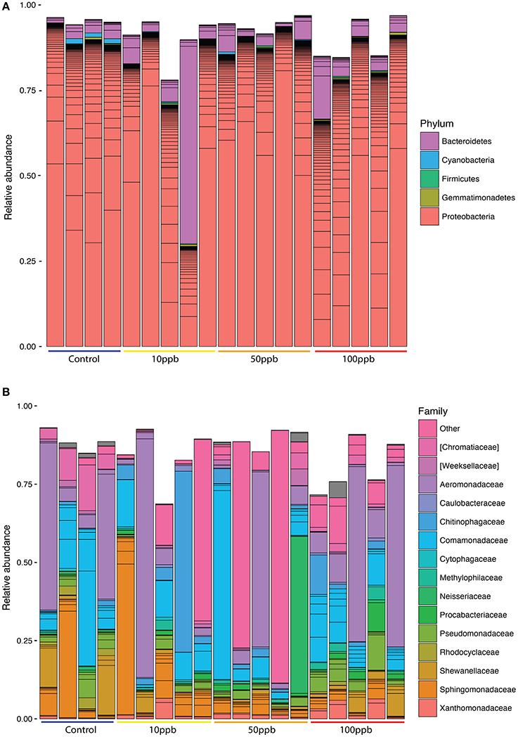
Figure 1. Relative abundance of taxa across treatments. (A) Phylum level classification, filtered to remove taxa below 0.001 abundance. (B) Family level classification, filtered to remove taxa below 0.003 abundance. Lines within same-colored bars are of different ASVs of the same classification. Category “Other” represents ASVs that were not classified to the family level. Grey areas are unclassified ASVs. Control n = 4; 10 ppb n = 5; 50 ppb n = 5; 100 ppb n = 5.
We identified 120 ASVs as part of the core microbiome among the four zebrafish sampled from the control population (Table 1; Table S1). Interestingly, we found that the core microbiome described in this study, even though extracted from the whole larvae, was compositionally similar to that of healthy zebrafish gut microbiomes either living in wild or laboratory conditions (Roeselers et al., 2011). This is most likely due to the fact that, as in animals (Sender et al., 2016), fish microbiota are most dense in the gastrointestinal tract.
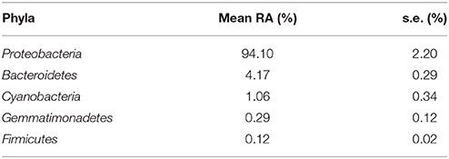
Table 1. Relative abundance (RA) for core phyla abundances ± s.e.
When comparing the composition of healthy fish microbiomes to that of fish exposed to arsenic, we found that a total of 78 ASVs significantly differed in abundance (DESeq2; adj-P ≤ 0.01; Figure 2; Table S2). ASVs that increased in abundance included genera such as Acinetobacter (base mean: 125), Sediminibacterium (base mean: 201) and Janthinobacterium (base mean: 315), whereas genera that decreased in abundance included Bdellovibrio (base mean: 110), and Pseudomonas (base mean: 131) among others. Interestingly a Legionellales ASV (ASV3) dominated the increase in the arsenic treatment (base mean: 8,464), but could not be identified at the genus level.
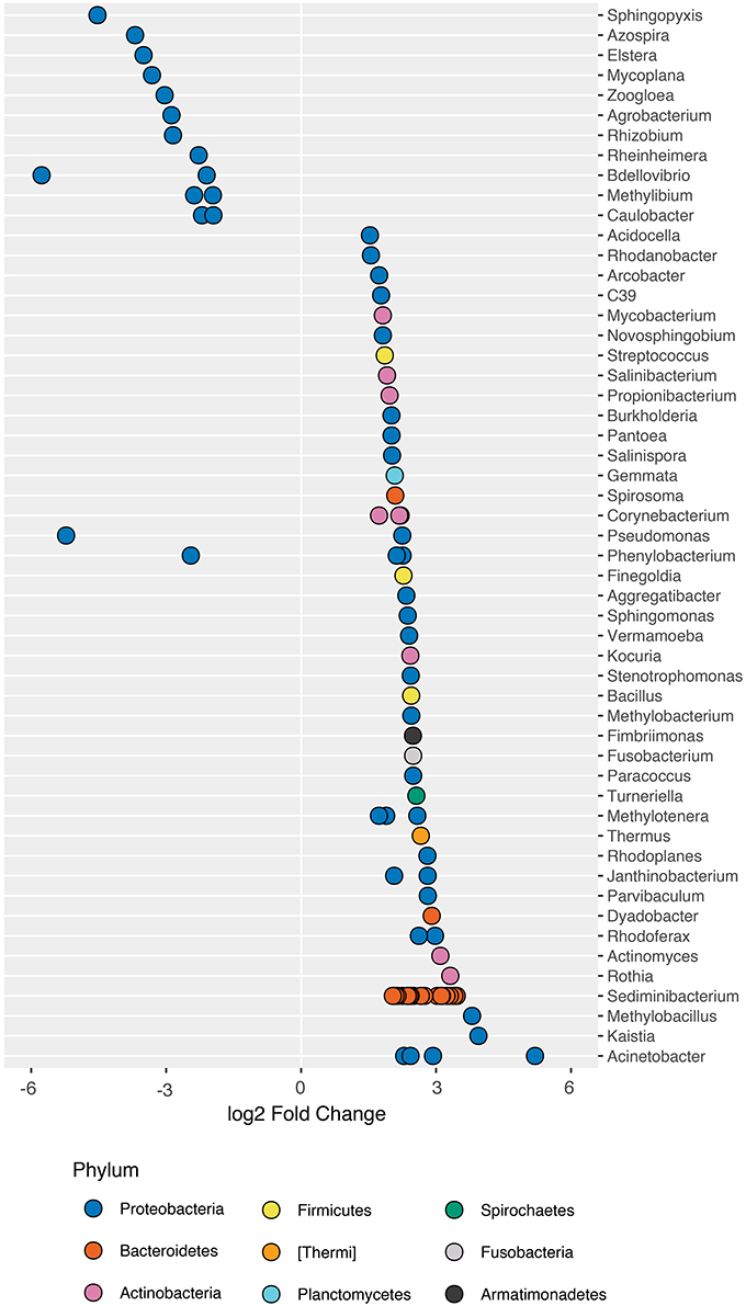
Figure 2. ASVs that significantly differ in abundance in the presence of arsenic. 186 ASVs were identified as significantly different in abundance in the presence of arsenic (DESeq2; adj-P < 0.05; ST1 for complete list of differentially abundance ASVs). 43 ASVs that increased in abundance and 38 that decreased were not assigned taxonomy to the genus level and were removed for plotting purposes. Genus-level classification is provided where available.
When considering a less conservative alpha-level (α = 0.05) for the detection of trends in taxa that changed in abundance in response to arsenic treatment, we found that 186 ASVs differed in abundance (DESeq2; adj-Ps < 0.05; Table S3). We found that ASVs within individual genera at times responded differently to arsenic exposure, indicative of phenotypic heterogeneity within the genus. For example, within Pseudomonas, variant ASV32 (base mean: 1,200) increased by a ~2.3x while ASV152 (base mean: 131.1) decreased in abundance by a ~5.2x under the effect of arsenic when compared to its abundance in control populations. Interestingly, certain species within the genus such as P. aeruginosa are associated with antimicrobial resistance (Perron et al., 2007).
We compared the overall taxonomic diversity across treatments using different metrics of alpha diversity that incorporate species richness and evenness (Table 2). We estimated observed richness (i.e., number of ASVs), as well as the Shannon index and Pielou's evenness metric from the ASV table rarefied to the lowest sampling depth (119,609 ASVs). Looking at richness, we found a quadratic relationship between the number of observed ASVs and arsenic concentrations (F(2, 15) = 9.83; P < 0.01; Figure 3A). More specifically, we found that the highest number of ASVs was observed at intermediate arsenic concentrations: the number of ASVs increasing by 1.9-fold from ~319.3 ASVs at 0 ppb to ~580.0 ASVs at 50 ppb before decreasing when exposed to ~393.2 ASVs 100 ppb. When considering both Shannon diversity and Pielou's evenness, however, we found that arsenic generally decreased diversity at 10 and 50 ppb and increased diversity at 100 ppb. Specifically, we found that, relative to the control, Shannon diversity decreased by 1.2x from 0 to 50 ppb, but increased by 1.1x when compared to the control at 100 ppb (F(2, 15) = 4.32, P = 0.03; Figure 3B). Similarly, Pielou's evenness decreased by 1.3x from 0 to 50 ppb and increased by 1.1x at 100 ppb (F(2, 15) = 6.04, P = 0.01; Figure 3C; Table S6). Because Shannon diversity index is calculated using both observed ASVs and their relative abundance, the lack of congruence between the latter and observed ASVs suggest that changes in ecosystem diversity are likely due to fluctuations in relative abundance of different ASVs following arsenic exposure. In other words, even though the number of ASVs observed in arsenic treatment is higher than in control population, the microbiome exposed to arsenic is likely dominated by relatively few taxa.
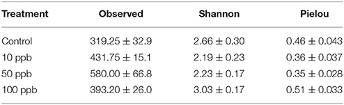
Table 2. Means for alpha diversity metrics of zebrafish microbial communities ± s.e.
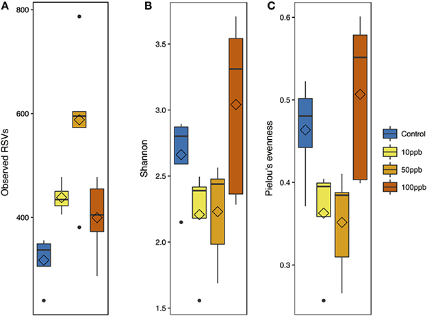
Figure 3. Alpha diversity metrics with arsenic exposure. (A) Observed ASV measurements [F(2, 15) = 9.83; adj-P < 0.01]. (B) Shannon diversity measurements [F(2, 15) = 4.32; adj-P = 0.03]. (C) Pielou's evenness measurements [F(2, 15) = 6.04; adj-P = 0.01]. All metrics are plotted against arsenic concentration gradient; with mean (diamond) and median (line), and hinges as first and third quartiles (25th and 75th percentiles).
We then tested whether arsenic exposure resulted in changes in overall microbial community composition, or beta-diversity. Using principle coordinate analysis (PCoA) on unweighted UniFrac scores, we found that the two highest-ranked dimensions, PCo1 and PCo2, explained 31.8 and 11.9% of variance respectively (Figure 4A) and that arsenic exposure itself explained 78.4% of the variance in bacterial composition, resulting in clustering of arsenic treated microbiomes compared to control microbiomes (ANOSIM; R = 0.784; adj-P < 0.01; perm = 999; ANOSIM; Figure 4A; Table S4). When treating arsenic concentration as a continuous variable, we found that UniFrac scores tended to diverge away from control microbiomes as concentration increased (ADONIS; R2 = 0.12; perm = 999; adj-P = 0.034; Figure 4B; Table S5). We found no significant differences in sample distances to their treatment centroids, suggesting homogeneity of variance (adj-Ps > 0.05; ANOVA; Tables S7,S8). Not surprisingly, many of the genera found to increase in abundance in the presence of arsenic (Figure 2), e.g., Sediminibacterium, Methylotenera, Sphingomonas, and Dyella, also clustered with arsenic samples.
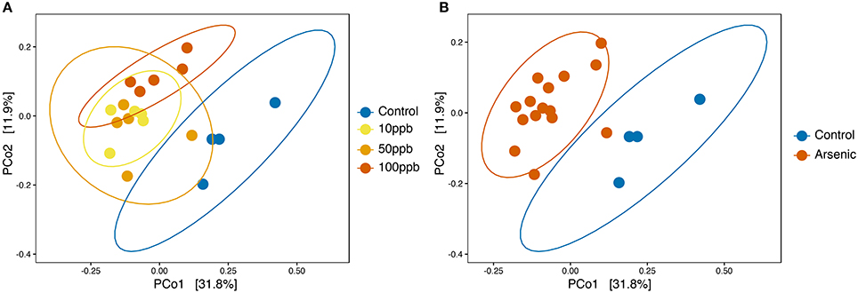
Figure 4. Principle coordinates analysis (PCoA) based on unweighted UniFrac scores of zebrafish microbiota. (A) PCoA on unweighted UniFrac scores with all treatments labeled. Significant dissimilarity between samples when arsenic was treated as a continuous variable (ADONIS; adj-P = 0.036; R2 = 0.124; perm = 999). (B) PCoA on unweighted UniFrac scores with arsenic pooled group and control. Significant dissimilarity between the pooled arsenic group and control group (ANOSIM; adj-P < 0.01, R = 0.786, perm = 999). Ellipses are drawn at 0.95 C.I.
We replicated the above analyses using the OTU table produced by QIIME defaults, utilizing a method based on the uclust algorithm which cluster reads into OTUs based on ≥97% similarity (see Datasheet 5 for unfiltered representative OTUs). On average, we observed five times more OTUs per sample (mean: 3,502 ± s.e. 125.9) than ASVs (mean: 598.0 ± s.e. 46.16.) (Wilcoxon test; W = 361; P < 0.01; Figure S1). We also observed higher variance in the number of OTUs identified between samples (Fligner-Killeen test; χ2 = 6.58; P = 0.0126). This difference was partly explained by the fact that 40.80% of OTUs were doubletons while only 5.55% of ASVs were doubletons, most likely due to DADA2 correcting probable sequencing errors in sequences at low abundance (Callahan et al., 2016).
When looking at species composition, we observed a similar pattern among OTUs distribution: 148 OTUs significantly differed in abundance in the presence of arsenic (DESeq2; adj-Ps ≤ 0.01; Figure S2; Table S10) with an important overlap between genera identified via OTUs and ASVs. However, we did not detect significant changes in alpha-diversity between arsenic and control treatments when using the rarefied OTU table (using a depth of 96,879 reads per sample; Figure S3). The differences between the two analyses are most likely due to fewer reads post-quality filtering in the QIIME pipeline (median: 245,577) compared to the DADA2 methods (median: 497,470) and increased spurious resolution when using OTUs rather than ASVs. Despite the lack of difference at the alpha-diversity level, we found that the presence of arsenic explained a high degree of the variance (54.7%) in overall bacterial composition when comparing arsenic and control sample measured as unweighted UniFrac scores, in accordance with DADA2 (ANOSIM; R = 0.547; P < 0.01; perm = 999; Figure S4). Finally, a Procrustes analysis revealed a high level of similarity between the two analyses (PROTEST; P < 0.01; Observation = 0.758; perm = 999), confirming that despite differences between OTU and ASV results, the two methods reveal similar taxonomic composition throughout the experiment.
Finally, following the observation that certain taxa increasing in abundance in the presence of arsenic were previously associated with antimicrobial resistance, we measured the relative abundance of int1 to investigate the possible link between arsenic exposure and the spread of integron-like structures in microbial communities. We found that that arsenic concentration had a significant effect on the relative abundance of int1: exposure to high arsenic concentrations correlated with an increase in int1 copies (F(2, 14) = 13.27; P < 0.01; Figure 5). The effect of arsenic was especially strong at the highest concentration where int1 relative copy number was 9 times higher than in control populations. Using a dissimilarity matrix comparing principle coordinate analysis (PCoA) to int1 abundance, we found that int1 did not significantly correlate with UniFrac clustering between the different samples (ADONIS; R = 0.056; P = 0.56; perm = 999; Figure S5), indicating that increases in int1 relative copy number was not likely associated with one or a few specific taxa.
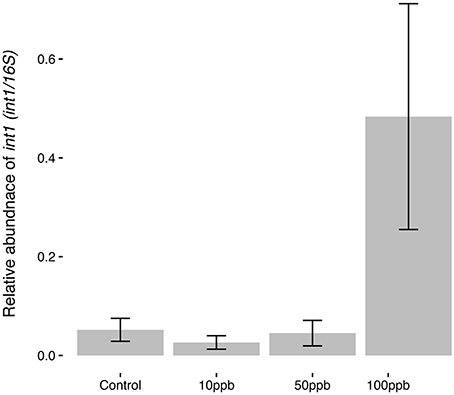
Figure 5. Int1 abundance in response to arsenic concentration. Relative abundance of int1 is taken as the proportion of int1 copies per 16S rRNA copies, where copy numbers are calculated using standard curves and qPCR. Error bars are ± standard error. Quadratic model suggests highest arsenic concentration (100 ppb) significantly increased int1 abundance (F(2,14) = 13.27; P < 0.01).
Mounting concern over arsenic contamination in drinking water has led to a better understanding of both arsenic's negative outcomes on human health (Smith et al., 2000) and its influence on microbial communities in the environment (Cai et al., 2009; Sarkar et al., 2012; Escudero et al., 2013; Bhadury, 2014; Farias et al., 2015; Paul et al., 2015; Sultana et al., 2017). In this study, we investigated whether environmental concentrations of arsenic can perturb microbiota in developing zebrafish larvae. We found that exposure to arsenic had a destabilizing effect on the zebrafish microbiome, leading to dysbiosis both in terms of microbial diversity and in terms of microbial composition between arsenic treatments. We also found that exposure to a high concentration of arsenic increased the frequency of int1, a genetic mechanism responsible for the horizontal transfer of resistance genes. To the best of our knowledge, this is the first study examining how arsenic affects developing zebrafish microbiomes.
Overall, we found that arsenic created distinguishable microbial communities comparable across the different level of arsenic exposure. We found that even a concentration as low as 10 ppb, considered safe by the World Health Organization, altered the larval microbiome. Interestingly, 12 out of the 15 most abundant families present in the core microbiome of healthy larvae significantly changed in abundance in the presence of arsenic. We also observed a reduction in taxa evenness at all levels of arsenic exposure and an increase in the total number of observed taxa at 10 and 50 ppb. While a decrease in evenness was associated with adverse health effects in premature infants with necrotizing enterocolitis (Chang et al., 2008; McMurtry, 2015), increases in taxa richness at low concentrations of arsenic were also observed in contaminated aquifers (Li et al., 2015). These observations agree with the perturbation theory of the subsidy-stress gradient, stating that low levels of stress can potentially enhance ecosystem productivity via the input of energy (Odum et al., 1979).
Many of the taxa that increased in abundance in the presence of arsenic were also observed in arsenic-contaminated environments (Oremland and Stolz, 2005; Liao et al., 2011; Costa et al., 2015). For example, Sediminibacterium and Acinetobacter, found in streams contaminated with arsenic (Costa et al., 2015; Li et al., 2015), were also found to harbor arsenic resistance genes (e.g., ars operon and ACR3 protein). Interestingly, while the 26 ASVs belonging to Sediminibacteirum significantly increased in abundance, this was not the case in other genera such as Phenylobacterium, where two ASVs increased in abundance and one decreased. These results suggest that the response of different taxa to environmental perturbations is not predictable from the phylogenetic relatedness of microbes alone. Not only bacterial lineages can vary a great deal within a genus or even within a species, but the spread of arsenic resistance genes such as ars can also be influenced by the horizontal transfer of genetic material (Sarkar et al., 2012).
Indeed, our results show that exposure to arsenic can increase the abundance of int1 genes in microbial communities, an important genetic factor responsible for the transmission of gene cassettes conferring resistance to heavy metals and different antibiotics (Naas et al., 2001; Fluit and Schmitz, 2004; Gaze et al., 2011; Koenig et al., 2011). Interestingly, while our data suggest that high concentrations of arsenic (i.e., 100 ppb) strongly increased int1 abundance, Guo et al. (2014) noted that type 1 integrons did not increase in the gut microbiota of mice exposed to 3,000 ppb of arsenic trioxide. Our study thus implies that arsenic contamination possibly exerts different selective pressures in different hosts or could be stronger in aquatic environments. Taken together, our results suggest that arsenic exposure not only select directly for taxa harboring resistance mechanisms, but also for strains of bacteria with high recombinogenic activity, possibly contributing to the presence of antibiotic resistance reservoirs in aquifers and animals (Baker-Austin et al., 2006; Gullberg et al., 2014; Chen et al., 2015).
The combination of two computational methods to analyze the effect of arsenic on microbial communities offered the opportunity to compare microbial diversity at different levels. While uclust's OTU picking method clusters similar sequences based on sequence differences at a fixed 97% similarity threshold (Caporaso et al., 2010b; Edgar, 2010), DADA2 integrates sequence variation, quality scores and an error model in order to infer exact sample sequences from amplicon reads (Callahan et al., 2016). The coarse-grain method favored by OTU-picking defined by uclust can be a good indicator of broad-scale patterns in microbial communities, especially when prior functional clades are known (Koeppel and Wu, 2013).
Crucially, the overall effect of arsenic on microbial composition remained a significant factor when considering both OTUs and ASVs, highlighting the fact that arsenic plays an important role in dysbiosis both at the fine and coarse-grain levels. Still, our results also suggest that sequence variants-based analyses are more powerful for detecting changes associated with final-scale microbial diversity. While OTU-based analyses revealed more taxa overall, such analyses failed to detect changes in abundance associated with important taxa. This result likely reflects the fact that uclust clusters polyphyletic reads into OTUs and misses variation at lower phylogenetic levels (Koeppel and Wu, 2013). The higher number of taxa observed in OTUs is likely due to the high false-positive rates associated with Illumina sequencing (Callahan et al., 2016), as supported by the fact that 40.8% of OTUs were doubletons and only 5.55% of ASVs were doubletons. In addition to providing a more accurate description of microbial communities, sequence variant-based analyses are likely to provide the opportunity to distinguish between similar sequences that can be crucial to revealing the difference between harmless and virulent strains as more information is gathered (McElroy et al., 2013).
Finally, even though we replaced media every 48 h to reduce cross-contamination between individual larvae, it is possible that the environment in which the fish were raised confounded the effect observed in this study. The fact that fish from separate treatments were raised in the same dishes could explain part of the variance observed among the different microbiomes, a phenomenon known as batch effect (Hurlbert, 1984). Still, microbiomes exhibited high inter-samples variability as expected with most 16S amplicon studies (Hiergeist et al., 2016), suggesting that the level of variability in our study was not limited by specific batches. Furthermore, we observed a strong overall effect of arsenic on the investigated microbiomes as well as linear trends in both int1 relative abundance and diversity measurements in relation to arsenic concentration, indicating that the data structure follows a gradient along arsenic concentration rather than being randomly assigned to batches. Taken together, these results suggest arsenic most likely exerted a predictable effect on the microbial communities.
In conclusion, our study demonstrates that concentrations of arsenic, even below maximum concentration limits mandated by local authorities, can alter the developing microbiota of zebrafish and increase the relative abundance of integron 1 genetic elements. The possible risk of pathogenicity associated with highly recombinogenic strains remains to be investigated in this system (Stecher et al., 2013). Future studies should also integrate multi-omics approaches with physiological studies to determine how arsenic exposure alters the microbiome functions in developing hosts with possible health consequences, as well as the consequences of these changes for the health of humans and other animals.
DD, FK, and GP designed the experiment. DD, GP, and BJ designed and conducted experimental procedures, including zebrafish arsenic exposures, gDNA extractions, MiSeq library preparation, and qPCR. RL led the sequencing project as well as provided bar-coded reverse primers. DD and GP conducted bioinformatic and statistical analyses. DD and GP were the major contributors in writing the manuscript. All authors read and approved the final manuscript.
The work conducted in this study was supported by the Biology Department of Bard College, and by a grant to Juniata College from the Howard Hughes Medical Institute (http://www.hhmi.org) through the Precollege and Undergraduate Science Education Program, and the National Science Foundation (www.nsf.org), NSF Award #DBI-1248096.
The authors declare that the research was conducted in the absence of any commercial or financial relationships that could be construed as a potential conflict of interest.
We thank Dr. William Gaze for his kind gift of E. coli SK4903 with IncPβ R751 and Dr. Benjamin J. Callahan for his guidance on the DADA2 analysis and reviewing the DADA2 workflow.
The Supplementary Material for this article can be found online at: https://www.frontiersin.org/articles/10.3389/fmicb.2018.01323/full#supplementary-material
Figure S1. Compared observed richness of ASVs and OTUs.
Figure S2. OTUs differentially abundant in the presence of arsenic (P ≤ 0.01).
Figure S3. Alpha diversity metrics, measured with OTUs, with arsenic exposure.
Figure S4. Principle coordinates analysis (PCoA) based on unweighted UniFrac scores of zebrafish microbiota, using OTU table.
Figure S5. PCoA based on unweighted UniFrac scores of ASVs from zebrafish microbiota amplified for int1.
Datasheet 1. R markdown file with scripts outlining DADA2 forward read processing.
Datasheet 2. R markdown file with scripts outlining community analyses.
Datasheet 3. Mapping file with sample data.
Datasheet 4. Fasta file with representative ASVs.
Datasheet 5. Fasta file with representative OTUs.
Table S1. Core microbiome in control population.
Table S2. ASVs differentially abundant in the presence of arsenic (P ≤ 0.01).
Table S3. ASVs differentially abundant in the presence of arsenic (P ≤ 0.05).
Tables S4, S5. ANOVA tables for PCoA comparisons.
Table S6. OTUs differentially abundant in the presence of arsenic.
Table S7. Genera counts of differentially abundant ASVs and OTUs.
Tables S8–S10. Polynomial regressions for effect of arsenic on OTU alpha diversity.
Andres, J., and Bertin, P. N. (2016). The microbial genomics of arsenic. FEMS Microbiol. Rev. 40, 299–322. doi: 10.1093/femsre/fuv050
Baker-Austin, C., Wright, M. S., Stepanauskas, R., and McArthur, J. V. (2006). Co-selection of antibiotic and metal resistance. Trends Microbiol. 14, 176–182. doi: 10.1016/j.tim.2006.02.006
Bhadury, P. (2014). Diversity of arsenite oxidizing bacterial communities in arsenic-rich deltaic aquifers in West Bengal, India. Front Microbiol. 5:602. doi: 10.3389/fmicb.2014.00602
Cai, L., Liu, G., Rensing, C., and Wang, G. (2009). Genes involved in arsenic transformation and resistance associated with different levels of arsenic-contaminated soils. BMC Microbiol. 9:4. doi: 10.1186/1471-2180-9-4
Callahan, B. J., McMurdie, P. J., Rosen, M. J., Han, A. W., Johnson, A. J., and Holmes, S. P. (2016). DADA2: high-resolution sample inference from Illumina amplicon data. Nat Methods 13, 581–583. doi: 10.1038/nmeth.3869
Caporaso, J. G., Bittinger, K., Bushman, F. D., DeSantis, T. Z., Andersen, G. L., and Knight, R. (2010a). PyNAST: a flexible tool for aligning sequences to a template alignment. Bioinformatics 26, 266–267. doi: 10.1093/bioinformatics/btp636
Caporaso, J. G., Kuczynski, J., Stombaugh, J., Bittinger, K., Bushman, F. D., Costello, E. K., et al. (2010b). QIIME allows analysis of high-throughput community sequencing data. Nat. Methods 7, 335–336. doi: 10.1038/nmeth.f.303
Caporaso, J. G., Lauber, C. L., Walters, W. A., Berg-Lyons, D., Huntley, J., Fierer, N., et al. (2012). Ultra-high-throughput microbial community analysis on the Illumina HiSeq and MiSeq platforms. ISME J. 6, 1621–1624. doi: 10.1038/ismej.2012.8
Chang, J. Y., Antonopoulos, D. A., Kalra, A., Tonelli, A., Khalife, W. T., Schmidt, T. M., et al. (2008). Decreased diversity of the fecal microbiome in recurrent clostridium difficile-associated diarrhea. J. Infect. Dis. 197, 435–438. doi: 10.1086/525047
Chen, S., Li, X., Sun, G., Zhang, Y., Su, J., and Ye, J. (2015). Heavy metal induced antibiotic resistance in bacterium LSJC7. IJMS 16, 23390–23404. doi: 10.3390/ijms161023390
Claus, S. P., Guillou, H., and Ellero-Simatos, S. (2016). The gut microbiota: a major player in the toxicity of environmental pollutants? NPG Biofilms Microb. 3:17001. doi: 10.1038/npjbiofilms.2017.1
Costa, P. S., Reis, M. P., Ávila, M. P., Leite, L. R., de Araújo, F. M., Salim, A. C., et al. (2015). Metagenome of a microbial community inhabiting a metal-rich tropical stream sediment. PLoS ONE 10:e0119465. doi: 10.1371/journal.pone.0119465
Costello, E. K., Stagaman, K., Dethlefsen, L., Bohannan, B. J., and Relman, D. A. (2012). The application of ecological theory toward an understanding of the human microbiome. Science 336, 1255–1262. doi: 10.1126/science.1224203
DeSantis, T. Z., Hugenholtz, P., Larsen, N., Rojas, M., Brodie, E. L., Keller, K., et al. (2006). Greengenes, a chimera-checked 16S rRNA gene database and workbench compatible with ARB. Appl. Environ. Microbiol. 72, 5069–5072. doi: 10.1128/AEM.03006-05
de Theije, C. G. M., Wopereis, H., Ramadan, M., van Eijndthoven, T., Lambert, J., Knol, J., et al. (2014). Brain, behavior, and immunity. Brain Behav Immun. 37, 197–206. doi: 10.1016/j.bbi.2013.12.005
Dheer, R., Patterson, J., Dudash, M., Stachler, E. N., Bibby, K. J., Stolz, D. B., et al. (2015). Arsenic induces structural and compositional colonic microbiome change and promotes host nitrogen and amino acid metabolism. Toxicol. Appl. Pharmacol. 289, 397–408. doi: 10.1016/j.taap.2015.10.020
Edgar, R. C. (2010). Search and clustering orders of magnitude faster than BLAST. Bioinformatics 26, 2460–2461. doi: 10.1093/bioinformatics/btq461
Edgar, R. C., Haas, B. J., Clemente, J. C., Quince, C., and Knight, R. (2011). UCHIME improves sensitivity and speed of chimera detection. Bioinformatics 27, 2194–2200. doi: 10.1093/bioinformatics/btr381
Escudero, L. V., Casamayor, E. O., Chong, G., Pedrós-Alió, C., and Demergasso, C. (2013). Distribution of microbial arsenic reduction, oxidation and extrusion genes along a wide range of environmental arsenic concentrations. PLoS ONE 8:e78890. doi: 10.1371/journal.pone.0078890
Farias, P., Espírito Santo, C., Branco, R., Francisco, R., Santos, S., Hansen, L., et al. (2015). Natural hot spots for gain of multiple resistances: arsenic and antibiotic resistances in heterotrophic, aerobic bacteria from marine hydrothermal vent fields. Appl. Environ. Microbiol. 81, 2534–2543. doi: 10.1128/AEM.03240-14
Fluit, A. C., and Schmitz, F. J. (2004). Resistance integrons and super-integrons. Clin. Microbiol. Infect. 10, 272–288. doi: 10.1111/j.1198-743X.2004.00858.x
Foxx-Orenstein, A. E., and Chey, W. D. (2012). Manipulation of the gut microbiota as a novel treatment strategy for gastrointestinal disorders. Am. J. Gastroenterol. Suppl. 1, 41–46. doi: 10.1038/ajgsup.2012.8
Fritz, J. V., Desai, M. S., Shah, P., Schneider, J. G., and Wilmes, P. (2013). From meta-omics to causality: experimental models for human microbiome research. Microbiome 1:14. doi: 10.1186/2049-2618-1-14
Gaze, W. H., Zhang, L., Abdouslam, N. A., Hawkey, P. M., Calvo-Bado, L., Royle, J., et al. (2011). Impacts of anthropogenic activity on the ecology of class 1 integrons and integron-associated genes in the environment. ISME J. 5, 1253–1261. doi: 10.1038/ismej.2011.15
Guha Mazumder, D. N., Haque, R., Ghosh, N., De, B. K., Santra, A., Chakraborty, D., et al. (1998). Arsenic levels in drinking water and the prevalence of skin lesions in West Bengal, India. Int. J. Epidemiol. 27, 871–877. doi: 10.1093/ije/27.5.871
Gullberg, E., Albrecht, L. M., Karlsson, C., Sandegren, L., and Andersson, D. I. (2014). Selection of a multidrug resistance plasmid by sublethal levels of antibiotics and heavy metals. MBio 5:e01918-14. doi: 10.1128/mBio.01918-14
Guo, X., Liu, S., Wang, Z., Zhang, X. X., Li, M., and Wu, B. (2014). Metagenomic profiles and antibiotic resistance genes in gut microbiota of mice exposed to arsenic and iron. Chemosphere 112, 1–8. doi: 10.1016/j.chemosphere.2014.03.068
Hang, J., Desai, V., Zavaljevski, N., Yang, Y., Lin, X., Satya, R. V., et al. (2014). 16S rRNA gene pyrosequencing of reference and clinical samples and investigation of the temperature stability of microbiome profiles. Microbiome 2, 31–15. doi: 10.1186/2049-2618-2-31
Hiergeist, A., Reischl, U., and Priority Program 1656 Intestinal Microbiota Consortium/quality assessment participants, Gessner, A. (2016). Multicenter quality assessment of 16S ribosomal DNA-sequencing for microbiome analyses reveals high inter-center variability. Int. J. Med. Microbiol. 306, 334–342. doi: 10.1016/j.ijmm.2016.03.005
Hughes, M. F., Beck, B. D., Chen, Y., Lewis, A. S., and Thomas, D. J. (2011). Arsenic exposure and toxicology: a historical perspective. Toxicol. Sci. 123, 305–332. doi: 10.1093/toxsci/kfr184
Hurlbert, S. H. (1984). Pseudoreplication and the design of ecological field experiments. Ecol. Monogr. 54, 187–211. doi: 10.2307/1942661
Jackson, D. A. (1995). PROTEST: a PROcrustean randomization TEST of community environment concordance. Ecoscience 2, 297–303. doi: 10.1080/11956860.1995.11682297
Koenig, J. E., Spor, A., Scalfone, N., Fricker, A. D., Stombaugh, J., Knight, R., et al. (2011). Succession of microbial consortia in the developing infant gut microbiome. Proc. Natl. Acad. Sci. U.S.A. 108, 4578–4585. doi: 10.1073/pnas.1000081107
Koeppel, A. F., and Wu, M. (2013). Surprisingly extensive mixed phylogenetic and ecological signals among bacterial operational taxonomic units. Nucleic Acids Res. 41, 5175–5188. doi: 10.1093/nar/gkt241
Kong, H. H., Oh, J., Deming, C., Conlan, S., Grice, E. A., Beatson, M. A., et al. (2012). Temporal shifts in the skin microbiome associated with disease flares and treatment in children with atopic dermatitis. Genome Res. 22, 850–859. doi: 10.1101/gr.131029.111
Kostic, A. D., Howitt, M. R., and Garrett, W. S. (2013). Exploring host-microbiota interactions in animal models and humans. Genes Dev. 27, 701–718. doi: 10.1101/gad.212522.112
Lawrence, C. (2007). The husbandry of zebrafish (Danio rerio): a review. Aquaculture 269, 1–20. doi: 10.1016/j.aquaculture.2007.04.077
Li, P., Wang, Y., Dai, X., Zhang, R., Jiang, Z., Jiang, D., et al. (2015). Microbial community in high arsenic shallow groundwater aquifers in hetao basin of inner Mongolia, China. PLoS ONE 10:e0125844. doi: 10.1371/journal.pone.0125844
Liao, V. H., Chu, Y. J., Su, Y. C., Hsiao, S. Y., Wei, C. C., Liu, C. W., et al. (2011). Arsenite-oxidizing and arsenate-reducing bacteria associated with arsenic-rich groundwater in Taiwan. J. Contam. Hydrol. 123, 20–29. doi: 10.1016/j.jconhyd.2010.12.003
Love, M. I., Huber, W., and Anders, S. (2014). Moderated estimation of fold change and dispersion for RNA-seq data with DESeq2. Genome Biol. 15, 31–21. doi: 10.1186/s13059-014-0550-8
Lozupone, C., and Knight, R. (2005). UniFrac: a new phylogenetic method for comparing microbial communities. Appl. Environ. Microbiol. 71, 8228–8235. doi: 10.1128/AEM.71.12.8228-8235.2005
Lu, K., Abo, R. P., Schlieper, K. A., Graffam, M. E., Levine, S., Wishnok, J. S., et al. (2014). Arsenic exposure perturbs the gut microbiome and its metabolic profile in mice: an integrated metagenomics and metabolomics analysis. Environ. Health Perspect. 3, 284–291. doi: 10.1289/ehp.1307429
Martinez, V. D., Vucic, E. A., Becker-Santos, D. D., Gil, L., and Lam, W. L. (2011). Arsenic exposure and the induction of human cancers. J. Toxicol. 2011, 431287–431213. doi: 10.1155/2011/431287
Mateos, L. M., Ordóñez, E., Letek, M., and Gil, J. A. (2006). Corynebacterium glutamicum as a model bacterium for the bioremediation of arsenic. Int. Microbiol. 9, 207–215.
Matthews, M., and Varga, Z. M. (2012). Anesthesia and Euthanasia in Zebrafish. ILAR J. 53, 192–204. doi: 10.1093/ilar.53.2.192
McDonald, D., Price, M. N., Goodrich, J., Nawrocki, E. P., DeSantis, T. Z., Probst, A., et al. (2011). An improved Greengenes taxonomy with explicit ranks for ecological and evolutionary analyses of bacteria and archaea. ISME J.6, 610–618. doi: 10.1038/ismej.2011.139
McElroy, K., Zagordi, O., Bull, R., Luciani, F., and Beerenwinkel, N. (2013). Accurate single nucleotide variant detection in viral populations by combining probabilistic clustering with a statistical test of strand bias. BMC Genomics 14:501. doi: 10.1186/1471-2164-14-501
McMurdie, P. J., and Holmes, S. (2013). phyloseq: an R package for reproducible interactive analysis and graphics of microbiome census data. PLoS ONE 8:e61217. doi: 10.1371/journal.pone.0061217
McMurdie, P. J., and Holmes, S. (2014). Waste not, want not: why rarefying microbiome data is inadmissible. PLoS Comput. Biol. 10:e1003531. doi: 10.1371/journal.pcbi.1003531
McMurtry, V. E. (2015). Bacterial diversity and Clostridia abundance decrease with increasing severity of necrotizing enterocolitis. Microbiome 3:11. doi: 10.1186/s40168-015-0075-8
Moon, Y., Kim, H. J., Kim, J. Y., Kim, H., Kim, W. R., and Sun, W. (2011). Different expression of human GFAP promoter-derived GFP in different subsets of astrocytes in the mouse brain. Animal Cells Syst. 15, 268–273. doi: 10.1080/19768354.2011.611254
Mukherjee, A. B., and Bhattacharya, P. (2001). Arsenic in groundwater in the bengal delta plain: slow poisoning in Bangladesh. Dossiers Environ. 9, 189–220. doi: 10.1139/a01-007
Naas, T., Mikami, Y., Imai, T., Poirel, L., and Nordmann, P. (2001). Characterization of In53, a class 1 plasmid- and composite transposon-located integron of Escherichia coli which carries an unusual array of gene cassettes. J. Bacteriol. 183, 235–249. doi: 10.1128/JB.183.1.235-249.2001
Narrowe, A. B. (2015). Perturbation and restoration of the fathead minnow gut microbiome after low-level triclosan exposure. Microbiome 3, 1–18. doi: 10.1186/s40168-015-0069-6
Navas-Acien, A., Silbergeld, E. K., Pastor-Barrusio, R., Guallar, E., and Silbergeld, E. K. (2008). Arsenic exposure and prevalence of type 2 diabetes in US adults. JAMA 300, 1–9. doi: 10.1001/jama.300.7.814
Neu, J., and Rushing, J. (2011). Cesarean versus vaginal delivery: long-term infant outcomes and the hygiene hypothesis. Clin. Perinatol. 38, 321–331. doi: 10.1016/j.clp.2011.03.008
Odum, E. P., Finn, J. T., and Franz, E. H. (1979). Perturbation theory and the subsidy-stress gradient. BioScience 29, 349–352. doi: 10.2307/1307690
Oksanen, J., Blanchet, G., Kindt, R., Legendre, P., Minchin, P. R., OHara, R. B., et al. (2015). vegan: Community Ecology Package. R Package Version 2.3.2. 1–285.
Olszak, T., An, D., Zeissig, S., Vera, M. P., Richter, J., Franke, A., et al. (2012). Microbial exposure during early life has persistent effects on natural killer T cell function. Science 336, 489–493. doi: 10.1126/science.1219328
Oremland, R. S., and Stolz, J. F. (2005). Arsenic, microbes and contaminated aquifers. Trends Microbiol. 13, 45–49. doi: 10.1016/j.tim.2004.12.002
Palmer, C., Bik, E. M., DiGiulio, D. B., Relman, D. A., and Brown, P. O. (2007). Development of the human infant intestinal microbiota. PLoS Biol. 5:e177. doi: 10.1371/journal.pbio.0050177
Paul, D., Kazy, S. K., Gupta, A. K., Pal, T., and Sar, P. (2015). Diversity, metabolic properties and arsenic mobilization potential of indigenous bacteria in arsenic contaminated groundwater of west Bengal, India. PLoS Biol. 10:e0118735. doi: 10.1371/journal.pone.0118735
Perron, G. G., Gonzalez, A., and Buckling, A. (2007). Source-sink dynamics shape the evolution of antibiotic resistance and its pleiotropic fitness cost. Proc. Biol. Sci. 274, 2351–2356. doi: 10.1098/rspb.2007.0640
Pielou, E. C. (1966). The measurement of diversity in different types of biological collection. J. Theor. Biol. 131–144. doi: 10.1016/0022-5193(66)90013-0
Price, M. N., Dehal, P. S., and Arkin, A. P. (2010). FastTree 2 – approximately maximum-likelihood trees for large alignments. PLoS Biol. 5:e9490. doi: 10.1371/journal.pone.0009490
Qin, J., Li, Y., Cai, Z., Li, S., Zhu, J., Zhang, F., et al. (2012). A metagenome-wide association study of gut microbiota in type 2 diabetes. Nature 490, 55–60. doi: 10.1038/nature11450
Raymann, K., and Moran, N. A. (2018). The role of the gut microbiome in health and disease of adult honey bee workers. Curr. Opin. Insect Sci. 26, 97–104. doi: 10.1016/j.cois.2018.02.012
Roeselers, G., Mittge, E. K., Stephens, W. Z., Parichy, D. M., Cavanaugh, C. M., Guillemin, K., et al. (2011). Evidence for a core gut microbiota in the zebrafish. ISME J. 5, 1595–1608. doi: 10.1038/ismej.2011.38
Saha, J. C., Dikshit, A. K., and Bandyopadhyay, M. (2003). A review of arsenic poisoning and its effects on human health. Crit. Rev. Environ. Sci. Technol. 29, 281–313. doi: 10.1080/10643389991259227
Sarkar, A., Kazy, S. K., and Sar, P. (2012). Characterization of arsenic resistant bacteria from arsenic rich groundwater of West Bengal, India. Ecotoxicology 22, 363–376. doi: 10.1007/s10646-012-1031-z
Sarkar, B., and Bhattacharya, P. (2002). Heavy Metals in the Environment. New York, NY: Marcel Decker.
Sender, R., Fuchs, S., and Milo, R. (2016). Revised estimates for the number of human and bacteria cells in the body. PLoS Biol. 14:e1002533. doi: 10.1371/journal.pbio.1002533
Smedley, P. L., and Kinniburgh, D. G. (2002). A review of the source, behaviour and distribution of arsenic in natural waters. Appl. Geochem. 17, 517–568. doi: 10.1016/S0883-2927(02)00018-5
Smith, A. H., Lingas, E. O., and Rahman, M. (2000). Contamination of drinking-water by arsenic in Bangladesh: a public health emergency. Bull. World Health Organ. 78, 1093–1103.
Spence, R., Fatema, M. K., Reichard, M., Huq, K. A., Wahab, M. A., Ahmed, Z. F., et al. (2006). The distribution and habitat preferences of the zebrafish in Bangladesh. J Fish Biol. 69, 1435–1448. doi: 10.1111/j.1095-8649.2006.01206.x
Stecher, B., Maier, L., and Hardt, W. D. (2013). “Blooming” in the gut: how dysbiosis might contribute to pathogen evolution. Nat. Rev Microbiol. 11, 277–284. doi: 10.1038/nrmicro2989
Sultana, M., Mou, T. J., Sanyal, S. K., Diba, F., Mahmud, Z. H., Parvez, A. K., et al. (2017). Investigation of arsenotrophic microbiome in arsenic-affected bangladesh groundwater. Groundwater 55, 736–746. doi: 10.1111/gwat.12520
Summers, A. O., Wireman, J., Vimy, M. J., Lorscheider, F. L., Marshall, B., Levy, S. B., et al. (1993). Mercury released from dental “silver” fillings provokes an increase in mercury- and antibiotic-resistant bacteria in oral and intestinal floras of primates. Antimicrob. Agents Chemother. 37, 825–834.
Thioulouse, J., and Dray, S. (2007). Interactive Multivariate data analysis in rwith the ade4 and ade4TkGUI packages. J. Stat. Softw. 22, 1–14. doi: 10.18637/jss.v022.i05
Torrazza, R. M., and Neu, J. (2011). The developing intestinal microbiome and its relationship to health and disease in the neonate. J. Perinatol. 31, S29–S34. doi: 10.1038/jp.2010.172
Turnbaugh, P. J., Ley, R. E., Mahowald, M. A., Magrini, V., Mardis, E. R., and Gordon, J. I. (2006). An obesity-associated gut microbiome with increased capacity for energy harvest. Nature 444, 1027–1131. doi: 10.1038/nature05414
Vallès, Y., Artacho, A., Pascual-García, A., Ferrús, M. L., Gosalbes, M. J., Abellán, J. J., et al. (2014). Microbial succession in the gut: directional trends of taxonomic and functional change in a birth cohort of spanish infants. PLoS Genet. 10:e1004406. doi: 10.1371/journal.pgen.1004406
Veldman, M. B., and Lin, S. (2008). Zebrafish as a developmental model organism for pediatric research. Pediatr. Res. 64, 470–476. doi: 10.1203/PDR.0b013e318186e609
Wang, Q., Garrity, G. M., Tiedje, J. M., and Cole, J. R. (2007). Naive Bayesian classifier for rapid assignment of rRNA sequences into the new bacterial taxonomy. Appl. Environ. Microbiol. 73, 5261–5267. doi: 10.1128/AEM.00062-07
Werner, J. J., Koren, O., Hugenholtz, P., DeSantis, T. Z., Walters, W. A., Caporaso, J. G., et al. (2011). Impact of training sets on classification of high-throughput bacterial 16s rRNA gene surveys. ISME J. 6, 94–103. doi: 10.1038/ismej.2011.82
World Health Organization (2001). Country Situation Report: Arsenic in Drinking Water in Bangladesh: A Challenge in Near Future.
Keywords: microbiome, zebrafish, heavy metals, arsenic, integrons, DADA2, QIIME
Citation: Dahan D, Jude BA, Lamendella R, Keesing F and Perron GG (2018) Exposure to Arsenic Alters the Microbiome of Larval Zebrafish. Front. Microbiol. 9:1323. doi: 10.3389/fmicb.2018.01323
Received: 07 March 2018; Accepted: 30 May 2018;
Published: 21 June 2018.
Edited by:
Robert Brucker, Rowland Institute at Harvard, United StatesReviewed by:
Ronald Oremland, United States Geological Survey, United StatesCopyright © 2018 Dahan, Jude, Lamendella, Keesing and Perron. This is an open-access article distributed under the terms of the Creative Commons Attribution License (CC BY). The use, distribution or reproduction in other forums is permitted, provided the original author(s) and the copyright owner are credited and that the original publication in this journal is cited, in accordance with accepted academic practice. No use, distribution or reproduction is permitted which does not comply with these terms.
*Correspondence: Gabriel G. Perron, Z3BlcnJvbkBiYXJkLmVkdQ==
†Present Address: Dylan Dahan, Department of Microbiology and Immunology, Stanford University School of Medicine, Stanford, CA, United States
Disclaimer: All claims expressed in this article are solely those of the authors and do not necessarily represent those of their affiliated organizations, or those of the publisher, the editors and the reviewers. Any product that may be evaluated in this article or claim that may be made by its manufacturer is not guaranteed or endorsed by the publisher.
Research integrity at Frontiers
Learn more about the work of our research integrity team to safeguard the quality of each article we publish.