- 1Hunan Provincial Key Laboratory of Protein Engineering in Animal Vaccines, College of Veterinary Medicine, Hunan Agricultural University, Changsha, China
- 2College of Animal Science and Veterinary Medicine, Heilongjiang Bayi Agricultural University, Daqing, China
- 3School of Basic Medical Sciences, Xiangya School of Medicine, Central South University, Changsha, China
- 4College of Veterinary Medicine, Northwest A&F University, Yangling, China
- 5Key Laboratory of Agricultural Ministry, State Key Laboratory of Special Economic Animal Molecular Biology, Institute of Special Animal and Plant Sciences of Chinese Academy of Agricultural Sciences, Changchun, China
- 6College of Animal Science and Technology, Changchun Sci-Tech University, Changchun, China
- 7College of Basic Medicine, Mudanjiang Medical College, Mudanjiang, China
Enterocytozoon bieneusi is the most important causative agent of microsporidiosis and can infect almost all vertebrate and invertebrate hosts, including minks (Neovison vison). In the present study, a total of 298 feces samples (including 79 from Heilongjiang province, 31 from Hebei province, 67 from Jilin province, 90 from Liaoning province, and 31 from Shandong province, Northern China) were examined by nested PCR amplification of the internal transcribed spacer (ITS) region of the rRNA gene. The overall prevalence of E. bieneusi in minks was 10.1%, with 10.5% in Jilin province, 32.3% in Hebei province, 8.9% in Liaoning province, 0% in Shandong province, and 6.3% in Heilongjiang province. Furthermore, multiple logistic regression analysis revealed that region was only risk factors associated with E. bieneusi infection in the investigated minks. Five E. bieneusi ITS genotypes (three known genotypes, namely D, Peru11, and EbpC; two novel genotypes, namely, NCM-1 and NCM-2) were found in the current study. Importantly, genotypes D, Peru11 and EbpC, previously identified in humans, were also found in minks, which suggested that minks are the potential sources of human microsporidiosis. To the best of our knowledge, this is the first report of E. bieneusi infection in minks worldwide. The results of the present survey have implications for the controlling E. bieneusi infection in minks, other animals and humans.
Introduction
Enterocytozoon bieneusi is the most frequently detected Microsporidia species, which composed of over 1300 named species, classified into 160 genera (da Cunha et al., 2016; Li et al., 2016a). It is ubiquitous in the environment and is responsible for over 90% of intestinal microsporidiosis in humans (Desportes et al., 1985). Transmission of E. bieneusi was mainly through fecal-oral route, such as ingestion of food and/or water contaminated by spores of E. bieneusi (Prasertbun et al., 2017). AIDS infection with this pathogen may cause life-threatening chronic diarrhea (Canning and Hollister, 1990). Although immunocompetent individual infection with E. bieneusi are usually asymptomatic, they can shed spores into the environment (Zhao et al., 2015).
To date, more than 240 E. bieneusi genotypes were defined based on the internal transcribed spacer (ITS) region of the rRNA gene (da Cunha et al., 2016), which were classified into nine groups by phylogenetic analyses. Among these groups, group 1 representing zoonotic potential phylogenetic group was responsible for the majority of human infections while the remainings (groups 2 to 9) were host-adapted phylogenetic groups, which more frequently recorded in specific hosts or water (Hu et al., 2014; Ma et al., 2015). Interestingly, several genotypes, such as I, J, and BEB4 were divided into phylogenetic groups 2, but they have also been found in humans (Jiang et al., 2015), indicating that these genotypes have zoonotic potential. Therefore, there are raised some questions in the genotypes identification of E. bieneusi.
Enterocytozoon bieneusi was firstly identified in pig feces (Deplazes et al., 1996). Many animals have been now recognized as its hosts (Du et al., 2015; Qi et al., 2015; Fiuza et al., 2016; Li et al., 2016b; Xu et al., 2016; Zhang et al., 2016a,b; da Cunha et al., 2017; Yue et al., 2017). Although E. bieneusi from other animals can be a potential source for human infections (Xu et al., 2016; Zhang et al., 2016a; Yue et al., 2017), no information is available about prevalence and genotypes of E. bieneusi in minks. Therefore, in the present study, a total of 298 feces samples from five provinces of Northern China were examined to estimate the E. bieneusi prevalence and to identify their genotypes. Our results should provide a foundation for the improved control of E. bieneusi infection in humans and animals in these regions and elsewhere in China.
Materials and Methods
Ethics Approval and Consent to Participate
This study was approved by the Animal Ethics Committee of Hunan Agricultural University. Minks used for the study were handled in accordance with good animal practices required by the Animal Ethics Procedures and Guidelines of the People’s Republic of China.
Specimen Collection
A total of 298 fecal samples were randomly collected from nine farmed minks (randomly collected) in Jilin province (41°∼46° N, 122°∼131° E), Liaoning province (38°∼43° N, 118°∼125° E), Heilongjiang province (43°26′∼53°33′ N, 121°11′∼135°05′ E), Hebei province (36°05′∼42°40′ N, 113°27′∼119°50′ E), and Shandong province (34°22.9′∼38°24.01′ N, 114°47.5′∼122°42.3′ E), Northern China in 2016 (Table 1). The numbers of minks reared on each farm ranged from 300 to 1800, approximately. From each farm, approximately 3% of animals was sampled. Before sampling, animals were subjected to clinical examination to determine their health status. Each fecal sample (approximately 50 g) was collected using sterile gloves immediately after the animal had defecated, and then was placed into ice boxes and quickly transported to the laboratory. Information about each mink, such as season (missing the spring), gender, geographic origin, age, and farm ID were collected.

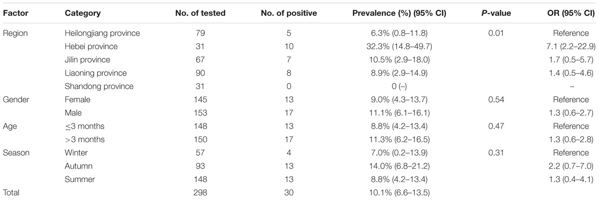
TABLE 1. Prevalence of Enterocytozoon bieneusi in minks in Heilongjiang, Hebei, Jilin, Liaoning, and Shandong provinces, Northern China.
DNA Extraction, PCR Amplification, and Sequencing
Genomic DNA was extracted using the E.Z.N.A.® Stool DNA Kit (Omega Bio-tek Inc., Norcross, GA, United States) (Xu et al., 2016). Then DNA samples were stored at -20°C until tested. The molecular identity and genotype of E. bieneusi of each specimen was detected by PCR-based sequencing of ITS locus using previous established methods (Xu et al., 2016; Zhang et al., 2016a). PCR products were send to Sangon Biotech Company (Shanghai, China) for sequencing from both directions.
Phylogenetic Analyses
The ITS locus sequences of the representative samples (representing different genotypes) were used for phylogenetic analyses. The obtained ITS sequences were aligned with the corresponding reference sequences in GenBank using ClustalX1.81 (Thompson et al., 1997). The neighbor-joining (NJ) were performed using Mega 5.0 (Tamura et al., 2011). The Kimura 2-parameter model were selected as the most suitable model. NJ tree were calculated based on 1,000 bootstrap replicates.
Statistical Analysis
The variation in prevalence of E. bieneusi-infected minks (y) of age (x1), gender (x2), different geographical location (x3), and season (x4) were analyzed by χ2 test using SAS version 9.1 (SAS Institute, Cary, NC, United States) (Meng et al., 2017; Zhang et al., 2017). Each of these variables was included in the binary logit model as an independent variable by multivariable regression analysis. When P < 0.05, the results were considered statistically significant. The adjusted odds ratio (OR) and 95% confidence interval (CI) for each variable were calculated with binary logistic regression and all risk factors entered simultaneously.
Results
Prevalence of E. bieneusi
The study showed that 30 (10.1%, 95% CI 6.6–13.5) of the 298 tested fecal samples were positive for E. bieneusi (Table 1). The prevalence of E. bieneusi in different region groups ranged from 0.0 to 32.3% (Table 1). Of the nine farms, six farms were detected E. bieneusi-positive, with the highest prevalence in farm 3 (32.3%) located in Hebei province (Table 2). Furthermore, prevalence of E. bieneusi in minks of less than 3 months old and minks of more than 3 months old was 8.8 and 11.3%, respectively (P = 0.47). The prevalence of E. bieneusi in female minks (9.0%) was lower than males (11.1%) (P = 0.54). Prevalence of E. bieneusi was 8.8% in summer, 14.0% in autumn and 7.0% in winter (P = 0.31). Moreover, a significant correlation between the investigated region and E. bieneusi infection (P = 0.01) was observed by logistic regression analysis.
Genetic Characterizations and Genotype Distribution of E. bieneusi in Minks
DNA sequence analysis of the ITS locus suggested five E. bieneusi ITS genotypes (three known genotypes and two novel genotypes) were identified in this study, namely, D, Peru11, EbpC, NCM-1, and NCM-2 (Table 2). Of these genotypes, D was the predominance genotype which is present in four farms (n = 12), was responsible for 40.0% of all infection; genotype EbpC (n = 7, 23.3%) and NCM-1 (n = 5, 16.7%) were all found in two farms; genotype Peru11 (n = 5, 16.7%) was found in three farms; NCM-2 (n = 1, 3.3%) was only identified in farm 8 located in Heilongjiang province. Moreover, a total of seven polymorphic sites were observed among the five genotypes (Table 3). Phylogenetic analysis of ITS sequences indicated that the three known genotypes sequences (accession nos: MF440664, MF440667, and MF440668) were identical to that of genotypes D (accession no. KT922238), Peru11 (accession no. JX994269), and EbpC (accession no. KR815517) sequences, respectively.

TABLE 3. Variations in the ITS nucleotide sequences among genotypes of the Enterocytozoon bieneusi in minks in Northern China.
Phylogenetic Relationship of E. bieneusi
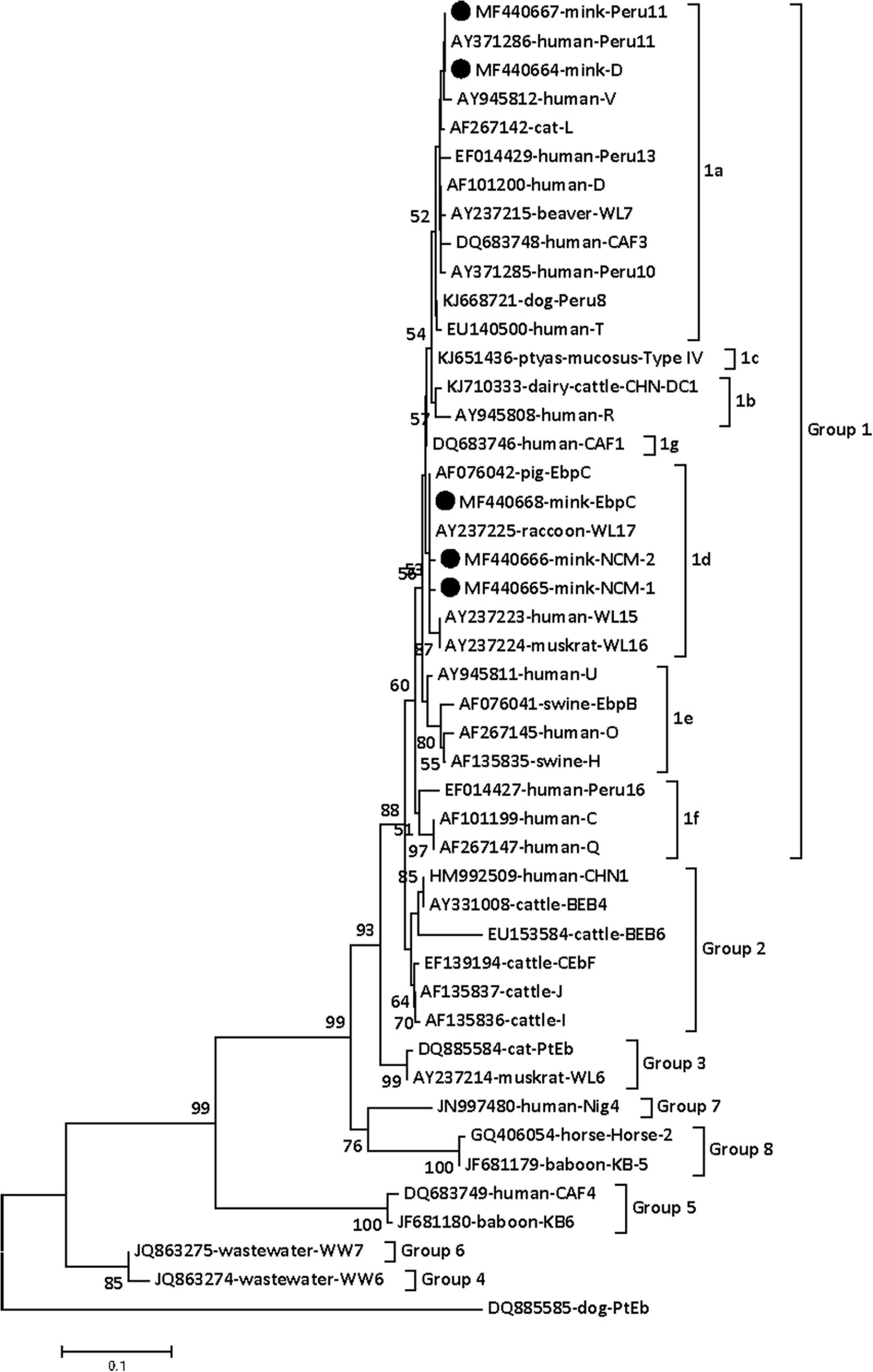
To ascertain the identity of the E. bieneusi isolates, phylogenetic relationship of E. bieneusi isolates were reconstructed base on the ITS sequences. In this tree, groups 1, 2, 3, 5 were in different clade, respectively; however, groups 7, 8 and groups 4, 6 were in two different clade, respectively (Figure 1). Group 1 and group 2 were more closely related than to other groups. These results indicated that all the E. bieneusi isolates in present study were classified to group 1 (Figure 1). Group 1 can be further divided into six subgroup (subgroups 1a–f). D and Peru11 were in supgroup 1a (Figure 1); however, EbpC, NCM-1, and NCM-2 were in subgroup 1d (Figure 1).

FIGURE 1. Phylogenetic analyses of Enterocytozoon bieneusi using neighbor-joining (NJ). E. bieneusi isolates identified in the present study are pointed out by solid circles.
Discussion
Enterocytozoon bieneusi is an important enteric pathogen. Humans acquired E. bieneusi infection may present with the symptoms of diarrhea and enteric diseases (Zhao et al., 2015). Various studies have been reported from E. bieneusi prevalence in different animals around the world (Santín and Fayer, 2011; Ma et al., 2015; Santin and Fayer, 2015; Prasertbun et al., 2017), but information about distribution of genotypes of E. bieneusi in captive animals in China is scarce, especially in minks.
In the present study, the overall prevalence of E. bieneusi in minks was 10.1% (30/298), which was higher than that in domestic rabbits (0.94%, 4/426) (Zhang et al., 2016b), raccoon dogs (4.1%, 2/49) (Zhao et al., 2015), pet chinchillas (3.6%, 5/140) (Qi et al., 2015), captive snakes (4.6%, 11/240) (Karim et al., 2014b), but was lower than that in captive golden snub-nosed monkeys (46.2%, 74/160) (Yu et al., 2017), and captive Asiatic black bears (27.4%, 29/106) (Yang et al., 2015) in other provinces of China. This is most likely due to the worse raising conditions and generally higher density of minks in investigated farms. We found that minks from Hebei province (32.3%, 95% CI 14.8–49.7, OR = 7.05, P = 0.01) have a higher prevalence of E. bieneusi compared with Jilin (10.5%, 95% CI 2.9–18.0, OR = 1.7), Heilongjiang (6.3%, 95% CI 0.8–11.8, OR = 1), Liaoning (8.9%, 95% CI 2.9–14.9, OR = 1.4), and Shandong provinces (0%). Moreover, the difference in prevalence may be related to other factors, such as animal welfares, climates, and animal husbandry practices.
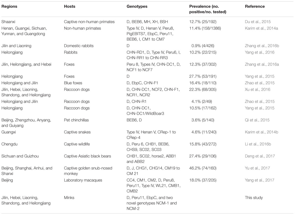
More than 50 E. bieneusi ITS genotypes have been identified in captive animals in China (Table 4). However, only five genotypes were identified in present study. These findings suggested that the five genotypes were endemic E. bieneusi in minks in Northern China. In the present study, the most prevalent genotype was D which has also been found in captive non-human primates, domestic rabbits, captive foxes, captive raccoon dogs, pet chinchillas, and many other captive wildlife (Karim et al., 2014a; Du et al., 2015; Qi et al., 2015; Yang et al., 2015, 2016, 2017; Zhao et al., 2015; Li et al., 2016b; Xu et al., 2016; Zhang et al., 2016a,b; Yu et al., 2017). In addition, the second most prevalent genotype was EbpC which was also more frequently found in some captive animals (captive foxes and captive non-human primates) (Karim et al., 2014a; Zhao et al., 2015; Yang et al., 2017); however, Peru11 was only found in captive non-human primates in China (Karim et al., 2014a; Yang et al., 2017). These findings also suggested that these genotypes of E. bieneusi might transmission among these captive animals. More importantly, three known genotypes of E. bieneusi from the present study (D, Peru11, and EbpC) have been also previously identified in humans in China (Wang et al., 2013; Ojuromi et al., 2016). Therefore, our results have indicated that mink might be a potential source of infection for humans. Moreover, seven polymorphic sites were observed among the five genotypes, implying the more genetic diversity of E. bieneusi in minks in Northern China.
Minks is one of the most important economic animals. They can provide thick fur for humans. Therefore, more and more minks were raised as an important source of income in many countries. Previous studies have indicated that many pathogens can infect minks (Chalmers et al., 2015; Gholipour et al., 2017; Xie et al., 2018). In the present study, we found that E. bieneusi can also infect minks which expend the hosts range of E. bieneusi. The results of the present survey have implications for the controlling E. bieneusi infection in minks, other animals and humans in China and elsewhere in the world.
Conclusion
This is the first record of E. bieneusi in minks, and the prevalence is associated with the region of investigated minks. These findings also suggest that D, Peru11, EbpC, NCM-1, and NCM-2 are endemic in minks in Northern China. The occurrence of zoonotic E. bieneusi genotypes in the feces of the minks suggests potential environmental contamination with E. bieneusi oocysts and may raise a public health concern. Moreover, effective measures should be implemented to avoid water-born microsporidiosis outbreaks.
Data Availability Statement
Representative nucleotide sequences were submitted to GenBank under accession numbers: MF440664–MF440668.
Author Contributions
G-HL and GH conceived and designed the study, and critically revised the manuscript. X-XZ, R-LJ, and J-GM performed the experiments. X-XZ and R-LJ analyzed the data. X-XZ drafted the manuscript. CX and QZ helped in study design, study implementation, and manuscript preparation. All authors read and approved the final manuscript.
Funding
This project was supported by the Scientific Research Fund of Hunan Provincial Education Department (Grant No. 16A102).
Conflict of Interest Statement
The authors declare that the research was conducted in the absence of any commercial or financial relationships that could be construed as a potential conflict of interest.
References
Canning, E. U., and Hollister, W. S. (1990). Enterocytozoon bieneusi (Microspora): prevalence and pathogenicity in AIDS patients. Trans. R. Soc. Trop. Med. Hyg. 84, 181–186. doi: 10.1016/0035-9203(90)90247-C
Chalmers, G., McLean, J., Hunter, D. B., Brash, M., Slavic, D., Pearl, D. L., et al. (2015). Staphylococcus spp., Streptococcus canis, and Arcanobacterium phocae of healthy Canadian farmed mink and mink with pododermatitis. Can. J. Vet. Res. 79, 129–135.
da Cunha, M. J., Cury, M. C., and Santín, M. (2016). Widespread presence of human-pathogenic Enterocytozoon bieneusi genotypes in chickens. Vet. Parasitol. 217, 108–112. doi: 10.1016/j.vetpar.2015.12.019
da Cunha, M. J. R., Cury, M. C., and Santín, M. (2017). Molecular identification of Enterocytozoon bieneusi, Cryptosporidium, and Giardia in Brazilian captive birds. Parasitol. Res. 116, 487–493. doi: 10.1007/s00436-016-5309-6
Deng, L., Li, W., Zhong, Z., Gong, C., Cao, X., Song, Y., et al. (2017). Multi-locus genotypes of Enterocytozoon bieneusi in captive Asiatic black bears in southwestern China: high genetic diversity, broad host range, and zoonotic potential. PLoS One 12:e0171772. doi: 10.1371/journal.pone.0171772
Desportes, I., Le Charpentier, Y., Galian, A., Bernard, F., Cochand-Priollet, B., Lavergne, A., et al. (1985). Occurrence of a new microsporidan: Enterocytozoon bieneusi n.g., n. sp., in the enterocytes of a human patient with AIDS. J. Protozool. 32, 250–254. doi: 10.1111/j.1550-7408.1985.tb03046.x
Deplazes, P., Mathis, A., Müller, C., and Weber, R. (1996). Molecular epidemiology of Encephalitozoon cuniculi and first detection of Enterocytozoon bieneusi in faecal samples of pigs. J. Eukaryot. Microbiol. 43:93S. doi: 10.1111/j.1550-7408.1996.tb05018.x
Du, S. Z., Zhao, G. H., Shao, J. F., Fang, Y. Q., Tian, G. R., Zhang, L. X., et al. (2015). Cryptosporidium spp., Giardia intestinalis, and Enterocytozoon bieneusi in captive non-human primates in Qinling Mountains. Korean J. Parasitol. 53, 395–402. doi: 10.3347/kjp.2015.53.4.395
Fiuza, V. R., Lopes, C. W., Cosendey, R. I., de Oliveira, F. C., Fayer, R., and Santín, M. (2016). Zoonotic Enterocytozoon bieneusi genotypes found in brazilian sheep. Res. Vet. Sci. 107, 196–201. doi: 10.1016/j.rvsc.2016.06.006
Gholipour, H., Busquets, N., Fernández-Aguilar, X., Sánchez, A., Ribas, M. P., De Pedro, G., et al. (2017). Influenza A virus surveillance in the invasive American mink (Neovison vison) from freshwater ecosystems, Northern Spain. Zoonoses Public Health 64, 363–369. doi: 10.1111/zph.12316
Hu, Y., Feng, Y., Huang, C., and Xiao, L. (2014). Occurrence, source, and human infection potential of Cryptosporidium and Enterocytozoon bieneusi in drinking source water in Shanghai, China, during a pig carcass disposal incident. Environ. Sci. Technol. 48, 14219–14227. doi: 10.1021/es504464t
Jiang, Y., Tao, W., Wan, Q., Li, Q., Yang, Y., Lin, Y., et al. (2015). Erratum for Jiang, Zoonotic and potentially Host-Adapted Enterocytozoon bieneusi Genotypes in Sheep and cattle in Northeast China and an increasing concern about the Zoonotic importance of previously considered Ruminant-Adapted Genotypes. Appl. Environ. Microbiol. 81:5278. doi: 10.1128/AEM.01928-15
Karim, M. R., Wang, R., Dong, H., Zhang, L., Li, J., Zhang, S., et al. (2014a). Genetic polymorphism and zoonotic potential of Enterocytozoon bieneusi from nonhuman primates in China. Appl. Environ. Microbiol. 80, 1893–1898. doi: 10.1128/AEM.03845-13
Karim, M. R., Yu, F., Li, J., Li, J., Zhang, L., Wang, R., et al. (2014b). First molecular characterization of enteric protozoa and the human pathogenic microsporidian, Enterocytozoon bieneusi, in captive snakes in China. Parasitol. Res. 113, 3041–3048. doi: 10.1007/s00436-014-3967-9
Li, J., Luo, N., Wang, C., Qi, M., Cao, J., Cui, Z., et al. (2016a). Occurrence, molecular characterization and predominant Genotypes of Enterocytozoon bieneusi in dairy cattle in Henan and Ningxia, China. Parasit. Vectors 9:142. doi: 10.1186/s13071-016-1425-5
Li, W., Deng, L., Yu, X., Zhong, Z., Wang, Q., Liu, X., et al. (2016b). Multilocus Genotypes and broad host-range of Enterocytozoon bieneusi in captive wildlife at zoological gardens in China. Parasit. Vectors 9:395. doi: 10.1186/s13071-016-1668-1
Ma, J., Li, P., Zhao, X., Xu, H., Wu, W., Wang, Y., et al. (2015). Occurrence and molecular characterization of Cryptosporidium spp. and Enterocytozoon bieneusi in dairy cattle, beef cattle and water buffaloes in China. Vet. Parasitol. 207, 220–227. doi: 10.1016/j.vetpar.2014.10.011
Meng, Q. F., Yao, G. Z., Qin, S. Y., Wu, J., Zhang, X. C., Bai, Y. D., et al. (2017). Seroprevalence of and risk factors for Neospora caninum infection in yaks (Bos grunniens) in China. Vet. Parasitol. 242, 22–23. doi: 10.1016/j.vetpar.2017.05.022
Ojuromi, O. T., Duan, L., Izquierdo, F., Fenoy, S. M., Oyibo, W. A., Del Aguila, C., et al. (2016). Genotypes of Cryptosporidium spp. and Enterocytozoon bieneusi in human immunodeficiency virus-infected patients in Lagos, Nigeria. J. Eukaryot. Microbiol. 63, 414–418. doi: 10.1111/jeu.12285
Prasertbun, R., Mori, H., Pintong, A. R., Sanyanusin, S., Popruk, S., Komalamisra, C., et al. (2017). Zoonotic potential of Enterocytozoon genotypes in humans and pigs in Thailand. Vet. Parasitol. 233, 73–79. doi: 10.1016/j.vetpar.2016.12.002
Qi, M., Luo, N., Wang, H., Yu, F., Wang, R., Huang, J., et al. (2015). Zoonotic Cryptosporidium spp. and Enterocytozoon bieneusi in pet chinchillas (Chinchilla lanigera) in China. Parasitol. Int. 64, 339–341. doi: 10.1016/j.parint.2015.05.007
Santín, M., and Fayer, R. (2011). Microsporidiosis: Enterocytozoon bieneusi in domesticated and wild animals. Res. Vet. Sci. 90, 363–371. doi: 10.1016/j.rvsc.2010.07.014
Santin, M., and Fayer, R. (2015). Enterocytozoon bieneusi, Giardia, and Cryptosporidium infecting white-tailed deer. J. Eukaryot. Microbiol. 62, 34–43. doi: 10.1111/jeu.12155
Tamura, K., Peterson, D., Peterson, N., Stecher, G., Nei, M., and Kumar, S. (2011). MEGA5: molecular evolutionary Genetics Analysis using maximum likelihood, evolutionary distance, and maximum parsimony methods. Mol. Biol. Evol. 28, 2731–2739. doi: 10.1093/molbev/msr121
Thompson, J. D., Gibson, T. J., Plewniak, F., Jeanmougin, F., and Higgins, D. G. (1997). The Clustal X windows interface: flexible strategies for multiple sequence alignment aided by quality analysis tools. Nucleic Acids Res. 25, 4876–4882. doi: 10.1093/nar/25.24.4876
Wang, L., Xiao, L., Duan, L., Ye, J., Guo, Y., Guo, M., et al. (2013). Concurrent infections of Giardia duodenalis, Enterocytozoon bieneusi, and Clostridium difficile in children during a cryptosporidiosis outbreak in a pediatric hospital in China. PLoS Negl. Trop. Dis. 7:e2437. doi: 10.1371/journal.pntd.0002437
Xie, X. T., Kropinski, A. M., Tapscott, B., Weese, J. S., and Turner, P. V. (2018). Prevalence of fecal Viruses and bacteriophage in Canadian farmed mink (Neovison vison). Microbiologyopen 10:e00622. doi: 10.1002/mbo3.622
Xu, C., Ma, X., Zhang, H., Zhang, X. X., Zhao, J. P., Ba, H. X., et al. (2016). Prevalence, risk factors and molecular characterization of Enterocytozoon bieneusi in raccoon dogs (Nyctereutes procyonoides) in five provinces of Northern China. Acta Trop. 161, 68–72. doi: 10.1016/j.actatropica.2016.05.015
Yang, H., Lin, Y., Li, Y., Song, M., Lu, Y., and Li, W. (2017). Molecular characterization of Enterocytozoon bieneusi isolates in laboratory macaques in North China: zoonotic concerns. Parasitol. Res. 116, 2877–2882. doi: 10.1007/s00436-017-5603-y
Yang, Y., Lin, Y., Li, Q., Zhang, S., Tao, W., Wan, Q., et al. (2015). Widespread presence of human-pathogenic Enterocytozoon bieneusi genotype D in farmed foxes (Vulpes vulpes) and raccoon dogs (Nyctereutes procyonoides) in China: first identification and zoonotic concern. Parasitol. Res. 114, 4341–4348. doi: 10.1007/s00436-015-4714-6
Yang, Z., Zhao, W., Shen, Y., Zhang, W., Shi, Y., Ren, G., et al. (2016). Subtyping of Cryptosporidium cuniculus and genotyping of Enterocytozoon bieneusi in rabbits in two farms in Heilongjiang Province, China. Parasite 23:52. doi: 10.1051/parasite/2016063
Yu, F., Wu, Y., Li, T., Cao, J., Wang, J., Hu, S., et al. (2017). High prevalence of Enterocytozoon bieneusi zoonotic genotype D in captive golden snub-nosed monkey (Rhinopithecus roxellanae) in zoos in China. BMC Vet. Res. 13:158. doi: 10.1186/s12917-017-1084-6
Yue, D. M., Ma, J. G., Li, F. C., Hou, J. L., Zheng, W. B., Zhao, Q., et al. (2017). Occurrence of Enterocytozoon bieneusi in donkeys (Equus asinus) in China: a public health concern. Front. Microbiol. 8:565. doi: 10.3389/fmicb.2017.00565
Zhang, X. X., Cong, W., Lou, Z. L., Ma, J. G., Zheng, W. B., Yao, Q. X., et al. (2016a). Prevalence, risk factors and multilocus genotyping of Enterocytozoon bieneusi in farmed foxes (Vulpes lagopus), Northern China. Parasit. Vectors 9:72. doi: 10.1186/s13071-016-1356-1
Zhang, X. X., Jiang, J., Cai, Y. N., Wang, C. F., Xu, P., Yang, G. L., et al. (2016b). Molecular characterization of Enterocytozoon bieneusi in Domestic Rabbits (Oryctolagus cuniculus) in Northeastern China. Korean J. Parasitol. 54, 81–85. doi: 10.3347/kjp.2016.54.1.81
Zhang, X. X., Shi, W., Zhang, N. Z., Shi, K., Li, J. M., Xu, P., et al. (2017). Prevalence and genetic characterization of Toxoplasma gondii in donkeys in Northeastern China. Infect. Genet. Evol. 54, 455–457. doi: 10.1016/j.meegid.2017.08.008
Keywords: Enterocytozoon bieneusi, prevalence, minks, genotyping, Northern China
Citation: Zhang X-X, Jiang R-L, Ma J-G, Xu C, Zhao Q, Hou G and Liu G-H (2018) Enterocytozoon bieneusi in Minks (Neovison vison) in Northern China: A Public Health Concern. Front. Microbiol. 9:1221. doi: 10.3389/fmicb.2018.01221
Received: 20 March 2018; Accepted: 18 May 2018;
Published: 12 June 2018.
Edited by:
John W. A. Rossen, University Medical Center Groningen, NetherlandsReviewed by:
Vikram Saini, All India Institute of Medical Sciences, IndiaYanmei Zhang, Fudan University, China
Copyright © 2018 Zhang, Jiang, Ma, Xu, Zhao, Hou and Liu. This is an open-access article distributed under the terms of the Creative Commons Attribution License (CC BY). The use, distribution or reproduction in other forums is permitted, provided the original author(s) and the copyright owner are credited and that the original publication in this journal is cited, in accordance with accepted academic practice. No use, distribution or reproduction is permitted which does not comply with these terms.
*Correspondence: Guangyu Hou, aGd5MTk2OTIwMDZAMTI2LmNvbQ== Guo-Hua Liu, bGl1Z3VvaHVhNTIwMjAwOEAxNjMuY29t
†These authors have contributed equally to this work.
 Xiao-Xuan Zhang
 Ruo-Lan Jiang3†
 Chao Xu
 Guo-Hua Liu
