94% of researchers rate our articles as excellent or good
Learn more about the work of our research integrity team to safeguard the quality of each article we publish.
Find out more
CORRECTION article
Front. Immunol. , 18 February 2020
Sec. T Cell Biology
Volume 11 - 2020 | https://doi.org/10.3389/fimmu.2020.00088
This article is a correction to:
Increased Expression of sST2 in Early HIV Infected Patients Attenuated the IL-33 Induced T Cell Responses
 Xian Wu1,2†
 Yao Li1,3†
 Cheng-Bo Song1,4,5,6†
 Ya-Li Chen1,4,5,6
 Ya-Jing Fu1,4,5,6
 Yong-Jun Jiang1,4,5,6
 Hai-Bo Ding1,4,5,6
 Hong Shang1,4,5,6*
 Zi-Ning Zhang1,4,5,6*
A Corrigendum on
Increased Expression of sST2 in Early HIV Infected Patients Attenuated the IL-33 Induced T Cell Responses
by Wu, X., Li, Y., Song, C.-B., Chen, Y.-L., Fu, Y.-J., Jiang, Y.-J., et al. (2018). Front. Immunol. 9:2850. doi: 10.3389/fimmu.2018.02850
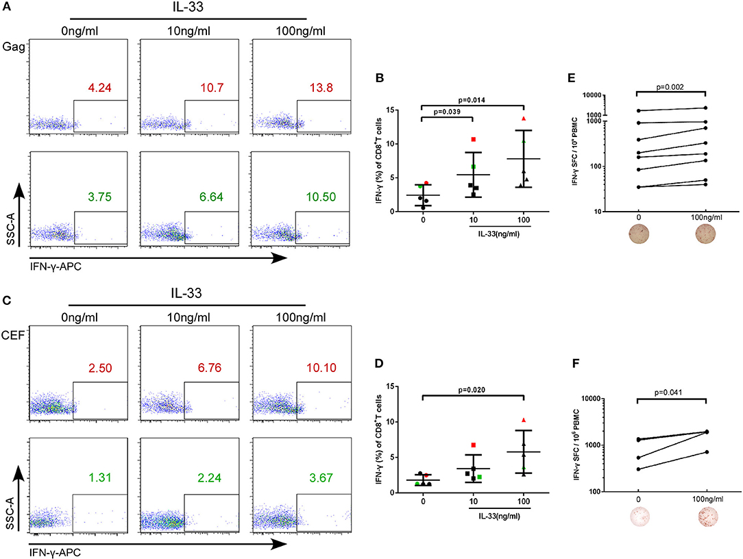
In the original article, there was a mistake in Figure 2C as published. The leftmost diagram at the bottom of Figure 2C was mistakenly duplicated from the third diagram at the bottom of Figure 2C during the figure preparation. The corrected Figure 2 appears below.

Figure 2. IL-33 increases the expression of IFN-γ by Gag and CEF stimulated CD8+ T cells. CD8+ T cells were isolated from HIV-1 individuals and treated with Gag peptide pools with rhIL-33 (10 ng/mL and 100 ng/mL) or without IL-33 (0 ng/mL). Intracellular IFN-γ expression was detected by flow cytometer and compared by paired t-test (0 ng/mL: 2.44 ± 1.53%; 10 ng/mL: 5.46 ± 3.30%; 100 ng/mL: 7.81 ± 4.20%). Representative flow cytometry dot plot (A) and summary data (B) were shown. CD8+ T cells were isolated from HIV-1 individuals and treated with CEF peptide pools with rhIL-33 (10 ng/mL and 100 ng/mL) or without IL-33 (0 ng/mL). Intracellular IFN-γ expression was detected by flow cytometer and compared by paired t-test (0 ng/mL: 1.81 ± 0.75%; 10 ng/mL: 3.44 ± 1.93%; 100 ng/mL: 5.80 ± 3.00%). Representative flow cytometry dot plot (C) and summary data (D) were shown. CD8+ T cells were stimulated with Gag peptide pools (E) or CEF peptides (F) and IFN-γ secretion was detected by ELISPOT assay. The numbers of spot forming cells (SFC) were log transformed and then compared by paired t-test. The number of SFC treated by 100 ng/mL IL-33 were compared with cells without IL-33 stimulation (0 ng/mL).
The authors apologize for this error and state that this does not change the scientific conclusions of the article in any way. The original article has been updated.
Keywords: IL-33, ST2, T cell response, IFN-γ, HIV infection
Citation: Wu X, Li Y, Song C-B, Chen Y-L, Fu Y-J, Jiang Y-J, Ding H-B, Shang H and Zhang Z-N (2020) Corrigendum: Increased Expression of sST2 in Early HIV Infected Patients Attenuated the IL-33 Induced T Cell Responses. Front. Immunol. 11:88. doi: 10.3389/fimmu.2020.00088
Received: 07 January 2020; Accepted: 14 January 2020;
Published: 18 February 2020.
Edited and reviewed by: Loretta Tuosto, Sapienza University of Rome, Italy
Copyright © 2020 Wu, Li, Song, Chen, Fu, Jiang, Ding, Shang and Zhang. This is an open-access article distributed under the terms of the Creative Commons Attribution License (CC BY). The use, distribution or reproduction in other forums is permitted, provided the original author(s) and the copyright owner(s) are credited and that the original publication in this journal is cited, in accordance with accepted academic practice. No use, distribution or reproduction is permitted which does not comply with these terms.
*Correspondence: Hong Shang, aG9uZ3NoYW5nMTAwQGhvdG1haWwuY29t; Zi-Ning Zhang, emlfbmluZzEwMUBob3RtYWlsLmNvbQ==
†These authors have contributed equally to this work
Disclaimer: All claims expressed in this article are solely those of the authors and do not necessarily represent those of their affiliated organizations, or those of the publisher, the editors and the reviewers. Any product that may be evaluated in this article or claim that may be made by its manufacturer is not guaranteed or endorsed by the publisher.
Research integrity at Frontiers

Learn more about the work of our research integrity team to safeguard the quality of each article we publish.