- 1Metabolomics Core Technology Platform, Centre for Organismal Studies (COS), Heidelberg University, Heidelberg, Germany
- 2Institute of Pharmacology, Heidelberg University, Heidelberg, Germany
- 3Division of Signal Transduction in Cancer and Metabolism, German Cancer Research Center (DKFZ), Heidelberg, Germany
- 4European Center for Angioscience (ECAS), Department of Vascular Biology and Tumor Angiogenesis, Medical Faculty Mannheim, Heidelberg University, Mannheim, Germany
- 5Department of Internal Medicine I and Clinical Chemistry, Heidelberg University Hospital, Heidelberg, Germany
- 6Heidelberg Institute for Stem Cell Technology and Experimental Medicine (HI-STEM GGmbH), Heidelberg, Germany
- 7Division of Stem Cells and Cancer, Deutsches Krebsforschungszentrum (DKFZ) and DKFZ-ZMBH Alliance, Heidelberg, Germany
- 8Faculty of Biosciences, Heidelberg University, Heidelberg, Germany
Metabolic profiling harbors the potential to better understand various disease entities such as cancer, diabetes, Alzheimer’s, Parkinson’s disease or COVID-19. To better understand such diseases and their intricate metabolic pathways in human studies, model animals are regularly used. There, standardized rearing conditions and uniform sampling strategies are prerequisites towards a successful metabolomic study that can be achieved through model organisms. Although metabolomic approaches have been employed on model organisms before, no systematic assessment of different conditions to optimize metabolite extraction across several organisms and sample types has been conducted. We address this issue using a highly standardized metabolic profiling assay analyzing 630 metabolites across three commonly used model organisms (Drosophila, mouse, and zebrafish) to find an optimal extraction protocol for various matrices. Focusing on parameters such as metabolite coverage, concentration and variance between replicates we compared seven extraction protocols. We found that the application of a combination of 75% ethanol and methyl tertiary-butyl ether (MTBE), while not producing the broadest coverage and highest concentrations, was the most reproducible extraction protocol. We were able to determine up to 530 metabolites in mouse kidney samples, 509 in mouse liver, 422 in zebrafish and 388 in Drosophila and discovered a core overlap of 261 metabolites in these four matrices. To enable other scientists to search for the most suitable extraction protocol in their experimental context and interact with this comprehensive data, we have integrated our data set in the open-source shiny app “MetaboExtract”. Hereby, scientists can search for metabolites or compound classes of interest, compare them across the different tested extraction protocols and sample types as well as find reference concentration values.
Introduction
Metabolomics, defined as the separation and subsequent measurement of small molecules in either a qualitative or quantitative way, enables the generation of metabolic profiles of any sample of interest. While genomics and transcriptomics are analyzed within the framework of a single organism and understood by the blueprint of genes or transcripts of the respective species, metabolomics encompasses all compounds that may be metabolized by an organism or its microbiome, or that are introduced by the environment (“exposome”) at a given time. Therefore, the metabolome incorporates the environmental influence as well as interactions with other organisms (Johnson et al., 2012). It can serve as a bridge between the organism, its interactions and any disease, e.g., between diet, the gut microbiome and metabolic disease (Pallister et al., 2017). While the complexity and dynamic nature of the metabolome is daunting from an analytical perspective, metabolomics harbors the potential to better understand as well as diagnose various disease entities such as diabetes (Arneth et al., 2019), kidney disease (Abbiss et al., 2019), Parkinson´s (Shao and Le, 2019), Alzheimer´s disease (Wilkins and Trushina, 2018) and most recently, COVID-19 (Sindelar et al., 2021).
The potential to understand the metabolic signatures of any given disease entity is tremendous, however, deciphering the intricate underpinnings of those in a human study requires costly, as well as time and work extensive population-wide association studies with several hundred participants per group (Nicholson et al., 2008). These broad studies may be successful in the discovery of new associations between a respective disease and the measured metabolites, i.e., biomarkers, but they are limited in their mechanistic explanations despite all efforts. While the dynamic nature of the metabolome provides incredibly powerful insights, it also highlights the challenges of metabolome analyses — its variability and associated pitfalls.
Variation and noise that are biologically inherent or are introduced at some point to the sample are complicating metabolic analyses, impairing the quality of the findings, limiting their validity and may even overshadow the effect size of the research question itself. Factors that introduce such variability range from intrinsic ones derived from the study organism (age or sex) (Brennan and Gibbons, 2020; Bell et al., 2021), to extrinsic factors (diet, lifestyle or medication) (Mellert et al., 2011; Adamski, 2016). Additionally, other factors, such as pre-analytical ones during sample collection (Yin et al., 2015; Lippi et al., 2020), or analytical factors deriving from the sample preparation, the extraction protocols or analytical approach used to conduct the measurement (Lin et al., 2007; Erben et al., 2021) are also influential and need to be accounted for.
Model organisms that are reared under controlled laboratory conditions and manipulated genetically to analyze a certain genotype address several of the challenges mentioned above. Combining the standardized rearing conditions and stringent sampling protocols with the already extensive knowledge accumulated from other “-omics” in models such as mice, Drosophila or zebrafish enhances the explanatory power of metabolomic studies tremendously while simultaneously reducing the number of samples needed to generate meaningful results. To ensure that the analytical phase, i.e., the extraction and measurement of metabolites, does not introduce biases and variability, an in-depth evaluation of such aspects is necessary.
Standardized metabolomic analyses are commercially available by companies such as Metabolon (www.metabolon.com) or Biocrates (www.biocrates.com). The latter has developed standardized and robust LC-MS/MS based kits which enable the absolute quantification of specific compound classes or more broadly, up to 630 metabolites in the case of the MxP Quant 500 kit (Biocrates). Within these 630 metabolites, the MxP Quant 500 kit covers 14 small molecule and 9 different lipid classes. Due to its standardized nature and compatibility with a multitude of LC-MS/MS platforms, data generated via such a kit-based approach enables inter- and intra-laboratory comparability (Siskos et al., 2017), as well as its integration from different experiments. Although these kits were initially developed for human biofluids, i.e., plasma and serum, they may be used for tissue samples (Zukunft et al., 2018) and other sample types such as cultured cells (Andresen et al., 2021) or supernatants likewise. However, there is no consensus on the optimal metabolite extraction procedure amongst the metabolomic community for the investigation of polar and non-polar metabolites covering that many chemical classes across different model organisms within one analysis.
In this study, this open question was addressed using the highly standardized targeted metabolomics kit (Biocrates MxP Quant 500) to evaluate seven extraction protocols designed to extract both, polar and non-polar metabolites, differing in their solvent composition and extraction mode (mono/biphasic) as well as handling complexity (Figure 1). We compared the metabolite coverage, concentration and robustness, i.e., the coefficient of variance (CV%) across three commonly used model organisms (mouse, zebrafish and Drosophila), focusing on either whole organisms as sample type (Drosophila) or specific organs (liver and kidney) of the respective model organism. Lastly, we integrated our data in the Shiny app “MetaboExtract” (Andresen et al., 2021) to provide a useful source of metabolite concentrations across model organisms and enable other scientists to search for an optimal extraction procedure for their metabolite or metabolite class of interest.
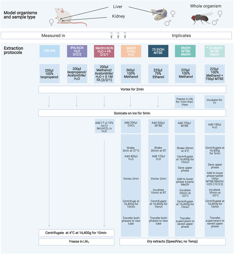
FIGURE 1. Overview of the seven extraction protocols used as well as the model organisms and sample types investigated. The protocols increase in handling effort and complexity from left to right. The color code indicates similarities amongst the protocols either through solvents or chemicals used. All extraction products were stored at −80°C until further processing. A list of abbreviations can be found above.
Materials and Methods
Chemicals
Chemicals were bought from Sigma-Aldrich (Germany). All solvents used for sample extractions and LC- or FIA-MS/MS analyses were of UHPLC-MS quality.
Model Organism Growth/Culturing Conditions
Mouse–Mus musculus
Nine-week-old C57Bl6N wildtype mice (Charles River, Germany) were anesthetized with isoflurane and blood was taken to generate EDTA-Plasma. Without regaining consciousness mice were killed by cervical dislocation. Livers and kidneys were excised rapidly and shortly rinsed in ice-cold 0.9% NaCl. Excess liquid was removed before whole organ weight was determined for later normalization and tissue was snap-frozen in liquid nitrogen. All procedures were approved by the Animal Care and Use Committee at the Regierungspräsidium Karlsruhe, Germany (T-40/20).
Fly - Drosophila melanogaster
W1118 Drosophila were acquired from Bloomington Drosophila Stock Center. For all metabolic measurements, Drosophila were grown under controlled conditions: Drosophila were allowed to lay eggs on apple plates for 12 h. First instar larvae hatching within a 4- or 6-h window were picked and seeded at a density of 60 animals per vial. Adult Drosophila of all genotypes enclosing within a 24-h time-window were separated by gender in groups of 30 Drosophila and aged for 10 days. Drosophila were grown and maintained on food consisting of the following ingredients for 30 L of food: 480 g agar, 660 g sugar syrup, 2400 g malt, 2400 g corn meal, 300 g soymeal, 540 g yeast, 72 g nipagin, 187 ml propionic acid and 18.7 ml phosphoric acid. At sample collection, Drosophila were pooled and snap-frozen for metabolic profiling.
Zebrafish - Danio rerio
Adult zebrafish were kept under a 13-h light/11-h dark cycle and feeding of zebrafish took places twice a day, freshly hatched Artemia salina in the morning and fish flake food in the afternoon. All experimental interventions on animals were approved by the local government authority, Regierungspräsidium Karlsruhe and by Medical Faculty Mannheim (I-19/02) and carried out in accordance with the approved guidelines. Age of adult male zebrafish was 9 months and both sexes were included. Zebrafish were sacrificed in ice water and livers were immediately dissected and frozen in liquid nitrogen and subsequently stored at −80°C. 7–10 mg of livers were used for further analysis.
Sample Preparation
To ensure sufficient input material across the model organisms 30 pooled Drosophila (w1118), 7–10 mg of zebrafish liver or 20–22 mg of mouse (C57Bl6N) liver and kidney pooled from three individuals respectively were used. All tissue samples were pulverized using a ball mill (MM400, Retsch) with precooled beakers and stainless-steel balls for 30 s at the highest frequency (30 Hz). The exact weight was determined for normalization of all measurements.
Metabolite Extraction Protocols
Here we evaluated six different extraction protocols that are described in Figure 1. We developed these protocols based on own preliminary experience and reviewing of general metabolomics literature addressing similar questions (Lisec et al., 2006; Rabinowitz and Kimball, 2007; Ivanisevic et al., 2013; Weir et al., 2013; Zukunft et al., 2018). The protocol “MeOH/MTBE”, noted with an asterisk, was applied twice with slight variations in mouse samples only. We are including this variation as an additional method (2xMeOH/MTBE) when we are referring to the seven extraction protocols.
Briefly, pulverized and frozen samples were extracted using the indicated solvents and subsequent steps of the respective protocol (Figure 1). After a final centrifugation step the solvent extract of the protocols 100IPA, IPA/ACN and MeOH/ACN were transferred into a new 1.5 ml tube (Eppendorf) and snap-frozen until kit preparation. The remaining protocols were dried using an Eppendorf Concentrator Plus set to no heat, stored at −80°C and reconstituted in 60 µL isopropanol (30 µL of 100% isopropanol, followed by 30 µL of 30% isopropanol in water) before the measurement.
Standardized Targeted Metabolic Profiling
After conducting the described seven extraction protocols, tissue extracts were processed following the manufacturer’s protocol of the MxP® Quant 500 kit (Biocrates). Briefly, 10 µL of the samples or blanks were pipetted on the 96 well-plate based kit containing calibrators and internal standards using an automated liquid handling station (epMotion 5075, Eppendorf) and subsequently dried under a nitrogen stream using a positive pressure manifold (Waters). Afterwards, 50 µL phenyl isothiocyanate 5% (PITC) was added to each well to derivatize amino acids and biogenic amines. After 1 h incubation time at room temperature, the plate was dried again. To resolve all extracted metabolites 300 µL of 5 mM ammonium acetate in methanol were pipetted to each filter and incubated for 30 min. The extract was eluted into a new 96-well plate using positive pressure. For the LC-MS/MS analyses 150 µL of the extract was diluted with an equal volume of water. Similarly, for the FIA-MS/MS analyses 10 µL extract was diluted with 490 µL of FIA solvent (provided by Biocrates). After dilution, LC-MS/MS and FIA-MS/MS measurements were performed in positive and negative mode. For chromatographic separation an UPLC I-class PLUS (Waters) system was used coupled to a SCIEX QTRAP 6500 + mass spectrometry system in electrospray ionization (ESI) mode. LC gradient composition and specific 50 × 2.1 mm column are provided by Biocrates. Data was recorded using the Analyst (Version 1.7.2 Sciex) software suite and further processed via MetIDQ software (Oxygen-DB110-3005). All metabolites were identified using isotopically labeled internal standards and multiple reaction monitoring (MRM) using optimized MS conditions as provided by Biocrates. For quantification either a seven-point calibration curve or one-point calibration was used depending on the metabolite class.
Data Processing and Analyses
Validation and Filtering
Data validation and quantification was performed using MetIDQ (Oxygen-DB110-3005). Here, metabolites were further categorized based on their quantitation ranges. Additional filtering per metabolite was based on the limit of detection (LOD), limit of quantification (LOQ) and concentration within the quantitative range (valid). To remove metabolites that were not present in any model organism and sample type, we considered a metabolite as detectable when at least 2 out of 3 replicates within a tested protocol were above LOD (see Figure 2). These metabolites are also visualized as Venn diagrams in Figure 5 for the extraction protocol EtOH/MTBE and for the remaining extraction protocols in Supplementary Figure S5. An overview of the LOD, LOQ and valid metabolite proportions are shown in Supplementary Figure S1. For all detectable metabolites, the coefficient of variation (CV) in percentage was calculated as well as the median absolute deviation (MAD) based on the concentrations.
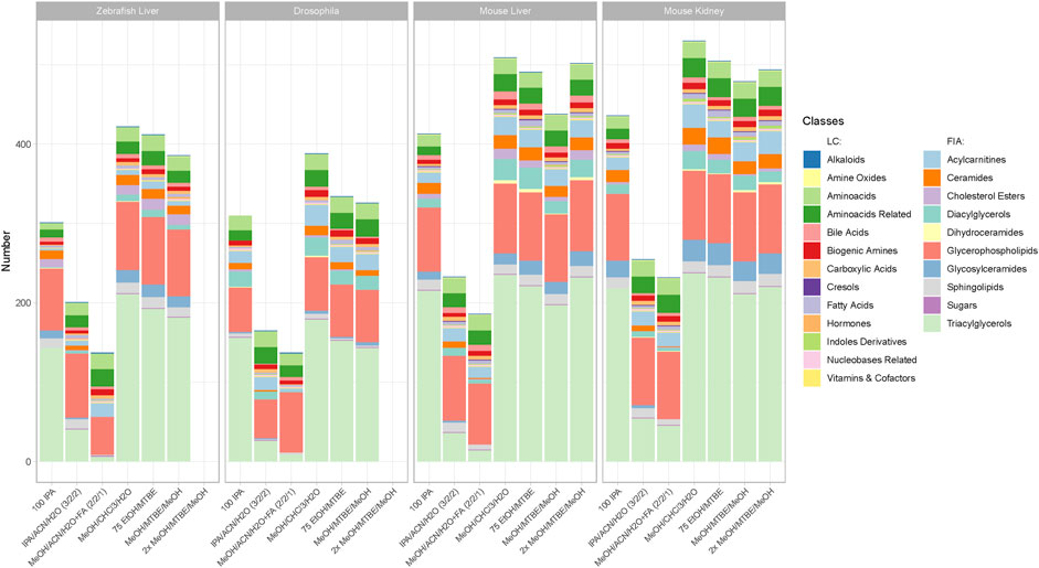
FIGURE 2. Metabolite coverage per extraction protocol across all sample types and model organisms. Indicated by color are the different metabolite classes measured. A metabolite was counted as detectable when at least 2 out of 3 replicates were >LOD within a given extraction protocol. The legend is categorized between compound classes measured via LC-MS/MS or FIA-MS/MS. Ordering from left to right follows the level of complexity and required time per extraction.
Statistical Analysis
To find the optimal protocol per model organism and sample type, we analyzed the concentration per metabolite achieved across the extraction protocols. For this comparison, missing values and zero values were imputed per metabolite with 20% of the minimal positive value of a given metabolite. Subsequently, to perform statistical analyses, the data was log2-transformed. We then performed an ANOVA per metabolite considering all metabolites that were detectable with at least a single method. Extraction protocols were used as categorical variables and concentration as dependent variables. A Tukey post-hoc test (alpha = 0.05) was used to determine the extraction protocols with the highest median concentration as well as non-significantly (p-adjusted > 0.05) lower concentrations. These extraction protocols were considered optimal, counted, and depicted in Figure 3. Conversely, metabolites that were significantly better extracted in a single extraction protocol are depicted in Supplementary Figure S4. We implemented and employed the R package “MetAlyzer” (https://CRAN.R-project.org/package=MetAlyzer), which provides an R-S4 object with methods to read output files from the MetIDQ software into R for convenient data processing, statistics and downstream analysis. It covers every step of filtering and analysis with the parameters used in this work to ensure the best possible reproducibility.
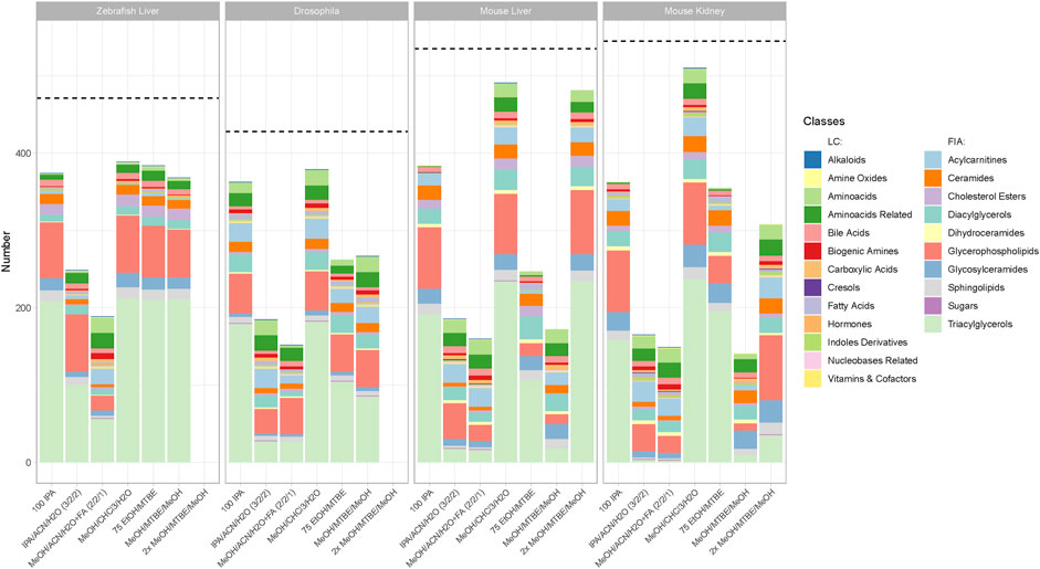
FIGURE 3. Number of metabolites per class with the highest concentration per extraction protocol across all sample types and model organisms. Metabolites that appear in the bar chart are only counted when they produce the highest or a non-significantly lower concentration (p > 0.05, see Material and Methods) than another tested extraction protocol. The dotted line shows the number of detectable metabolites for each sample type. Indicated by color are the different metabolite classes measured. The legend is categorized between the LC-MS/MS and FIA-MS/MS measurements. A list of the metabolites per extraction protocol can be viewed online in “MetaboExtract”. Of note, the metabolite classes that were best suited for a single extraction protocol are depicted in Supplementary Figure S4.
R Shiny app
Data can be explored and downloaded using the Shiny app “MetaboExtract” which is available at http://www.metaboextract.shiny.dkfz.de. The underlying code is also available at https://github.com/andresenc/MetaboExtract (Andresen et al., 2021). Figures 2, 3 as well as S3 were extracted from the Shiny app.
Results
The aim of this study was the comparison of seven extraction protocols (Figure 1) across three model organisms to determine the optimal extraction procedure with regards to metabolite coverage, their absolute concentration and robustness (CV%). In total, we analyzed 630 metabolites, however, after filtering for low concentrated metabolites below the limit of detection (LOD), we continued the analyses using this processed data. Supplementary Figure S1 shows the ratio of LOD, LOQ and valid measurements per extraction protocol across all sample types and model organisms.
Biphasic Extractions Generate the Highest Coverage and Concentration
The metabolic profiling kit (Biocrates MxP Quant 500) used for this study quantifies polar as well as non-polar metabolites across 14 small molecule and nine different lipid classes. Therefore, an extraction procedure is required that enables solubilization ranging from very polar metabolites (e.g., carbohydrates and amino acids) to very non-polar metabolites such as triacylglycerols (Figure 2). While the maximum coverage between the different model organisms is expected to be variable, the general performance of the respective protocol remained similar. Figure 2 shows the detected metabolites per extraction protocol across all sample types and model organisms. The ordering of the protocols from left to right also indicates the level of complexity and time required for the protocol (Figure 1).
Clear performance trends between the monophasic (100IPA, IPA/ACN/H2O, MeOH/ACN/H2O + FA) and biphasic (MeOH/CHCl3/H2O, 75EtOH/MTBE, (2x)MeOH/MTBE) extractions were apparent. The protocol using MeOH/CHCl3/H2O resulted in the highest metabolite coverage in all sample types and across all organisms (zebrafish liver (422), Drosophila (388), mouse liver (509), mouse kidney (530)). Similarly, 75EtOH/MTBE, as well as both MeOH/MTBE protocols, produced a broad coverage across all metabolite classes. In other words, all biphasic extractions performed well and were comparable regarding their metabolite coverage.
While 100IPA, a rapid and simple single solvent extraction protocol, produced fair coverage, the remaining protocols, both containing acetonitrile, achieved the lowest coverage regardless of the sample type or organism. Comparison of the different metabolite classes reveal that these monophasic extraction protocols failed to extract several lipids, such as di- and triacylglycerols as well as ceramides or cholesterol esters.
Although the coverage of a given extraction is essential, the concentration of a metabolite may vary across the different extraction procedures. Here, we consider an extraction protocol as better when higher concentration of metabolites can be achieved. To scrutinize the extraction protocols regarding this criterion we performed an ANOVA (see material and method part) counting the metabolites that reached the highest or a non-significantly lower concentration in a given extraction protocol per model organism (Figure 3). Therefore, high counts of metabolites in Figure 3. indicate that a given protocol extracted the highest concentration. Vice versa, lower counts in Figure 3 indicate that other protocols extracted significantly higher concentrations generating an overview and elucidate trends in performance. A list of the metabolites that are extracted with the highest concentration (or a non-significantly lower concentration that another protocol) is available online in “MetaboExtract”.
In Figure 3 a similar pattern compared to the metabolite coverage (Figure 2) emerged. The protocol using MeOH/CHCl3/H2O resulted in the highest concentrations of metabolites measured within each metabolite class across all organisms (zebrafish liver (392), Drosophila (379), mouse liver (493), mouse kidney (510)). The remaining biphasic extraction protocols performed comparable, apart from MeOH/MTBE in mouse liver and kidney, showing significantly lower concentrations per metabolite than the other protocols with a strong reduction in triacylglycerols. Within the group of MTBE protocols the combination with EtOH was superior to the MeOH extraction resulting in higher or comparable metabolite concentrations. Comparing both MeOH variations, in mice, 2xMeOH/MTBE resulted in higher concentrations than the MeOH/MTBE extraction. Of note, in mouse liver, 2xMeOH/MTBE generated the highest concentrations within the group of MTBE protocols indicating strong differences between the sample types. In-depth comparison of several metabolites using “MetaboExtract” shows that while not counted in Figure 3, MTBE containing protocols perform comparable across most model organisms and sample types.
Similarly to the coverage of metabolites, both acetonitrile-containing protocols performed worse across all model organisms and sample types overall with the exception of very few metabolite classes. For example, MeOH/ACN/H2O + FA extracted amino acids and their related metabolites at higher concentrations across all model organisms. Lastly, the rapid 100IPA protocol produced comparable or higher concentrations than the MTBE protocols across most metabolite classes.
Extraction Protocol Variability as an Essential Quality Parameter
While coverage and concentrations are important to determine the optimal extraction protocol for the broadest range of metabolite classes, the variability or the coefficient of variance (CV%) of each metabolite between the analysis of biological triplicates informs about the robustness of a protocol. To better understand the variability across the different protocols and compare it alongside the coverage we plotted both as a spider plot in Figure 4. There, the variability of the measurements between the triplicates per metabolite in CV% ranges from 0–10% (= excellent), 11–20% (= good), 21–30% (= acceptable) and >30% (= not acceptable). The percentage ranges were calculated from the total of 630 possible metabolites, elucidating the variability of a method but also the number of detectable metabolites per method. For example, in mouse kidney, 75EtOH/MTBE results in 202 (32.1%) metabolites with an excellent CV, 252 (40%) metabolites with a good CV, 21 (3.3%) metabolites with a CV that is acceptable CV and finally, 30 (4.8%) metabolites with a high CV that is not acceptable. Similarly, 100IPA appears as a well performing choice in this sample type with most measurements in a CV% range from 0–10% (= excellent).
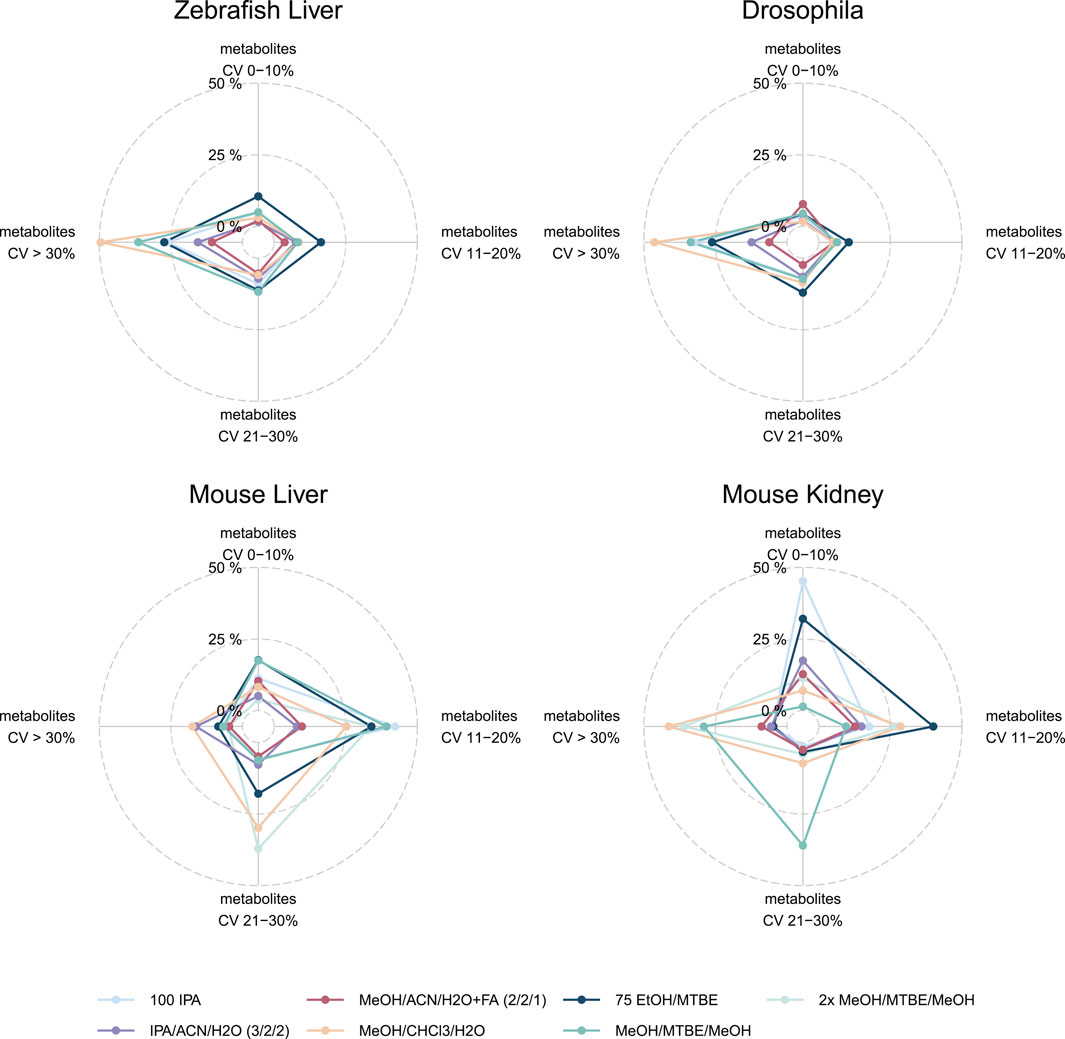
FIGURE 4. Variability of extraction procedures across all sample types and model organisms. Indicated by color are the different extraction protocols used. The CV% was generated based on the triplicate analyses. For each of the CV% categories the percentage of the total 630 metabolites. Note that 2xMeOH/MTBE/MeOH was only used for mice sample types. Alternative visualizations of the CV% and MAD are shown in the (Supplementary Figure S3, S4).
This visualization enables the comparison of several extraction protocols across all sample types regarding their robustness and coverage at once (Figure 4). The protocol using MeOH/CHCl3/H2O which resulted in the highest coverage and concentration performed the worst across all sample types and model organisms with most of the metabolites with a CV of >30% (zebrafish liver (312), Drosophila (292), mouse liver (110), mouse kidney (260). Similarly, 2xMeOH/MTBE generates a high variability but also a high coverage as well as concentrations in mouse sample types. Amongst the other model organisms (Drosophila and zebrafish), a single extraction with MeOH/MTBE resulted in a high portion of CV >30% (= not acceptable) compared to 75EtOH/MTBE. Yet, differences could be seen in mouse sample types, where 75EtOH/MTBE performed better in mouse kidney than mouse liver and conversely, for MeOH/MTBE.
75EtOH/MTBE resulted in acceptable levels of variance across all sample types and model organisms (<30%). An alternative visualization of the CV% across the different extraction protocols can be found in Supplementary Figure S2. The median and median absolute deviation (MAD) of the coefficient of variation (CV) across the seven extraction protocols is depicted in the Supplementary Figure S3. Both visualizations strengthen the conclusion described above. Notably, Drosophila and zebrafish samples show overall higher variability in the measurements.
As a next step, we used the 75EtOH/MTBE extraction protocol as an example to visualize the overlap and the uniquely determined metabolites across the different sample types and model organisms in a Venn diagram (Figure 5).
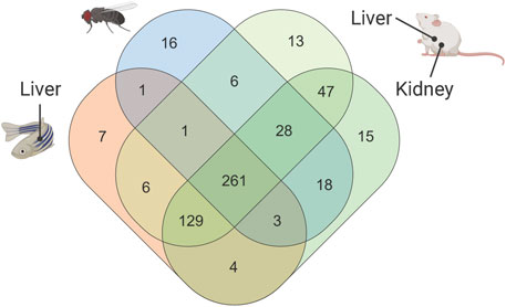
FIGURE 5. Venn diagram showing number of metabolites that are common or unique across all sample types and model organisms within the 75EtOH/MTBE extraction protocol. A comparison of the remaining extraction protocols can be found in the supplement (see Supplementary Figure S5). A list of the metabolites that are unique and overlapping can be found in the supplementary data file (see supplementary Data File S1).
Strong Overlap of Detectable Compounds Between Analyzed Sample Types Using 75EtOH/MTBE
Figure 5 shows the common and uniquely extracted metabolites per model organism within the 75EtOH/MTBE protocol. Overall, the 75EtOH/MTBE protocol resulted in adequate coverage and metabolite concentrations of the evaluated protocols while also showing excellent to acceptable levels of variance between the measurements. Within this protocol 261 common metabolites out of 630 possible compounds could be extracted across all matrices. Additional 129 metabolites were shared between the liver and kidney samples (mouse and zebrafish). Only very few metabolites were unique to each sample type, highlighting the broad coverage of the 75EtOH/MTBE extraction protocol. Venn diagrams for the remaining extraction protocols are provided in Supplementary Figure S5 together with the list of shared or unique metabolites in supplementary Data File S1. There, MeOH/CHCl3/H2O is once more the protocol producing the highest coverage across all sample types and model organism (304 metabolites).
“MetaboExtract” - An Interactive Resource to Explore Metabolite Extractions and Baseline Concentrations of Model Organisms
The presented data provides an attempt to inform about the optimal extraction protocol for a standardized profiling assay. However, it harbors further information such as baseline concentrations for sample types and whole model organisms. To access this information researchers may explore the data set via the easy-to-use interactive R/shiny app “MetaboExtract” (Andresen et al., 2021). There, the already present metabolite data on human tissue and cells was expanded by our data set focused on model organisms. Since all data was generated using the standardized MxP Quant 500 assay it is highly comparable. Organisms, tissues, extraction methods and classes of metabolites may be (de)selected to focus on the data of interest that are then provided in comprehensive and interactive visualizations. The data presented in Figure 2 and in Supplementary Figure S4 were generated using MetaboExtract. Its standardized nature provides the potential for further expansion via additional MxP Quant 500 assay measurements.
Discussion
Model organisms enable standardized laboratory-controlled handling, sampling and experiments. This level of standardization in the pre-analytical phase benefits metabolic profiling due to the dynamic nature of the metabolome, rapid turnover rates of metabolites and the influence of the environment (Edison et al., 2016; Saoi and Britz-mckibbin, 2021).
Here, we focused on the extraction and processing of diverse sample types, one of the most important aspects in the analytical phase requiring strict standardization for reproducibility of data. To this end, we used a targeted metabolic profiling approach (Biocrates MxP Quant 500) quantifying up to 630 metabolites and validated it across three commonly used model organisms (Drosophila, mouse and zebrafish) to find a robust, easy-to-use extraction protocol yielding a comprehensive coverage of the target analytes (Figure 1). This metabolic profiling assay quantifies polar as well as non-polar metabolites across 14 hydrophilic and 9 different lipid classes. Hence, it requires the extraction of a chemically diverse range of metabolites from solid samples. Besides broad metabolite coverage and high concentrations with little loss during the extraction, we evaluated the robustness of a given method as well as the practicability and effort of performing the protocols.
Biphasic Extractions are Superior to Monophasic Extractions
In our study, biphasic extractions (MeOH/CHCl3/H2O, 75EtOH/MTBE, (2x)MeOH/MTBE) resulted in superior coverage and concentrations across all model organisms and sample types. Here, the complementary phases, composed of an organic lipid-rich phase and an aqueous phase containing primary and secondary metabolites, were combined and dried in the final step of each protocol allowing for a greater coverage as compared to monophasic extractions. Chloroform based biphasic extractions by Bligh and Dyer (1959) have been dominantly used over the years due to the focus on the lipid fraction, however, MTBE (methyl tert-butyl ether, i.e., TBME) is more frequently used as a non-toxic and non-carcinogenic alternative to chloroform (Matyash et al., 2008; Furse et al., 2015). Here, both strong hydrophobic solvents in combination with another organic solvent of lower hydrophobicity such as ethanol or methanol resulted in comparable metabolite coverage. Importantly, the chloroform-based extraction resulted in the highest concentrations as well as broadest coverage, however, substituting it with MTBE resulted in similar but less variable measurements (Figures 3, 4).
Monophasic extractions (100IPA, IPA/ACN and MeOH/ACN) require less solvent and are performed more rapidly as compared to biphasic ones, which is a big advantage when processing large numbers of samples. While the robustness of monophasic extractions was comparable to that of the other well performing biphasic extractions, i.e., 75EtOH/MTBE, they provided lower compound coverage due to the lack of certain lipids that were poorly extracted, with the exception of 100IPA, which provided adequate coverage and concentrations in most cases. This easy-to-use and rapid protocol achieved adequate lipid coverage and reproducibility in most model organisms and sample types. However, the concentration for amino acids and amino acid related metabolites were lower as compared to biphasic extraction. Several other lipidomic studies concluded that isopropanol is an adequate alternative to more complex and time-consuming biphasic extractions. There, utilizing isopropanol in a ratio to water, e.g., 90:10 v/v or 75:25 v/v, performed well and was regarded as excellent alternative for lipidomic analyses (Calderón et al., 2019). Although such monophasic extractions are in general well suited for lipidomic approaches, the broad nature of the standardized metabolic profiling assay requires a trade-off between coverage, extracted concentration as well as reproducibility across all metabolite classes. The latter criterion was recently highlighted by Ghorasaini et al. (2021) in an interlaboratory assessment of extraction protocols for lipidomic analyses. The authors showed that the extraction with MeOH/MTBE performed better and was more practical than the comparable Bligh and Dyer extraction.
In line with this notion and matching the discussed criteria, we suggest the protocol 75EtOH/MTBE as a suitable broadly applicable biphasic extraction with MTBE. Importantly, similar conclusions could be drawn for other sample types. Erben et al. (2021) compared several extraction protocols for metabolic profiling of human stool samples via MxP Quant 500 and Andresen et al. (2021) of human cells from different tissues (human liver and bone marrow) or cell lines (adherent: HEK and non-adherent: HL60). Both studies concluded that protocols including methanol or ethanol with MTBE are suitable for these sample types confirming our findings.
No one Size Fits all Approach
The biphasic MTBE extractions achieve a broad coverage, high concentrations and little variability in between extractions. These attributes make it a versatile extraction method suitable for the different model organisms and sample types tested.
However, the extraction protocol of choice depends highly on the target as well as the sample type. There is no universal extraction protocol that is optimal in all instances. While our study aimed to find the most versatile protocol, it also showed that other protocols extracted certain metabolite classes more efficiently than the broader biphasic extractions. For instance, MeOH/ACN/H2O + FA was superior in the extraction of amino acids in zebrafish and of glycerophospholipids in Drosophila as compared to the other protocols (Supplementary Figure S4). Due to the fact that the 75EtOH/MTBE protocol is quite time and labor consuming, the rapid 100IPA protocol may provide an adequate alternative for large sample cohorts where high-throughput is required. Hence, the most suitable extraction protocol highly depends on the type of analysis (quantitative or qualitative assessment), the coverage needed (a small set of targets within one metabolite class or a broad screening), as well as the size, type, and number of samples to process.
To find the most adequate protocol for a given scenario we included this data in a publicly available shiny app - “MetaboExtract” (Andresen et al., 2021). This open access resource is expandable and makes use of the comparability of standardized assays such as MxP Quant 500. MetaboExtract enables users to review and explore standardized extractions and infer baseline concentrations of metabolites across a variety of sample types and organisms. There, users can search for a metabolite or metabolite class of interest, review or compare measured concentrations following a variety of mono- or biphasic extraction protocols across human cells, cell lines and tissue and, now, model organisms.
Data Availability Statement
The custom R scripts (R version 4.0.4) that were used for analysis and visualization are accessible at https://github.com/nilsmechtel/MO_extractions and https://CRAN.R-project.org/package=MetAlyzer. The underlying code for “MetaboExtract” is available at https://github.com/andresenc/MetaboExtract (Andresen et al., 2021).
Ethics Statement
The animal study was reviewed and approved by Regierungspräsidium Karlsruhe, Germany (T-40/20) Regierungspräsidium Karlsruhe and by Medical Faculty Mannheim (I-19/02).
Author Contributions
Conceptualization, HG, EH, NM, RH, and GP.; sample generation, AW, FC, and KB.; sample processing and analysis, EH and NM.; data analysis, NM, HG, and GP.; writing–original draft preparation, HG, NM, RH, and GP.; writing–original draft preparation, HG, NM, RH, and GP.; writing–review and editing, all authors; visualization, NM and HG.; project administration, RH and GP.; funding acquisition, TF, MF, AT, JK, RH and GP. All authors contributed to the article and approved the submitted version.
Funding
HG and parts of the study were funded by the German Federal Ministry of Education and Research within the SMART-CARE consortium under the funding code 161L0212. EH and NM. and parts of the study were funded by grants from Deutsche Forschungsgemeinschaft (CRC 1118 and IRTG 1874/2 DIAMICOM). The Metabolomics Core Technology Platform is supported by the Excellence cluster “CellNetworks” (Heidelberg University) and the Deutsche Forschungsgemeinschaft (Grant ZUK 40/2010-3009262). CA. is supported by an Add-on Fellowship of the Joachim Herz Foundation. The responsibility for the content of this publication lies with the authors.
Conflict of Interest
The authors declare that the research was conducted in the absence of any commercial or financial relationships that could be construed as a potential conflict of interest.
Publisher’s Note
All claims expressed in this article are solely those of the authors and do not necessarily represent those of their affiliated organizations, or those of the publisher, the editors and the reviewers. Any product that may be evaluated in this article, or claim that may be made by its manufacturer, is not guaranteed or endorsed by the publisher.
Acknowledgments
The authors gratefully acknowledge the data storage service SDS@hd supported by the Ministry of Science, Research and the Arts Baden-Württemberg (MWK) and the German Research Foundation (DFG) through grant INST 35/1314-1 FUGG and INST 35/1503-1 FUGG. We are grateful for support by the Zebrafish Core Unit Mannheim. Additionally, the authors would like to thank the technicians of the MCTP, Nina Kunze-Rohrbach, Eva-Maria Kaeshammer-Lorenz and Michael Schulz for their excellent work in the laboratory. Lastly, we would like to note that Figure 1 was created using BioRender.com.
Supplementary Material
The Supplementary Material for this article can be found online at: https://www.frontiersin.org/articles/10.3389/fchem.2022.869732/full#supplementary-material
Abbreviations
LC-MS/MS, Liquid chromatography tandem mass spectrometry; FIA-MS/MS, Flow injection analysis and tandem mass spectrometry; ESI, Electrospray ionization; MRM, Multiple reaction monitoring; EtOH, Ethanol; MeOH, Methanol; MTBE, Methyl tert-butyl ether; ACN, Acetonitrile; IPA, Isopropanol; FA, Formic acid; PITC, Phenylisothiocyanate (or Edman´s Reagent); LN2, Liquid nitrogen, LOD, Limit of detection; LOQ, Limit of quantification; CV%, Coefficient of variation in percentage; ANOVA, Analysis of variance; MAD, Median absolute deviation.
References
Abbiss, H., Maker, G., and Trengove, R. (2019). Metabolomics Approaches for the Diagnosis and Understanding of Kidney Diseases. Metabolites 9 (2), 34. doi:10.3390/metabo9020034
Adamski, J. (2016). Key Elements of Metabolomics in the Study of Biomarkers of Diabetes. Diabetologia 59 (12), 2497–2502. Springer Verlag. doi:10.1007/s00125-016-4044-y
Andresen, C., Boch, T., Gegner, H. M., Mechtel, N., Narr, A., Birgin, E., et al. (2021). Comparison of Extraction Methods for Intracellular Metabolomics. BioRxiv. doi:10.1101/2021.12.15.470649
Arneth, B., Arneth, R., and Shams, M. (2019). Metabolomics of Type 1 and Type 2 Diabetes. Int. J. Mol. Sci. 20 (10), 2467. doi:10.3390/ijms20102467
Bell, J. A., Santos Ferreira, D. L., Fraser, A., Soares, A. L. G., Howe, L. D., Lawlor, D. A., et al. (2021). Sex Differences in Systemic Metabolites at Four Life Stages: Cohort Study with Repeated Metabolomics. BMC Med. 19 (1), 1–13. doi:10.1186/s12916-021-01929-2
Bligh, E. G., and Dyer, W. J. (1959). Canadian Journal of Biochemistry and Physiology. Can. J. Biochem. Physiol. 37 (8), 911–917.
Brennan, L., and Gibbons, H. (2020). Sex Matters: a Focus on the Impact of Biological Sex on Metabolomic Profiles and Dietary Interventions. Proc. Nutr. Soc. 79 (2), 205–209. doi:10.1017/S002966511900106X
Calderón, C., Sanwald, C., Schlotterbeck, J., Drotleff, B., and Lämmerhofer, M. (2019). Comparison of Simple Monophasic versus Classical Biphasic Extraction Protocols for Comprehensive UHPLC-MS/MS Lipidomic Analysis of Hela Cells. Analytica Chim. Acta 1048, 66–74. doi:10.1016/j.aca.2018.10.035
Edison, A., Hall, R., Junot, C., Karp, P., Kurland, I., Mistrik, R., et al. (2016). The Time Is Right to Focus on Model Organism Metabolomes. Metabolites 6 (1), 8. doi:10.3390/metabo6010008
Erben, V., Poschet, G., Schrotz-King, P., and Brenner, H. (2021). Evaluation of Different Stool Extraction Methods for Metabolomics Measurements in Human Faecal Samples. BMJ Nutr. Prev. Health 4, 374–384. doi:10.1136/bmjnph-2020-000202
Furse, S., Egmond, M. R., and Killian, J. A. (2015). Isolation of Lipids from Biological Samples. Mol. Membr. Biol. 32 (3), 55–64. doi:10.3109/09687688.2015.1050468
Ghorasaini, M., Mohammed, Y., Adamski, J., Bettcher, L., Bowden, J. A., Cabruja, M., et al. (2021). Cross-Laboratory Standardization of Preclinical Lipidomics Using Differential Mobility Spectrometry and Multiple Reaction Monitoring. Anal. Chem. 93, 16369–16378. doi:10.1021/acs.analchem.1c02826
Ivanisevic, J., Zhu, Z.-J., Plate, L., Tautenhahn, R., Chen, S., O’Brien, P. J., et al. (2013). Toward 'Omic Scale Metabolite Profiling: A Dual Separation-Mass Spectrometry Approach for Coverage of Lipid and Central Carbon Metabolism. Anal. Chem. 85 (14), 6876–6884. doi:10.1021/ac401140h
Johnson, C. H., Patterson, A. D., Idle, J. R., and Gonzalez, F. J. (2012). Xenobiotic Metabolomics: Major Impact on the Metabolome. Annu. Rev. Pharmacol. Toxicol. 52, 37–56. doi:10.1146/annurev-pharmtox-010611-134748
Lin, C. Y., Wu, H., Tjeerdema, R. S., and Viant, M. R. (2007). Evaluation of Metabolite Extraction Strategies from Tissue Samples Using NMR Metabolomics. Metabolomics 3 (1), 55–67. doi:10.1007/s11306-006-0043-1
Lippi, G., Von Meyer, A., Cadamuro, J., and Simundic, A.-M. (2020). PREDICT: A Checklist for Preventing Preanalytical Diagnostic Errors in Clinical Trials. Clin. Chem. Lab. Med. 58 (4), 518–526. doi:10.1515/cclm-2019-1089
Lisec, J., Schauer, N., Kopka, J., Willmitzer, L., and Fernie, A. R. (2006). Gas Chromatography Mass Spectrometry-Based Metabolite Profiling in Plants. Nat. Protoc. 1 (1), 387–396. doi:10.1038/nprot.2006.59
Matyash, V., Liebisch, G., Kurzchalia, T. V., Shevchenko, A., and Schwudke, D. (2008). Lipid Extraction by Methyl-Tert-Butyl Ether for High-Throughput Lipidomics. J. Lipid Res. 49 (5), 1137–1146. doi:10.1194/jlr.D700041-JLR200
Mellert, W., Kapp, M., Strauss, V., Wiemer, J., Kamp, H., Walk, T., et al. (2011). Nutritional Impact on the Plasma Metabolome of Rats. Toxicol. Lett. 207 (2), 173–181. doi:10.1016/j.toxlet.2011.08.013
Nicholson, J. K., Holmes, E., and Elliott, P. (2008). The Metabolome-Wide Association Study: A New Look at Human Disease Risk Factors. J. Proteome Res. 7 (9), 3637–3638. doi:10.1021/pr8005099
Pallister, T., Jackson, M. A., Martin, T. C., Glastonbury, C. A., Jennings, A., Beaumont, M., et al. (2017). Untangling the Relationship between Diet and Visceral Fat Mass through Blood Metabolomics and Gut Microbiome Profiling. Int. J. Obes. 41 (7), 1106–1113. doi:10.1038/ijo.2017.70
Rabinowitz, J. D., and Kimball, E. (2007). Acidic Acetonitrile for Cellular Metabolome Extraction from Escherichia C. Anal. Chem. 79 (16), 6167–6173. doi:10.1021/ac070470c
Saoi, M., and Britz-mckibbin, P. (2021). New Advances in Tissue Metabolomics: A Review. Metabolites 11 (10), 672. doi:10.3390/metabo11100672
Shao, Y., and Le, W. (2019). Recent Advances and Perspectives of Metabolomics-Based Investigations in Parkinson's Disease. Mol. Neurodegeneration 14 (1), 1–12. doi:10.1186/s13024-018-0304-2
Sindelar, M., Stancliffe, E., Schwaiger-Haber, M., Anbukumar, D. S., Adkins-Travis, K., Goss, C. W., et al. (2021). Longitudinal Metabolomics of Human Plasma Reveals Prognostic Markers of COVID-19 Disease Severity. Cel Rep. Med. 2 (8), 100369. doi:10.1016/j.xcrm.2021.100369
Siskos, A. P., Jain, P., Römisch-Margl, W., Bennett, M., Achaintre, D., Asad, Y., et al. (2017). Interlaboratory Reproducibility of a Targeted Metabolomics Platform for Analysis of Human Serum and Plasma. Anal. Chem. 89 (1), 656–665. doi:10.1021/acs.analchem.6b02930
Weir, T. L., Manter, D. K., Sheflin, A. M., Barnett, B. A., Heuberger, A. L., and Ryan, E. P. (2013). Stool Microbiome and Metabolome Differences between Colorectal Cancer Patients and Healthy Adults. PLoS ONE 8 (8), e70803. doi:10.1371/journal.pone.0070803
Wilkins, J. M., and Trushina, E. (2018). Application of Metabolomics in Alzheimer's Disease. Front. Neurol. 8 (JAN), 1–20. doi:10.3389/fneur.2017.00719
Yin, P., Lehmann, R., and Xu, G. (2015). Effects of Pre-Analytical Processes on Blood Samples Used in Metabolomics Studies. Anal. Bioanal. Chem. 407 (17), 4879–4892. doi:10.1007/s00216-015-8565-x
Keywords: metabolomics, LC-MS/MS, extraction protocol, model organisms, drosophila, mouse, zebrafish, MxP Quant 500
Citation: Gegner HM, Mechtel N, Heidenreich E, Wirth A, Cortizo FG, Bennewitz K, Fleming T, Andresen C, Freichel M, Teleman AA, Kroll J, Hell R and Poschet G (2022) Deep Metabolic Profiling Assessment of Tissue Extraction Protocols for Three Model Organisms. Front. Chem. 10:869732. doi: 10.3389/fchem.2022.869732
Received: 04 February 2022; Accepted: 05 April 2022;
Published: 25 April 2022.
Edited by:
Roberto Romero González, University of Almeria, SpainReviewed by:
Claudio Luchinat, University of Florence, ItalyOsmar Prestes, Federal University of Santa Maria, Brazil
Copyright © 2022 Gegner, Mechtel, Heidenreich, Wirth, Cortizo, Bennewitz, Fleming, Andresen, Freichel, Teleman, Kroll, Hell and Poschet. This is an open-access article distributed under the terms of the Creative Commons Attribution License (CC BY). The use, distribution or reproduction in other forums is permitted, provided the original author(s) and the copyright owner(s) are credited and that the original publication in this journal is cited, in accordance with accepted academic practice. No use, distribution or reproduction is permitted which does not comply with these terms.
*Correspondence: Gernot Poschet, gernot.poschet@cos.uni-heidelberg.de
†These authors have contributed equally to this work
 Hagen M. Gegner
 Nils Mechtel
 Elena Heidenreich1
 Angela Wirth
 Thomas Fleming
 Carolin Andresen
 Marc Freichel
 Rüdiger Hell
 Gernot Poschet