Introduction: Magnetic particle imaging (MPI) is a real-time, quantitative and clinically safe imaging technique, potentially applicable for future clinical applications such as cancer imaging, cardiovascular imaging or stem cell labeling and tracking[1],[2]. MPI performance (i.e. spatial resolution and sensitivity) is highly dependent on nanoparticles (NPs) size distribution, magnetization and environment[3],[4]. Therefore, NPs MPI signal is different after distribution into tissues or binding to the cells. Here, using in vitro and in vivo studies, we present the potential capabilities of MPI for tissue targeted imaging applications such as cancer and atherosclerosis imaging.
Materials and Methods: Monodispersed iron oxide nanoparticles (NPs) optimized for MPI were synthesized by thermal decomposition of iron oleate in the presence of oleic acid and transferred to water according to our previous reports[5]-[9]. Nanoparticles were functionalized using PMAO-PEG co-polymers and different PEG derivatives such as NH2-PEG-NH2 and NH2-PEG-SH (MWs: 2-20kDa). Cy5.5 near infra-red fluorescent (NIRF) molecules were conjugated to the NPs for multimodal (MPI/NIRF) imaging. For targeting experiments, the relevant targeting peptides (e.g. lactoferrin for targeting the C6 glioma xenografts in mice) were conjugated to the NPs[10]. All NPs formulations were characterized using dynamic light scattering (DLS), Zeta potential measurements, vibrating sample magnetometer (VSM) and transmission electron microscope (TEM). The biodistribution, pharmacokinetics and delivery of the NPs to organs and tissues (e.g. tumors and heart lesions) were evaluated using different groups of mice with brain or prostate cancers or aorta atherosclerosis. A home-built magnetic particle spectrometer (MPS) was used to evaluate the MPI performance of the NPs in tissues[4]. MPI, magnetic resonance imaging (MRI) and NIRF (IVIS) were used to monitor the biodistribution of the NPs in mice. Histology (H & E and Prussian Blue staining) were used to investigate tissue distribution and side-effects of the NPs.
Results and Discussions: First, we used two preliminary experimental models to predict the MPI performance of the tracers in a tissue equivalent environment and an acidic lysosome-like solution[4],[7]. Then, for imaging of the reticuloendothelial system (RES) organs, cancers or heart lesions, we introduced functional groups (e.g. amine, -NH2) to the surface of the optimized NPs and used them as conjugation sites for bonding of the NIRF molecules (Cy5.5) and specific targeting peptides[8],[10]. We investigated the biodistribution (Fig. 1) of these NPs after their intravenous injection and evaluated their MPI performance after their accumulation in RES organs (e.g. liver and spleen) and other targeted tissues[5],[6]. Also, blood samples were collected by retro-orbital blood drawing and used for pharmacokinetic studies and evaluation of the NPs blood half-life. Our results show that MPI can be used as a potential whole-body imaging technique for a wide range of biomedical imaging applications. Also, NIRF has a high tracer mass sensitivity and reveals more details of microstructural distribution of these optimized MPI tracers in organs and tissues (e.g. cancers and heart lesions)[11]. Histology results showed no apparent side-effects due to accumulation of the NPs in organs.
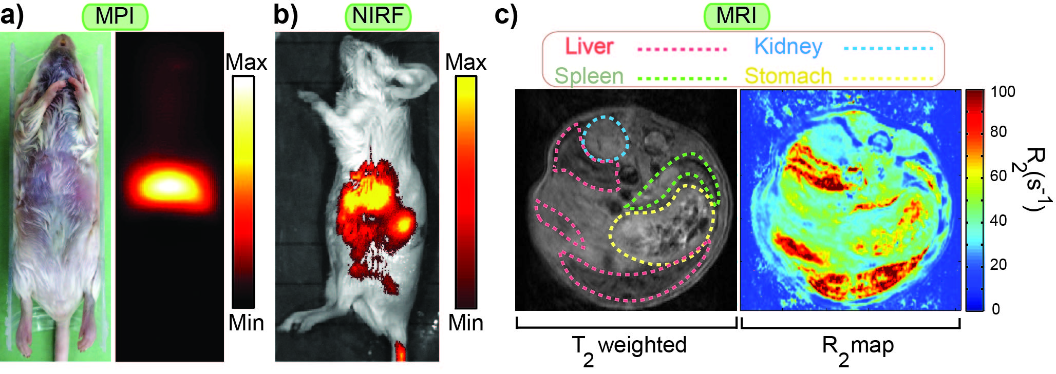
Fig.1. Multimodal MPI/NIRF/MRI imaging of the mice enabled us to map the biodistribution of the MPI tracers[5].
Conclusions: MPS and MPI enabled us to track our optimized MPI tracers in organs and targeted tissues effectively. Additional imaging modalities (NIRF and MRI) helped us to study the targeting ability, biodistribution and pharmacokinetics of the NPs in mice more accurately. These results pave the ways for future applications of MPI in pre-clinical tissue-targeted magnetic imaging and diagnostic applications.
This work was supported by NIH grants 1RO1EB013689-01/NIBIB, 2R42EB013520-02A1 and 1R41EB013520-01.
References:
[1] Bauer LM, Situ SF, Griswold MA, Samia ACS. Magnetic Particle Imaging Tracers: State-of-the-Art and Future Directions. The Journal of Physical Chemistry Letters 2015;6:2509-17.
[2] Goodwill PW, Saritas EU, Croft LR, Kim TN, Krishnan KM, Schaffer DV, et al. X-Space MPI: Magnetic Nanoparticles for Safe Medical Imaging. Advanced Materials 2012;24:3870-7.
[3] Ferguson RM, Khandhar AP, Arami H, Hua L, Hovorka O, Krishnan KM. Tailoring the magnetic and pharmacokinetic properties of iron oxide MPI tracers. Biomedical Engineering 2013;58:493-507.
[4] Arami H, Ferguson RM, Khandhar AP, Krishnan KM. Size-dependent ferrohydrodynamic relaxometry of magnetic particle imaging tracers in different environments. Medical Physics 2013;40:071904.
[5] Arami H, Khandhar AP, Tomitaka A, Yu E, Goodwill PW, Conolly SM, et al. In vivo multimodal magnetic particle imaging (MPI) with tailored magneto/optical contrast agents. Biomaterials 2015;52:251-61.
[6] Khandhar AP, Ferguson RM, Arami H, Krishnan KM. Monodisperse magnetite nanoparticle tracers for in vivo magnetic particle imaging. Biomaterials 2013;34:3837-45.
[7] Arami H, Krishnan KM. Intracellular performance of tailored nanoparticle tracers in magnetic particle imaging. Journal of Applied Physics 2014;115:17B306.
[8] Arami H, Krishnan KM. Highly Stable Amine Functionalized Iron Oxide Nanoparticles Designed for Magnetic Particle Imaging (MPI). IEEE Transactions on Magnetics 2013;49:3500-3.
[9] Hufschmid R, Arami H, Ferguson RM, Gonzales M, Teeman E, Brush LN, et al. Synthesis of phase-pure and monodisperse iron oxide nanoparticles by thermal decomposition. Nanoscale 2015;7:11142-54.
[10] Tomitaka A, Arami H, Gandhi S, Krishnan KM. Lactoferrin conjugated iron oxide nanoparticles for targeting brain glioma cells in magnetic particle imaging. Nanoscale 2015:In Press.
[11] Arami H, Khandhar A, Liggitt D, Krishnan KM. In vivo delivery, pharmacokinetics, biodistribution and toxicity of iron oxide nanoparticles. Chemical Society Reviews 2015:In Press.