Introduction: QHREDGS is a novel peptide derived from angiopoietin-1 (Ang1) and has shown generic protective effects on cardiac cells, endothelial cells, osteoblasts, and human induced pluripotent cells[1]-[6]. In this study, we examined its effect on human primary epidermal keratinocytes (HEKs) and hypothesized that QHREDGS peptide would accelerate diabetic wound healing by promoting re-epithelialization.
Materials and Methods: Low and High amount of QHREDGS peptide was conjugated to chitosan using method previously described[7]. Chitosan-collagen coating solution was prepared with 0.05 mg/ml collagen and chitosan (control, Low-peptide, High-peptide) in 0.02 N acetic acid. Culture plates were coated overnight at 4°C and rinsed before use.
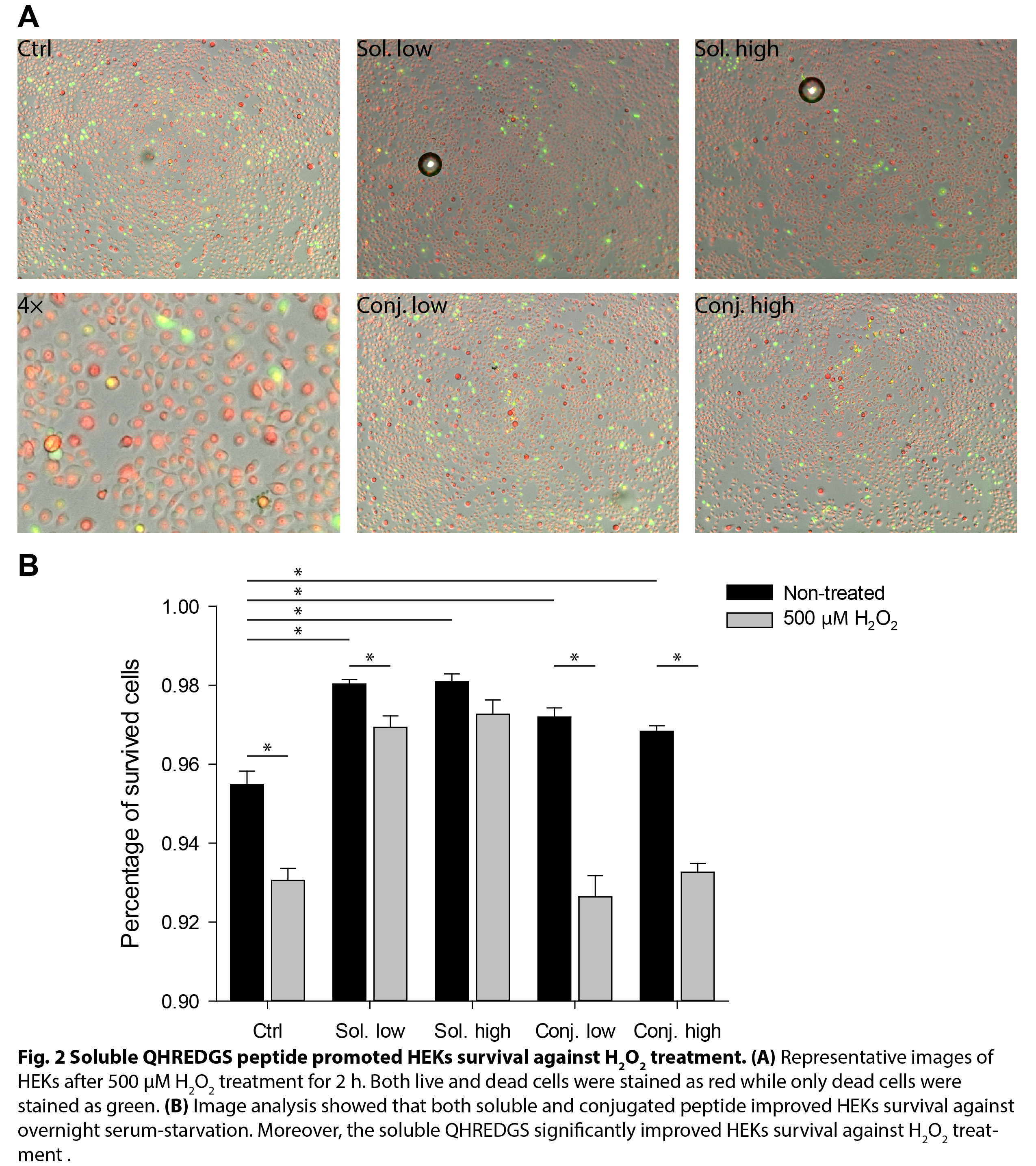
HEKs were seeded and allowed for attaching for 2 h in supplemented EpiLife medium, and then serum-starved in basal medium overnight. Apoptotic cells were detected by TUNEL assay afterwards. Alternatively, the serum-starved HEKs were treated with 500 µM H2O2 for 2 h and quantified using EarlyToxTM Cell Integrity Kit.
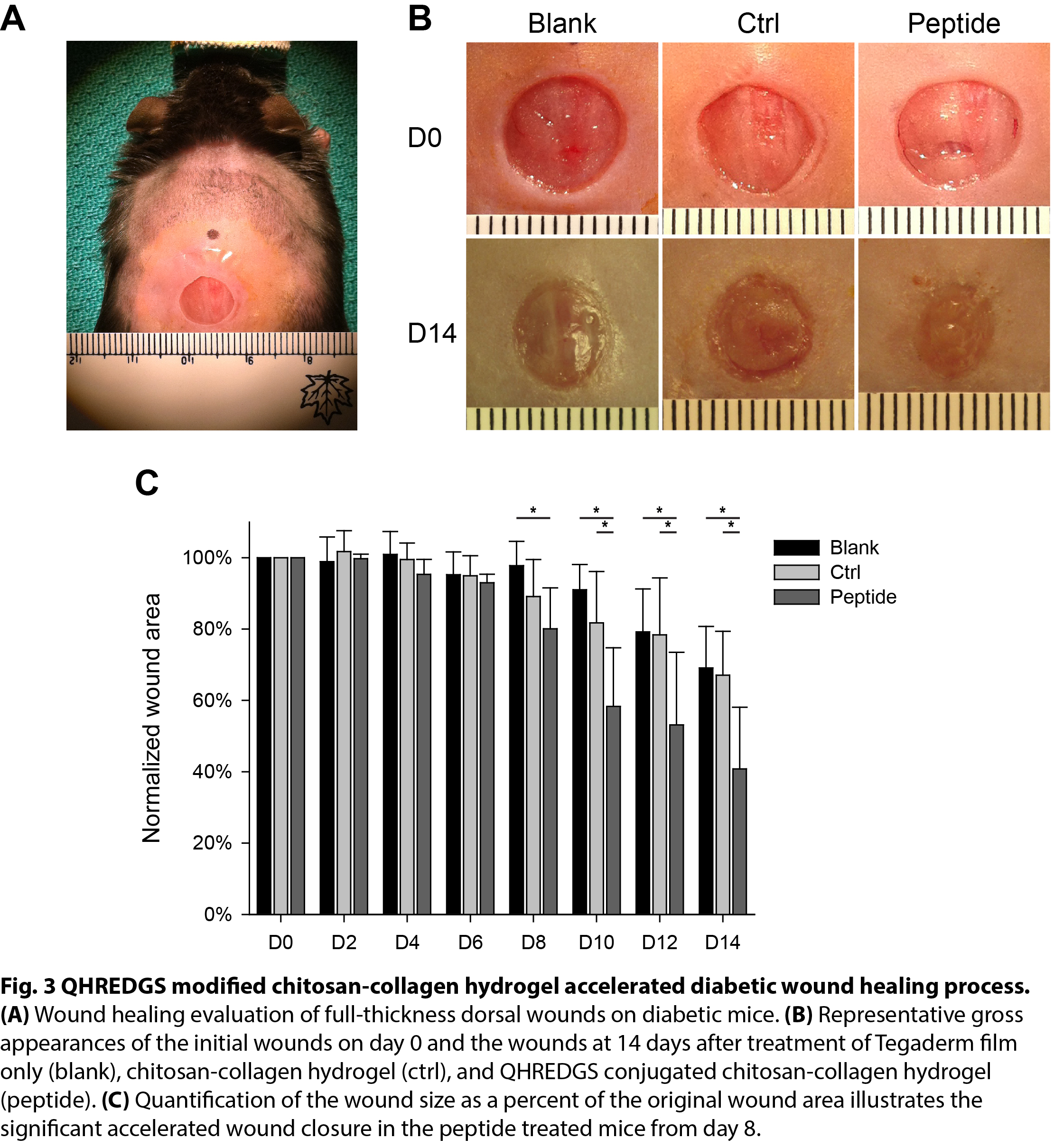
Full-thickness 8-mm wounds were created on eight weeks old, male BKS.Cg-Dock7m +/+ Leprdb/J mice (db/db). Chitosan-collagen hydrogel was prepared as previously described[7]. Either 50 µL control hydrogel without conjugated QHREDGS peptide or 50 µL hydrogel with conjugated Low-peptide was applied topically to the wound sites, or the wounds were left untreated as blank. All the wounds were then covered with TegardermTM film. Digital photographs of wounds were taken at the same distance by a digital camera every two days until the animals were sacrificed on day 14.
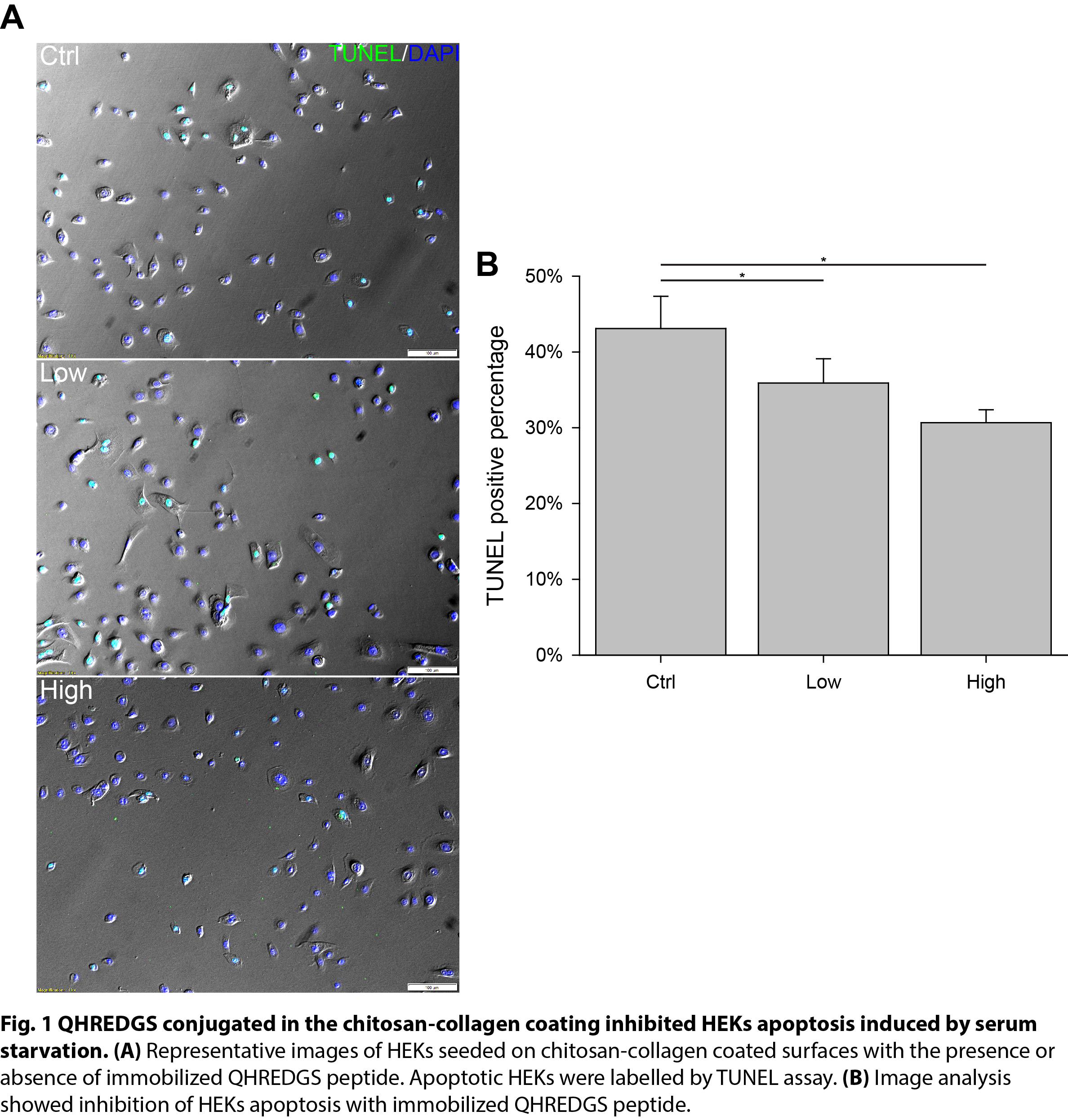
Resuts: As characterized by TUNEL assay, the presence of immobilized QHREDGS peptide in the coating inhibited HEKs apoptosis induced by serum-starvation in a dose dependent manner (Fig. 1). Moreover, HEKs survival against H2O2 stress was promoted with the presence of soluble QHREDGS peptide (Fig. 2).


The animal study showed that chitosan-collagen hydrogel with immobilized QHREDGS peptide accelerated wound closure on diabetic mice starting from day 8 as characterized (Fig. 3). The control chitosan-collagen hydrogel did not accelerate wound closure compared to the blank group.

Discussion: Ang1 has been recognized as an important regulator of vascular protection[8], cardiac remodeling[9],[10], inflammation[11], and wound healing[12]. For wound healing applications, various Ang1 derivatives have been developed due to the poor solubility of Ang1 itself. Here we decribed a water-soluble peptide, QHREDGS, which consists of seven amino acids. Because of its short sequence, the peptide can be modified using simple chemistry and has better stability.
Our results demonstrated the protective effect of QHREDGS peptide, in both soluble and immobilized form, on HEKs against serum-starvation and H2O2 stress. Nutrient depletion due to lack of vasculature and excessive reactive oxygen species are the characteristics of diabete-impaired wounds. The clinical relevance was further demonstrated on a db/db mice model, which suggested that the QHREDGS peptide is a potential candidate for diabetic wound healing therapies targeting at the epithelium.
Conclusion: The QHREDGS peptide promoted human keratinocytes survival against both serum-starvation and H2O2 stress, and accelerated diabetic wound healing by promoting re-epithelialization.
Canadian Institutes of Health Research (CIHR) Operating Grant (MOP-126027); Heart and Stoke Foundation GIA T6946; NSERC-CIHR Collaborative Health Research, Grant (CHRPJ 385981-10)
References:
[1] Rask, F., et al., Hydrogels modified with QHREDGS peptide support cardiomyocyte survival in vitro and after sub-cutaneous implantation. Soft Matter, 2010. 6(20): p. 5089-5099.
[2] Reis, L.A., et al., A peptide-modified chitosan-collagen hydrogel for cardiac cell culture and delivery. Acta biomaterialia, 2012. 8(3): p. 1022-36.
[3] Miklas, J.W., et al., QHREDGS Enhances Tube Formation, Metabolism and Survival of Endothelial Cells in Collagen-Chitosan Hydrogels. PLoS ONE, 2013. 8(8): p. e72956-e72956.
[4] Feric, N., et al., Angiopoietin-1 peptide QHREDGS promotes osteoblast differentiation, bone matrix deposition and mineralization on biomedical materials. Biomater Sci, 2014. 2(10): p. 1384-1398.
[5] Dang, L.T., et al., Inhibition of apoptosis in human induced pluripotent stem cells during expansion in a defined culture using angiopoietin-1 derived peptide QHREDGS. Biomaterials, 2014. 35(27): p. 7786-99.
[6] Reis, L.A., et al., Hydrogels With Integrin-Binding Angiopoietin-1-Derived Peptide, QHREDGS, for Treatment of Acute Myocardial Infarction. Circ Heart Fail, 2015. 8(2): p. 333-41.
[7] Xiao Y, et al., Modifications of biomaterials with immobilized growth factors or peptides for tissue engineering applications. Methods 2015;84:44-52.
[8] Brindle, N.P., et al., Signaling and functions of angiopoietin-1 in vascular protection. Circ Res, 2006. 98(8): p. 1014-23.
[9] Dallabrida, S.M., et al., Angiopoietin-1 promotes cardiac and skeletal myocyte survival through integrins. Circulation Research, 2005. 96(4).
[10] Dallabrida, S.M., et al., Integrin binding angiopoietin-1 monomers reduce cardiac hypertrophy. The FASEB Journal, 2008. 22(8): p. 3010-3023.
[11] Gamble, J.R., et al., Angiopoietin-1 is an antipermeability and anti-inflammatory agent in vitro and targets cell junctions. Circulation Research, 2000. 87(7): p. 603-607.
[12] Bitto, A., et al., Angiopoietin-1 gene transfer improves impaired wound healing in genetically diabetic mice without increasing VEGF expression. Clinical Science, 2008. 114(11-12): p. 707-718.