Introduction: Bone graft substitutes are needed to reduce or replace the reliance on autograft. Calcium phosphates (CaPs) have shown high potential for such applications[1], and further development of these materials is justified. Bioinorganics, with their natural presence in bone mineral, can be incorporated into the CaP’s crystal structure. Many promising bioinorganics have been individually identified; however, more research is needed to provide a deeper understanding of their efficacy, mechanisms of action, and possible combinatorial effects. Here, a combination of cobalt and fluoride was studied to address the major mechanisms of bone healing, being angiogenesis and osteogenesis.
Materials and Methods: CaP coatings incorporating cobalt and/or fluoride were prepared using a two-step biomimetic coating method on tissue culture plastic. Human mesenchymal stromal cells (hMSCs) were cultured in basic or osteogenic media, either on coatings, or in cobalt and/or fluoride supplemented media. At 7 and 14 days, DNA content and alkaline phosphatase (ALP) activity were measured with respective assay kits, while the expression of osteogenic or angiogenic related genes were measured using qPCR.
Results and Discussion: Cobalt added to media increased VEGF and CD31 expression at 14 days, which corresponds with previous results[2]. However, this effect was not as strongly noted when cobalt was incorporated into the CaP coatings. This may be due to the lower actual release from the coating into the media. Fluoride increased osteogenic gene expression at 14 days, aligning with previous results[3] .In combination, the higher dose of cobalt negated the positive osteogenic effect from the fluoride. Interestingly, the lower dose of cobalt provided similar angiogenic response compared to the higher cobalt dose, without strong adverse effects on the osteogenic responses promoted by the fluoride. This suggests that low doses of cobalt combined with fluoride can improve angiogenesis, while maintaining important osteogenic responses. These results demonstrate the potential benefits of combining bioinorganics; however, combinations can be difficult to manage considering the varied needs of the bone healing environment. Higher throughput methods will be needed in order to more rapidly screen combinations. Additionally, potential confounding factors need to be accounted for, and include changes in topography, toxicity doses, synergism with calcium and inorganic phosphate ions, or mechanical properties changes. A greater focus on these parameters would help elucidate mechanisms and aid future development.
Conclusion: Bioinorganics incorporated into CaPs have high potential for bone healing[4]. Fluoride appears beneficial for osteogenesis, and a low dose of cobalt has the potential to improve angiogenesis, while retaining osteogenic potential. This study supports the use of cobalt and fluoride incorporation into CaPs for use as more complete bone graft substitutes.

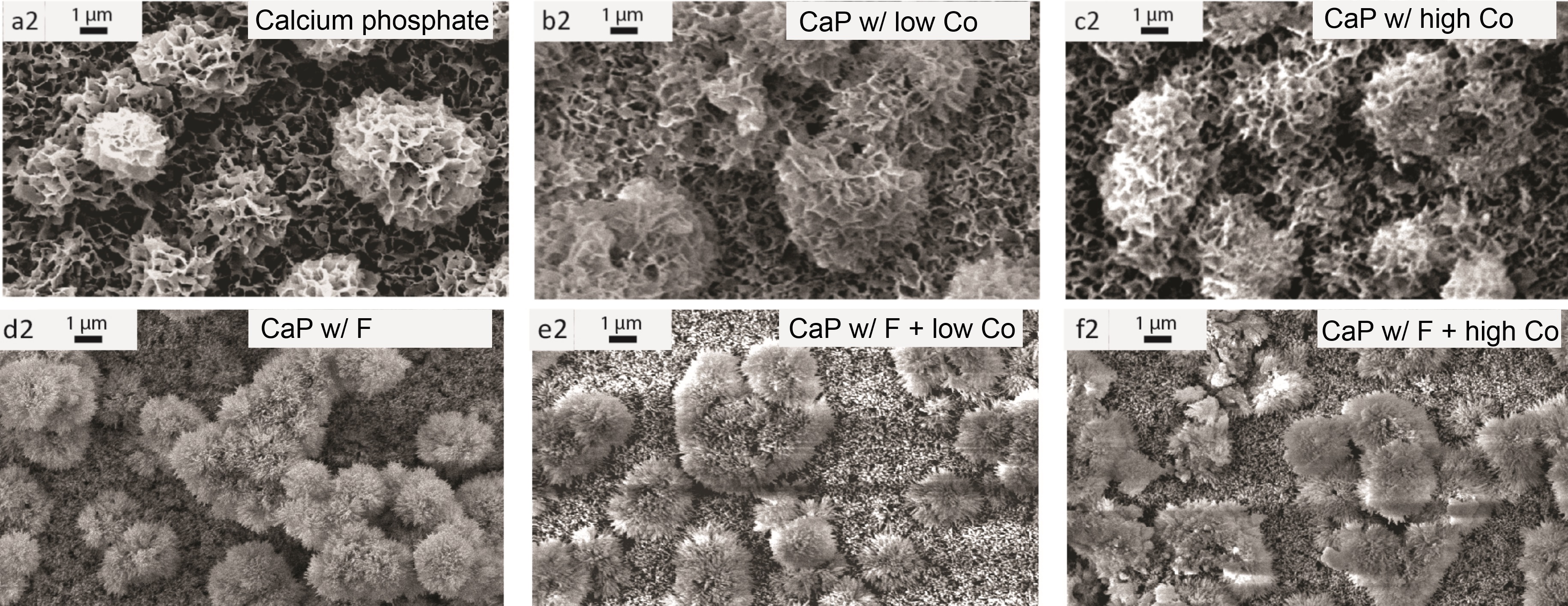
Figure 1. Calcium phosphate (CaP) coatings on the surface of tissue culture well plates. The presence of cobalt (Co) did not substantially change morphology, whereas fluoride (F) changed the crystal morphology from plate-like to rod-like structures.
This research forms part of the Project P2.04 BONE-IP of the research program of the BioMedical Materials institute, co-funded by the Dutch Ministry of Economic Affairs, Agriculture and Innovation. PH acknowledges The Netherlands Science Organization (NWO) for financial support (Aspasia premium number 015.008.039). This research has been in part made possible with the support of the Dutch Province of Limburg.
References:
[1] Bohner, M., L. Galea, and N. Doebelin, Calcium phosphate bone graft substitutes: Failures and hopes. Journal of the European Ceramic Society, 2012. 32(11): p. 2663-2671
[2] Barralet, J., et al., Angiogenesis in calcium phosphate scaffolds by inorganic copper ion release. Tissue Eng Part A, 2009. 15(7): p. 1601-9
[3] Yang, L., et al., The effects of inorganic additives to calcium phosphate on in vitro behavior of osteoblasts and osteoclasts. Biomaterials, 2010. 31(11): p. 2976-89
[4] Bose, S., et al., Understanding of dopant-induced osteogenesis and angiogenesis in calcium phosphate ceramics. Trends Biotechnol, 2013. 31(10): p. 594-605