Introduction: Bioactive glasses offer an important route for metal ions delivery systems due to their controllable dissolution within physiological fluids. Sol-gel bioactive glasses release ions to precipitate apatite inducing a strong bond with the host bone and the growth of new bone[1]. Cobalt (Co) ions are a well-established inducer of a hypoxia-like response to induce angiogenesis, which would be of great interest for applications in bone tissue engineering[2]. Co+2 in the glass network will determine the network connectivity and macroscopic properties, such as ion release rates and apatite formation[3]. A high local concentration of Co could to cause cell toxicity. Therefore, a controlled ion release rate is necessary for tissue engineering. Apatite formation and ion release from sol-gel glasses with different amounts of Co have not studied.
Materials and Methods: Sol-gel bioactive glass microparticles (58S, 60SiO2, 36CaO, 4P2O4, mol%) doped with 1, 2 y 4% of CoO were synthetized as described elsewhere[4]. Effect of the incorporation of Co on the apatite formation in simulated body fluid (SBF) was evaluated by X-ray diffraction (XRD) and scanning electron microscopy (SEM). Co+2 release until 14 days was measured using ICP-OES. Cell tests are in progress.
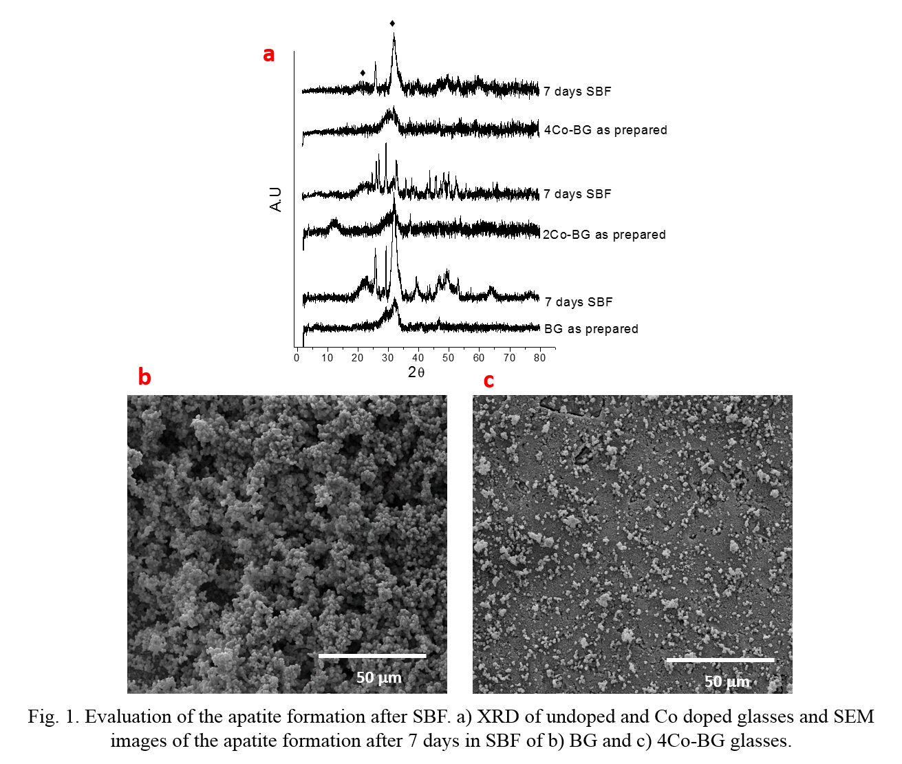
Results and Discussion: Fig. 1a shows the XRD for the apatite formation after 7 days in SBF for pure glass and Co-doped glasses. Pure glass developed the apatite peaks at 2θ=25.9 ° and 32°, showing high bioactivity. The apatite formation was diminished with the incorporation of Co, showing a greater effect to higher concentration of Co. This is due to the increase of the glass network connectivity diminishing the glass dissolution and ion release, which is necessary for the formation of apatite[5]. SEM images (Fig. 1b and c) also showed the inhibition of apatite formation by the incorporation of Co.
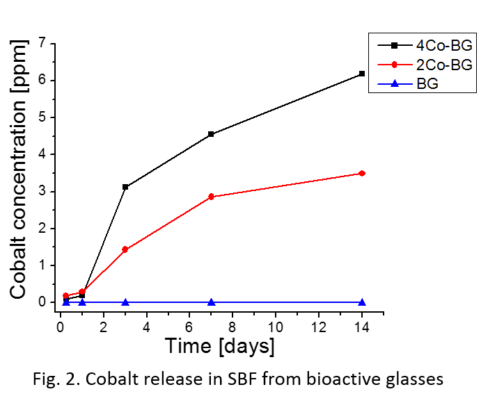
To evaluate the ion release is important to control the bioactivity and the cell behavior. Fig. 2 shows the Co+2 ion release in SBF. Cobalt doped glasses released ions with a gradual increase up to 14 days, reaching maximal cumulative values of 3.5 and 6.2 ppm for 2Co-BG and 4Co-BG samples, respectively. According to other studies these levels of ion concentration are appropriate to stimulate angiogenesis without citotoxicity.[5]
Conclusions: Cobalt doped glasses were synthetized by sol-gel method with different levels of apatite formation and metal release. The incorporation of cobalt diminished the capacity of apatite formation and the Co+2 ions were released from the glasses.
Comisión Nacional de Investigación Científica y Tecnológica (CONICYT). Project Fondecyt Nº 1110078. Government of Chile
References:
[1] Jones, J. R. Acta Biomater. 9, 4457–86 (2013).
[2] Azevedo, M. M. et al. J. Mater. Chem. 20, 8854 (2010).
[3] Smith, J. M., Martin, R. a., Cuello, G. J. & Newport, R. J. J. Mater. Chem. B 1, 1296 (2013).
[4] Bejarano, J., Caviedes, P. & Palza, H. Biomed. Mater. 10, 025001 (2015).
[5] Azevedo, M. M. et al. J. Mater. Chem. 20, 8854 (2010).