Introduction: Rapid and controlled vascularization of engineered tissues remains one of the key limitations in tissue engineering applications[1],[2]. While vascular endothelial growth factor (VEGF) which regards as an angiogenic regulator holds greatest potential to promote angiogenesis[3]. However, soluble VEGF will be quickly degraded by enzymes in vivo and clinical applications with high dose of VEGF are often associated with severe side effects[4],[5]. Thus, reducing the dosage of VEGF and enhancing its activity becomes particularly important. 2-N,6-O-sulfated chitosan (26SCS) as a synthetic sulfated polysaccharide has been demonstrated that it could efficiently associate with VEGF to promote angiogenesis in vitro during our previous work[6]. Nevertheless, the mechanism of the interaction between 26SCS and VEGF is not clear. This study aimed at the synergy of 26SCS with VEGF as well as its pro-angiogenesis in vitro and in vivo.
Materials and Methods: Human umbilical vein endothelial cells (HUVECs) which purchased from the American Type Culture Collection was used as a cell model. A series concentrations of 26SCS was used to characterize the interaction with VEGF (2 ng/mL) on cell viability. Subsequently, two experimental groups which named as 40 SCS/VEGF (26SCS, 40 ng/mL) and 160 SCS/VEGF (26SCS, 160 ng/mL) respectively were chosen to investigate the cell migration, sprouting and qRT-PCR analysis in vitro.
Furthermore, we employed the corresponding concentration ratio of 26SCS and VEGF to further investigate the pro-angiogenesis activity in vivo. Subcutaneous implantation of mice (VEGF, 100 ng) and chicken chorioallantoic membrane (CAM) assay (VEGF, 20 ng) were used as experimental models, respectively.
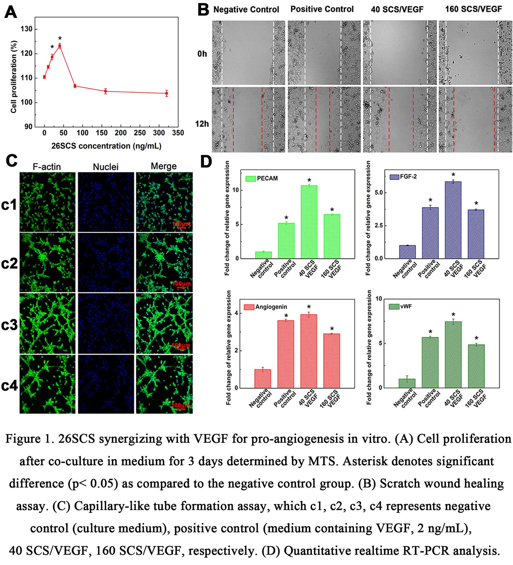
Results and Discussion: The pro-angiogenesis of 26SCS synergizing with VEGF was found and mainly studied. As shown in Figure 1, 26SCS synergizing with VEGF could directly improve HUVECs cell proliferation through dose-dependently regulating the expression of relevant angiogenic genes, and the group of 40 SCS/VEGF exhibited optimal results. Moreover, the repaired area of 40 SCS/VEGF was almost 1.5 folds compared to negative control in scratch wound healing assay, and 40 SCS/VEGF generated more branch points and forming tubes in capillary-like tube formation assay. In addition, using gelatin sponge loaded with VEGF and different content of 26SCS as samples implanted in subcutaneous model of mice, we investigated the influence of 26SCS and VEGF facilitate vascularization in vivo at day 7 (Figure 2A). The results of histological analysis showed that 26SCS and VEGF could significantly promote the repair rate of vascular network at the defect site (Figure 2B). Through the CAM assay by implanting filter paper containing different ratios of 26SCS and VEGF compounds into fertilized eggs (Figure 2C), the results further revealed that 26SCS synergizing VEGF could signally promote angiogenesis.

Conclusions: In this study, 26SCS was demonstrated that synergizing with VEGF could significantly promote angiogenesis in vitro and in vivo. This findings evidence that 26SCS have the potential to improve the VEGF activity and will be promising prospects as a synergistic factor of VEGF for vascularization in clinical applications.
The National Basic Research Program of China (973 Program, No. 2012CB933600); The National Natural Science Foundation of China (Grant Nos. 31271011, 31330028, and 31470923); The New Century Excellent Talents in University (Grant No. NCET-12-0856); The National Science and Technology Support Program (Grant No. 2012BAI17B02)
References:
[1] Tonnesen MG, Feng X, Clark RA. Angiogenesis in wound healing. Journal of Investigative Dermatology Symposium Proceedings: Nature Publishing Group; 2000. p. 40-6.
[2] Del Gaudio C, Baiguera S, Boieri M, Mazzanti B, Ribatti D, Bianco A, et al. Induction of angiogenesis using VEGF releasing genipin-crosslinked electrospun gelatin mats. Biomaterials. 2013;34:7754-65.
[3] Ferrara N, Chen H, Davis-Smyth T, Gerber H-P, Nguyen T-N, Peers D, et al. Vascular endothelial growth factor is essential for corpus luteum angiogenesis. Nature Medicine. 1998;4:336-40.
[4] Tayalia P, Mooney DJ. Controlled growth factor delivery for tissue engineering. Advanced Materials. 2009;21:3269-85.
[5] Chung Y-I, Kim S-K, Lee Y-K, Park S-J, Cho K-O, Yuk SH, et al. Efficient revascularization by VEGF administration via heparin-functionalized nanoparticle-fibrin complex. Journal of Controlled Release. 2010;143:282-9.
[6] Yu Y, Chen J, Chen R, Cao L, Tang W, Lin D, et al. Enhancement of VEGF-Mediated Angiogenesis by 2-N, 6-O-Sulfated Chitosan-Coated Hierarchical PLGA Scaffolds. ACS Applied Materials & Interfaces. 2015;7:9982-90.