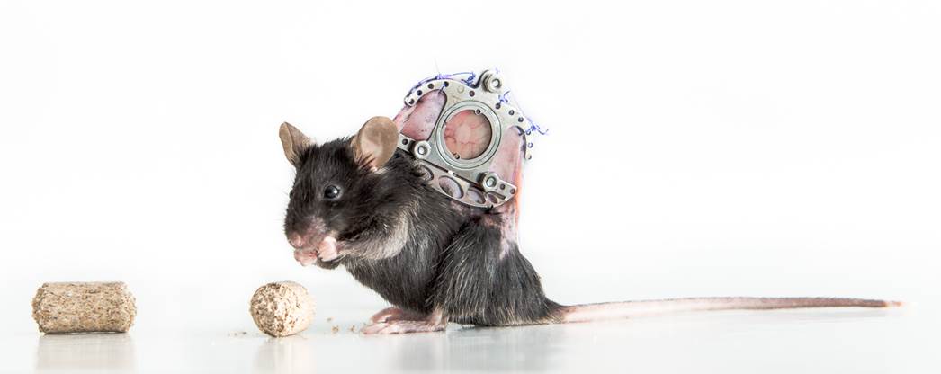
The Dorsal Skin Fold Chamber (DSFC) model is a useful tool that allows in vivo monitoring of vascularization of biomaterials.
Angiogenesis is indispensable in wound healing. Delayed wound healing is associated with impaired new blood vessel formation, which calls for the development of pro-angiogenic wound dressings. We have recently shown that poly(ethylene glycol) (starPEG)-heparin hydrogels can deliver the angiogenic factors FGF-2 and VEGF and encourage angiogenesis in a CAM assay[1]. However, whether these hydrogels promote angiogenesis during wound healing and improve the wound healing outcome needs to be shown. We have therefore established the DSFC as a wound healing model that allows real-time in vivo imaging of angiogenesis and a dynamic characterization of inflammation and re-epithelialization.
Dorsal skin of C57BL6 mice was shaved and depilated prior to DSFC implantation. Within the microscopy window, a 5 mm full-thickness wound was created. The chamber was filled with NaCl and closed with a transparent coverslip. At day 1, exudate was gently removed to receive a clear observation window for intravital microscopy (IVM).
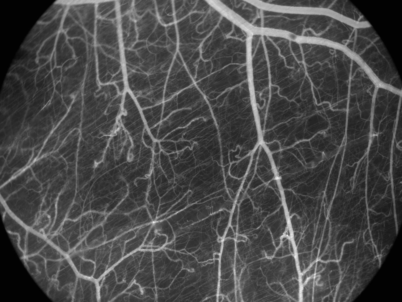
A starPEG-heparin hydrogel was applied onto the wound and the chamber was closed again. IVM was performed on day 1, 3, 6, 8, 10, 13, and 19 after wounding using FITC/dextran (i.v. injection) to visualize vessels in the wound area. Angiogenesis was analyzed with ZEN software (Zeiss), measuring the functional vessel density (defined as total blood vessel length per area) in the wound and wound edges. To correlate angiogenic events with the wound healing outcome inflammation, granulation tissue formation and re-epithelialization were analyzed in wound biopsies post mortem in the same kinetic as IVM using qRT-PCR and immunohistochemistry.
Wound healing in the DSFC is delayed as seen by a late wound closure at day 13. The DSFC impairs the contraction of the M. panniculus carnosus that mediates murine wound closure. Instead, wounds close with new tissue formed by proliferating myofibroblasts and keratinocytes. Delayed wound healing in the DSFC is characterized by prolonged inflammation with a slow increase on infiltrating immune cells, reaching a peak at day 13 and resolving till day 19. Using IVM, angiogenesis could be observed during the whole study. New capillaries resulted in formation of tubular-structured blood vessels. The application of hydrogels onto wounds does not impair the wound healing process.
We have adapted the DSFC as a useful model for standardized testing of angiogenic biomaterials during wound healing. Advantages of this model imply in vivo imaging of angiogenesis up to 3 weeks in a human-like wound healing model without muscle induced wound contraction that allows direct correlation of angiogenic effects with wound healing events. We are currently analyzing the impact of star-PEG-heparin hydrogels loaded with pro-angiogenic factors on angiogenesis and wound healing outcome using our DSFC wound healing model.
References:
[1] Zieris A. et al., J. Control Release 156:28-36, 2011