Introduction: The native extracellular matrix (ECM) has a complex composition. Tissue engineering matrices need to possess multiple bioactive signaling molecules in order to adequately mimic the ECM. In the central nervous system, hyaluronic acid (HA) is a regulator of cellular migration, proliferation, differentiation, and angiogenesis. In these experiments, a di-functional HA (diHA), which possesses thiol and azide groups for tethering of bioactive molecules at independent concentrations, is characterized to and initial biological testing is conducted on the unfunctionalized materials using mouse embryonic stem cells (mES).
Material and Methods: 60,000 kDa HA was functionalized to create diHA and methacrylated HA (mHA). The thiol group on di-HA was characterized using 1H-NMR, and 2,4,6-trinitrobenzene sulfonic acid (TNBS) assay. The azide group was characterized using 13C-NMR, Fourier transform infrared spectroscopy (FT-IR) spectrum, and radioTLC of 68Ga chelating agent binding. Photopolymerized hydrogels of diHA and mHA of varying compositions ratios were fabricated using Irgacure 2959 and 2.3 mJ cm-2 UVA light for 5 min. The swelling ratio, water contents, and mechanical properties (young’s and shear modulus) of 1:2 (diHA: mHA) and 1:5 (diHA: mHA) hydrogels was determined. D3 mES were encapsulated in the HA hydrogel at a density of 1 X 10⁶ cells/mL. mES in 3D HA hydrogels were examined proliferation and differentiation potential by MTT assay and b-tubulin III (TUJ1) staining, respectively.
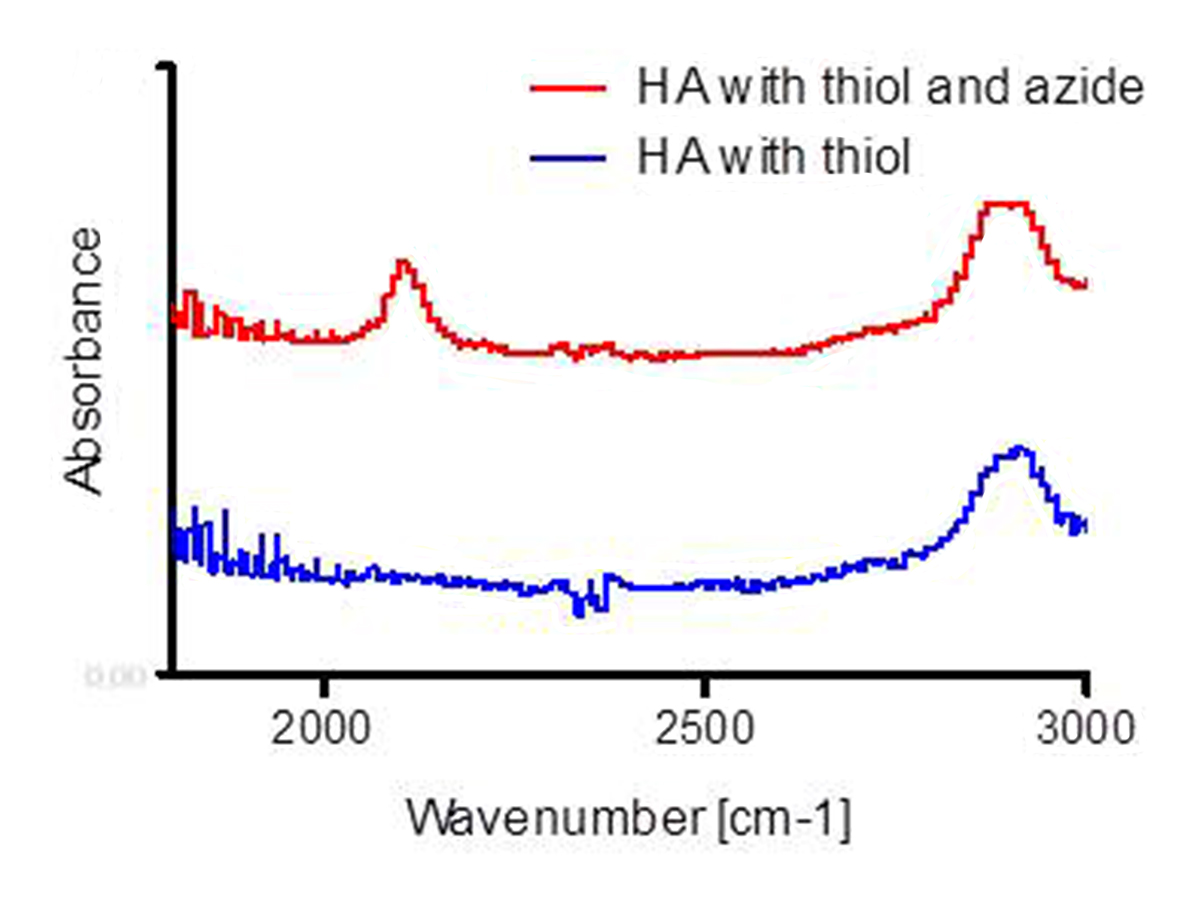
Results Discussion: On 1H-NMR, the thiol peak was observed at 2.9 ppm for thiol functionalized samples with and without azide functionalization. A positive staining for thiol was also observed by TNBS assay for these sample groups. An azide peak was observed at 2096 cm-1 on the FT-IR spectrum (Figure 1), and at 50.1 ppm on the 13C-NMR spectrum for only HA samples functionalized with both thiol and azide groups.
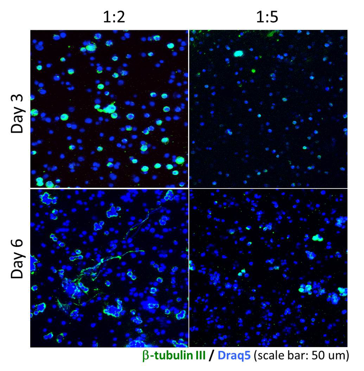
The average number of chelating moieties capping the azide group in HA was 3.9 ± 0.09 on radio TLC for diHA samples. A higher ratio of mHA (1:5) increased the young’s modulus, shear modulus, swelling ratio, and water content, significantly. Encapsulated mES were proliferated on 1:2 and 1:5 ratio of diHA:mHA hydrogel. After 3 and 6 days of differentiation, 44%±7% and55%±12% of mES in 1:2 (diHA: mHA) hydrogels expressed TUJ1, respectively. While mES in 1:5 (diHA: mHA) hydrogels expressed 39%±20% and 40%±14% after 3 and 6 days of differentiation, respectively (Figure 2). mES cultured in 1:2 (diHA: mHA) showed mature neural extension on day 6 of differentiation.
Conclusion: These results indicate that thiol and azide groups were present on the modified HA backbone. Encapsulated mES were able to proliferate and differentiate in base matrix without additional bioactive signaling. diHA is a valuable tool to enhance bioactivity and control of bioactive signal concentrations in HA matrices developed to treat central nervous system injuries.
The Bentsen Stoke Center University of Texas Medical School at Houston; William Stamp Farish Fund