Injectable hydrogel consisting of bisphosphonate-functionalized hyaluronan and doxorubicin@magnesium silicate nanoparticles for anti-cancer therapy
-
1
Uppsala University, Chemistry-Ångström, Sweden
Introduction: Hydrogels are promising drug delivery system (DDS) for localized anti-cancer therapy. Non-invasive administration of drug-loaded hydrogels implies injectability of such hydrogels which can be more reliably achieved with self-healing networks made from reversible bonds. In this study, we report on a novel hydrogel material cross-linked through metal-ligand coordination bonds between magnesium silicate (MgSiO3) nanoparticles and bisphosphonate-functionalized hyaluronan (HA-BP) biopolymer. Mixing-triggered gel formation, self-healing properties of the material as well as recognition of HA by cancer cells through HA-CD44 receptor binding makes this hydrogel a promising DDS for cancer therapy.

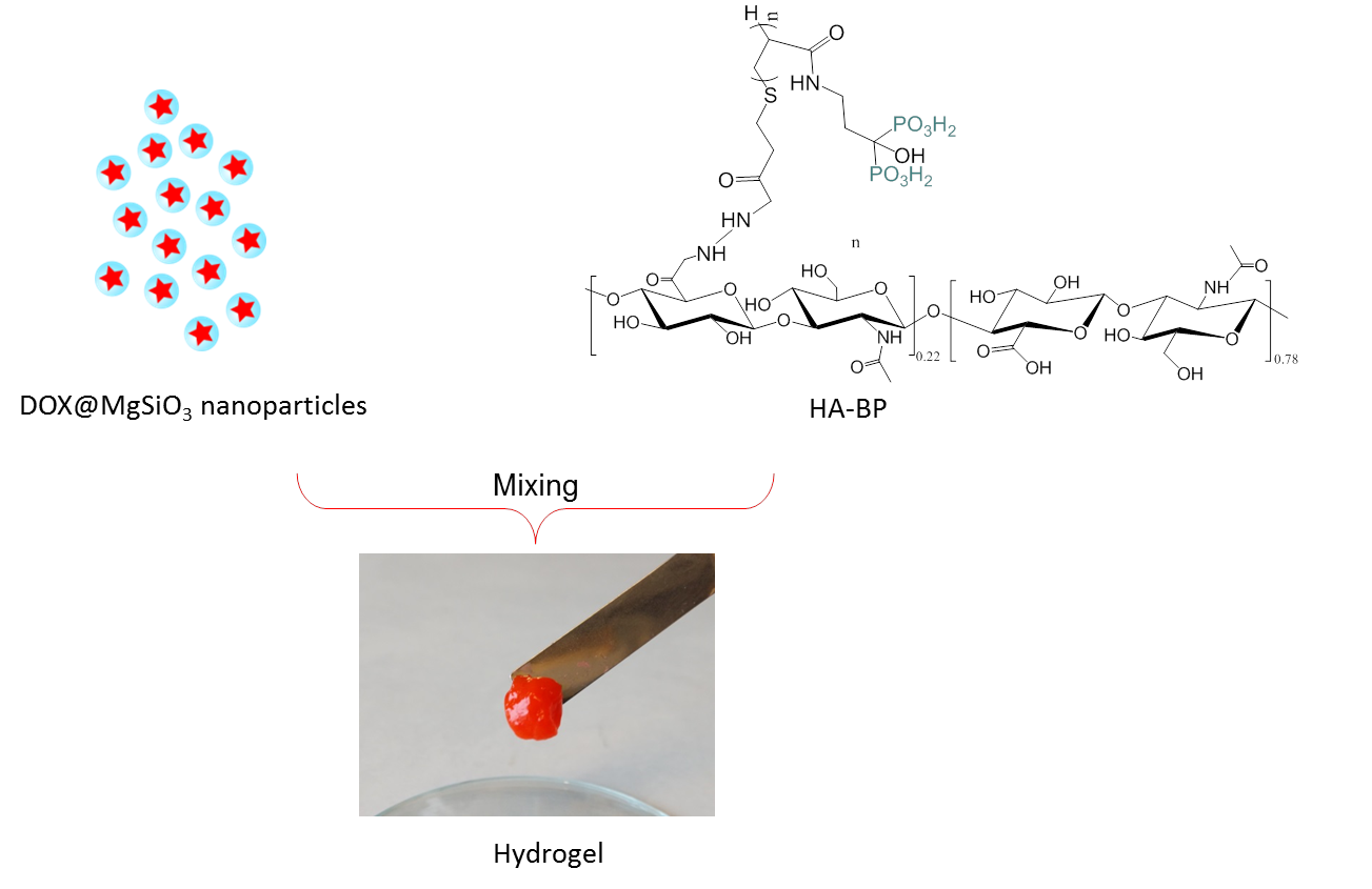
Materials and Methods: MgSiO3 nanoparticles were prepared from silica nanoparticles by hydrothermal treatment of SiO2 with ammonia in the presence of MgCl2[1]. The resulting MgSiO3 nanoparticles were loaded with Doxorubicin (Dox) by incubation in the drug solution at room temperature. The drug-loaded MgSiO3 nanoparticles (DOX@MgSiO3) were purified from the free drug by centrifugation at 4000 rpm. Hyaluronan (HA) was functionalized with bisphosphonate groups through thiol-ene photoreaction between thiolated-hyaluronan and acrylated bisphosphonate according to our previous report[2]. Hydrogel was formed by mixing equal volumes of 4% (w/v) HA-BP solution and 12% (w/v) dispersion of DOX@MgSiO3 nanoparticles in PBS buffer. Hydrogel was incubated with 3 mL of PBS at different pH (5.0, 6.5 and 7.4). At determined time points, PBS buffer was collected and refreshed with the fresh one. Fluorescence emission spectrum was recorded (490 nm excitation wavelength) to obtain a release profile of Dox. Solutions with the released Dox were applied to MCF-7 cells to examine cytotoxicity using MTS method.
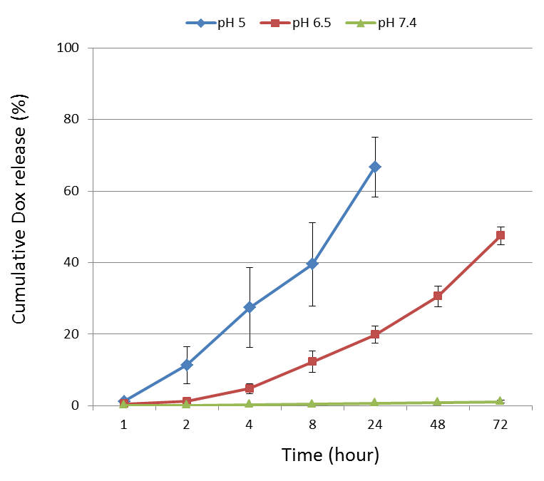
Results and Discussion: MgSiO3 nanoparticles were 350nm in diameter and had mesoporous structure. We found that BP groups of HA-BP and Mg2+ ions on the surface of MgSiO3 nanoparticles were two key factors to form hydrogel via coordination bonds. DOX release profile was pH-dependent, showing 67% Dox released already after 24 hours of incubation under pH 5.0 and 47% Dox released after 72 hours of incubation at pH 6.5. Almost no release was detected at neutral pH environment. This result can be explained by protonation of BP groups and subsequent braking of coordination bonds with Mg2+ ions. This leads to the disassembly of the polymer-nanoparticles network. Our observation that no Dox was released under neutral conditions pointed out that the drug could be only released from the nanoparticles after their liberation from the network. Additionally, faster Dox release was observed at more acidic conditions in support to our suggested mechanism of the network formation and disassembly. In vitro cytotoxicity experiments showed 20% viable MCF-7 cells after treatment with the release solutions. Importantly, this level of cytotoxicity was maintained over 3 days for the Dox-loaded hydrogel while the control group of Dox-free hydrogel showed more than 80% viability of MCF-7 cells.
Conclusion: We have developed a new DDS based on reversible and pH dependent hydrogel formation between HA-BP and MgSiO3 nanoparticles which is useful in anti-cancer therapy.

This work was supported by the European Community’s Seventh Framework Programme (Biodesign project) and Chinese Scholarship Council
References:
[1] Wang B, Meng W, Bi M, ect. Dalton Trans. 2013, 42(24):8918-8925.
[2] Nejadnik MR, Yang X, Bongio M, ect. Biomaterials, 2014 , 35(25):6918-6929.
Keywords:
Hydrogel,
Drug delivery,
composite,
Environmental response
Conference:
10th World Biomaterials Congress, Montréal, Canada, 17 May - 22 May, 2016.
Presentation Type:
New Frontier Oral
Topic:
Biomaterials for therapeutic delivery
Citation:
Shi
L,
Hiborn
J and
Ossipov
D
(2016). Injectable hydrogel consisting of bisphosphonate-functionalized hyaluronan and doxorubicin@magnesium silicate nanoparticles for anti-cancer therapy.
Front. Bioeng. Biotechnol.
Conference Abstract:
10th World Biomaterials Congress.
doi: 10.3389/conf.FBIOE.2016.01.01460
Copyright:
The abstracts in this collection have not been subject to any Frontiers peer review or checks, and are not endorsed by Frontiers.
They are made available through the Frontiers publishing platform as a service to conference organizers and presenters.
The copyright in the individual abstracts is owned by the author of each abstract or his/her employer unless otherwise stated.
Each abstract, as well as the collection of abstracts, are published under a Creative Commons CC-BY 4.0 (attribution) licence (https://creativecommons.org/licenses/by/4.0/) and may thus be reproduced, translated, adapted and be the subject of derivative works provided the authors and Frontiers are attributed.
For Frontiers’ terms and conditions please see https://www.frontiersin.org/legal/terms-and-conditions.
Received:
27 Mar 2016;
Published Online:
30 Mar 2016.
*
Correspondence:
Dr. Liyang Shi, Uppsala University, Chemistry-Ångström, Uppsala, Sweden, Email1
Dr. Dmitri A Ossipov, Uppsala University, Chemistry-Ångström, Uppsala, Sweden, dmitri.ossipov@kemi.uu.se