Super resolution imaging of supramolecular biomaterials: Shining a light on biomaterials in vitro and in cells
-
1
Institute for Complex Molecular Systems, Netherlands
-
2
University of Ghent, Belgium
-
3
Institute for Bionegineering of Catalonia, Spain
The use of nanocarriers for intracellular delivery of therapeutic moieties is a key application in the field of biomaterials. Supramolecular materials, i.e. materials based on self-assembly, such as micells, liposomes self-assembled nanoparticles and nanofibers plays a pivotal role in this framework. A crucial factor limiting the design of effective materials is the lack of understanding about material-cell interactions that hampers the rational design of nanosized carriers. This is particularly relevant for supramolecular materials as their complex structure pose several unanswered questions.
Here we discuss the use of super resolution microscopy to image materials in vitro and in mammalian cells. This novel technique had a dramatic impact in cell biology, however its use in the field of biomaterials is poorly explored. Super resolution microscopy offers nanometric resolution (down to 20nm compared to the 250-300nm of a confocal microscope) and multicolor ability; therefore it is an ideal tool to study nano-sized supramolecular assemblies of multiple components in vitro and in cells, unveiling materials behavior that was impossible to study before due to lack of suitable techniques.
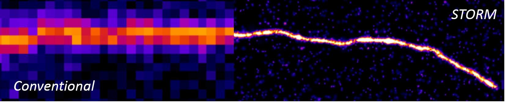
We employed Stochastic Optical Reconstruction Microscopy (STORM) to image biomaterials, with special emphasis on supramolecular polymers, in vitro unveiling novel information on materials structure and dynamics, a key issue of supramolecular materials[1] (See Figure below).
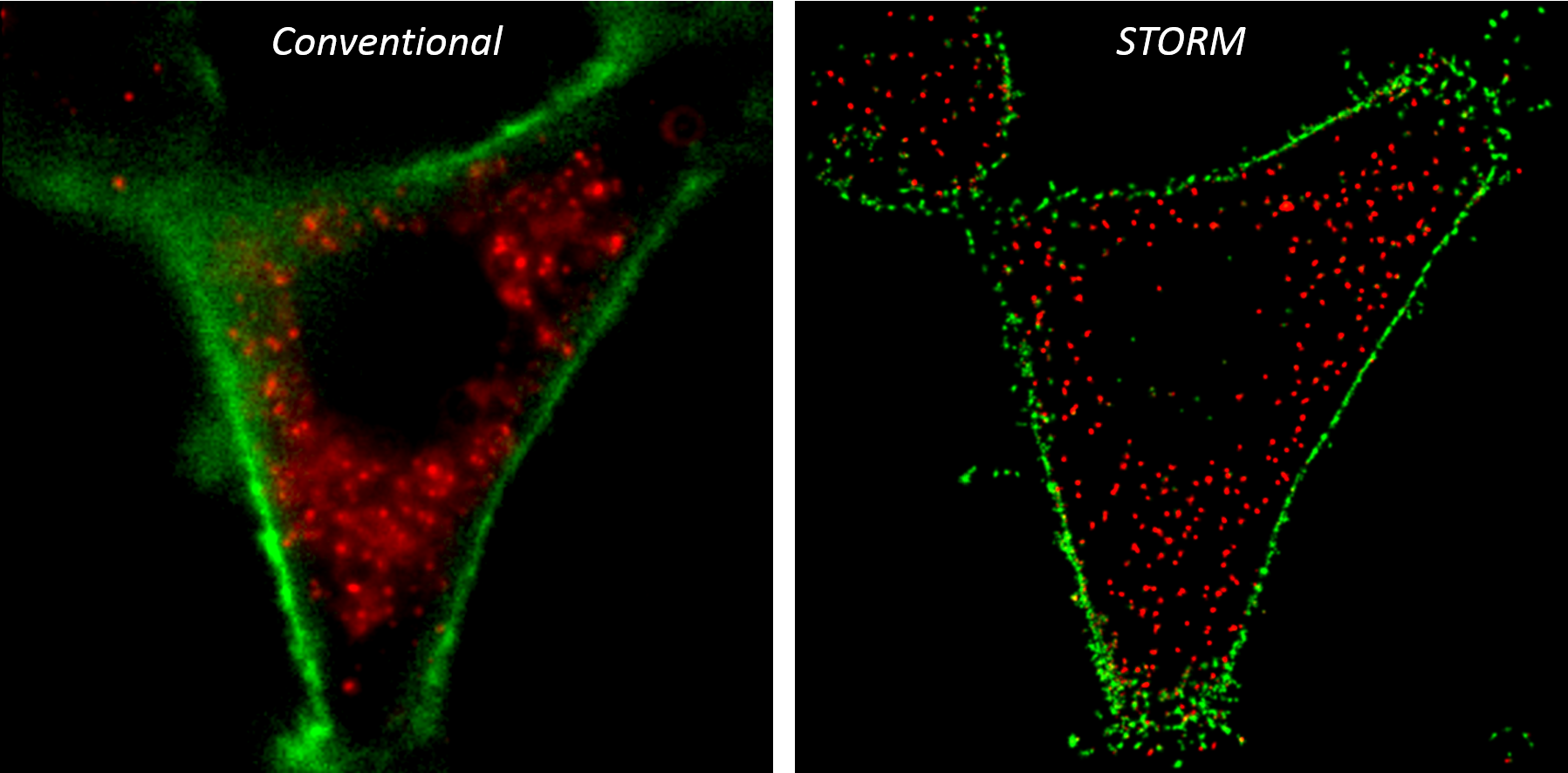
Moreover we propose a methodology to image nano-sized materials in cells, tracking them during their membrane targeting, cell uptake and intracellular targeting. We show how 2-color STORM can be used to perform nanometric-accurate colocalization, unveiling materials-cell interactions at the molecular level (see Figure below for a comparison between storm and conventional microscopy)[2].
The use of the information obtained thanks to these novel microscopy methodologies for the “STORM-guided” design of novel biomaterials will be discussed.
References:
[1] Albertazzi et al, Science (2014)
[2] Albertazzi et al, Submitted
Keywords:
Drug delivery,
nanofiber,
nanoparticle,
Imaging method
Conference:
10th World Biomaterials Congress, Montréal, Canada, 17 May - 22 May, 2016.
Presentation Type:
New Frontier Oral
Topic:
Biomaterials for therapeutic delivery
Citation:
Van Der Zwaag
D,
Wijnands
S,
Vanparijs
N,
Meijer
E,
De Geest
BG and
Albertazzi
L
(2016). Super resolution imaging of supramolecular biomaterials: Shining a light on biomaterials in vitro and in cells.
Front. Bioeng. Biotechnol.
Conference Abstract:
10th World Biomaterials Congress.
doi: 10.3389/conf.FBIOE.2016.01.01367
Copyright:
The abstracts in this collection have not been subject to any Frontiers peer review or checks, and are not endorsed by Frontiers.
They are made available through the Frontiers publishing platform as a service to conference organizers and presenters.
The copyright in the individual abstracts is owned by the author of each abstract or his/her employer unless otherwise stated.
Each abstract, as well as the collection of abstracts, are published under a Creative Commons CC-BY 4.0 (attribution) licence (https://creativecommons.org/licenses/by/4.0/) and may thus be reproduced, translated, adapted and be the subject of derivative works provided the authors and Frontiers are attributed.
For Frontiers’ terms and conditions please see https://www.frontiersin.org/legal/terms-and-conditions.
Received:
27 Mar 2016;
Published Online:
30 Mar 2016.
*
Correspondence:
Dr. Ew Meijer, Institute for Complex Molecular Systems, Eindhoven, Netherlands, e.w.meijer@tue.nl